Sarcoidosis is a systemic infiltrative disease characterized by non-caseating granuloma formation, and its cardiac phenotype is an important prognostic factor.1 Sarcoidosis affecting the heart without extracardiac involvement is termed isolated cardiac sarcoidosis (CS). Isolated CS was once thought to be a rare manifestation of sarcoidosis because of the diagnostic challenge arising from the low sensitivity of endomyocardial biopsy (EMB).2 Considering that the importance of early diagnosis and treatment are critical in managing CS,3 the Japanese Circulation Society (JCS) updated its guidelines to allow a clinical diagnosis of CS using multimodal imaging techniques, such as fluorine-18 fluorodeoxyglucose positron emission tomography (FDG-PET), even in the absence of histological evidence of non-caseating granulomas.4 Under the situation in which isolated CS can be clinically diagnosed without histological findings, we should pay more attention to differentiating isolated CS from other cardiomyopathies. Here, we describe a 50-year-old male who was clinically diagnosed with isolated CS and prescribed with prednisolone without much effect, and subsequent genetic analysis revealed pathogenic variants in LMNA and TNNT2, representative genes for cardiomyopathy. A 48-year-old man with hypertension and dyslipidaemia presented to a hospital with advanced atrioventricular block (AVB) pointed out during his annual health check-up. He had no family history of heart disease or sudden death, and his electrocardiogram (ECG) 2 years earlier showed a first-degree AVB and left axis deviation (Figure 1A). ECG in the first hospital presentation shows high-degree AVB and multiple morphologically distinct premature ventricular contractions (Figure 1B), and ambulatory ECG monitoring revealed advanced AVB with the longest pause being 1.57 seconds accompanied by escape rhythms. Transthoracic echocardiography (TTE) demonstrated normal cardiac chamber size and function without any regional wall motion abnormalities. He remained asymptomatic and declined further investigation or regular outpatient follow-up. At the age of 50, he complained of mild dyspnoea on exertion, categorized as New York Heart Association Class II, and was diagnosed with atrial fibrillation and complete AVB and referred to our hospital (Figure 1C). He started receiving edoxaban 60 mg/day. Blood test revealed elevated levels of B-type natriuretic peptide at 290.5 pg/mL and high-sensitivity cardiac troponin I at 30.9 pg/mL. Soluble interleukin-2 receptor and angiotensin-converting enzyme levels were within normal ranges. His chest X-ray indicated cardiomegaly. TTE showed a reduced left ventricular ejection fraction of 38% and hypokinesis of the septal and posterior walls of the left ventricle, without regional wall thinning or ventricular aneurysms (Figure 2A and Video S1). Non-contrast computed tomography did not depict any enlarged lymph nodes, pulmonary nodules or other organ abnormalities, and ophthalmologic and dermatologic examinations were unremarkable. Ambulatory ECG detected non-sustained ventricular tachycardia (NSVT). Coronary computed tomographic angiography showed intact coronary arteries. Cardiac magnetic resonance imaging demonstrated a linear pattern of late gadolinium enhancement (LGE) in the mid-wall of the septum, with a reduced ejection fraction and a dilated left ventricle (Figure 2B and Videos S2 and S3). T2-weighted imaging revealed a high signal in the mid-wall of the septum, corresponding to the region of LGE, indicative of myocardial inflammation (Figure S1). Subsequently, a whole-body FDG-PET scan after 18 h of fasting showed a focal FDG uptake in the ventricular septum, typical pattern for CS, but no uptake in other organs (Figure 2C). The multimodal imaging findings and the presence of arrhythmia such as AVB and NSVT were typical for CS, and we clinically diagnosed the patient with isolated CS without performing EMB, as the patient met criteria (a), (c), (d) and (e) in the JCS guideline4 (Table 1). Following his admission, we initiated prednisolone 30 mg (0.5 mg/kg)/day, but continuous ECG monitoring showed frequent NSVTs with multiple morphologies (Figure 2D). The heart team concluded that semi-urgent defibrillator implantation was necessary, and he received a cardiac resynchronization therapy device with defibrillator (CRT-D) while taking prednisolone 25 mg daily and was discharged 7 days after the implantation. However, 10 days after discharge, the wound was partly detached and bleeding, forcing the patient to be readmitted to the hospital. He was treated with prophylactic antibiotics and surgical site compression for 7 days and was discharged with prednisolone 15 mg/day. Despite the initiation of prednisolone therapy, his symptoms and cardiac dysfunction did not improve much, and NSVTs continued to occur. We performed whole-exome sequencing analysis to evaluate the risk of inherited cardiomyopathies and identified two missense variants in TNNT2 (chr1:201364335(hg38), NM_001001430.3, c.422G>A, p.Arg141Gln) and LMNA (chr1:156136096(hg38), NM_170707.4, c.1132A>C, p.Lys378Gln) (Figure 3). The TNNT2 variant has been previously reported as pathogenic in multiple patients with idiopathic cardiomyopathies (ClinVar accession number: VCV000043637.14). In contrast, the LMNA variant is novel and located in exon 6, a known hot-spot region. This region corresponds to part of the coil 2 domain within the central rod domain of lamin A/C and is related to multiple pathogenic missense variants.5 This variant has not been identified in the general population according to the gnomAD and Tohoku Medical Megabank databases and was predicted to be pathogenic by multiple in silico analyses with a Combined Annotation Dependent Depletion score of 26.6 and a Polymorphism Phenotyping v2 score of 0.999. According to the American College of Medical Genetics and Genomics guidelines,6 LMNA p.Lys378Gln was classified as likely pathogenic (PM1, PM2, PP2 and PP3). Therefore, we hypothesized that these variants were involved in the progressive conduction defects and worsened cardiac function. We describe a patient initially diagnosed with isolated CS using a multimodality imaging approach and treated with prednisolone. He had not improved with steroid therapy and was later found to have two likely pathogenic variants in cardiomyopathy-causing LMNA and TNNT2 genes. Although the possibility of an overlap of CS and genetic cardiomyopathy cannot be excluded, this case underscores the importance of early genetic analysis to reconsider an optimal therapeutic strategy for treatment-resistant patients. As patients with CS are at high risk of malignant arrhythmias,7 delays in diagnosis and intervention might worsen the patient's prognosis.3 Despite this, the diagnosis of isolated CS, characterized by the absence of extracardiac involvement,8 is basically challenging due to the low sensitivity of EMB, which is only up to 20%.2 This difficulty had been hindering early and appropriate management of isolated CS. To address this issue, the JCS guidelines proposed clinical diagnostic criteria for isolated CS without histological evidence,4 which enables timely diagnosis and treatment. Since then, clinical data based on the guideline-recommended diagnostic criteria have been accumulated.9 However, like the present case, there are patients fulfilling the clinical criteria for isolated CS, but the presence of pathogenic variants in cardiomyopathy-related genes might actually cause cardiac dysfunction or be a risk factor for exacerbation of the clinical condition. We clinically diagnosed this case as isolated CS without performing EMB because of concerns about possible procedural complications and the fact that a negative biopsy result would not change the management strategy. His clinical course and multimodal imaging findings, including FDG-PET, were strongly suggestive of isolated CS. However, previous studies have shown that cardiomyopathies, such as arrhythmogenic cardiomyopathy, can exhibit FDG uptake in the heart.10 Lal et al. reported that cardiomyopathy-related genetic variants can be identified in patients previously diagnosed with CS.11 Considering the steroid-unresponsiveness and the fact that cardiomyopathy caused by pathogenic LMNA variant can also produce a variety of abnormalities,12 the appropriate diagnosis for this case was dilated cardiomyopathy rather than CS. The CS diagnostic criteria in JCS guidelines4 contribute to improved sensitivity in the diagnosis; however, recent reports, including this case, have highlighted that the criteria might sacrifice diagnostic specificity. To prevent overdiagnosis of CS with improved diagnostic accuracy, genetic analysis could bridge the gap. In this case, high disease activity estimated from high FDG uptake and frequent NSVTs led to early implantation of CRT-D while taking high-dose prednisolone. Unfortunately, this patient suffered from the surgical site complication. He was at high risk of postoperative pocket haematoma due to the need for both CRT-D implantation and anticoagulant.13 Additionally, haematoma and steroid administration are significant risk factors for device infection, which could lead to serious adverse events.14 Although he recovered with conservative management, he remained exposed to a high risk of device infection. Early genetic assessment prior to the strategic decision-making might have provided a rationale for precision medicine, potentially preventing surgical complications. Furthermore, as laminopathy caused by pathogenic LMNA variants has high penetrance, genetic cascade screening can profoundly contribute to high-quality management of family members. TNNT2 encodes the cardiac isoform of troponin T, and LMNA encodes lamin A/C, which provides structural support with the nucleus. Both genes are strongly associated with genetic cardiomyopathy. Because his cardiac phenotypes such as progressive conduction disturbance and atrial fibrillation are often observed in laminopathy,12 his cardiac abnormalities might be caused by the LMNA variant. FDG uptake has also been reported in some cases of LMNA-related cardiomyopathy,11, 15 but not in those with TNNT2 variants. The mechanism of FDG uptake in laminopathy still remains elusive but is possibly explained by (1) myocardial inflammation and associated apoptosis reflecting rapid disease progression and/or (2) activated glucose metabolism in failing myocardium, which warrants further clarification. In conclusion, caution should be exercised when diagnosing isolated CS clinically without a myocardial biopsy, and genetic analysis might be useful for achieving an accurate diagnosis. We thank R. Nakanishi, I. Sakamoto, N. Matsuzaki, T. Miyoshi, Y. Kaneko, Y. Yokota, Y. Chiba, K. Akiba, A. Okamoto and M. Yoshitake for providing support with analysis. None declared. This work was supported by grants from the SENSHIN Medical Research Foundation (to S.N.), the Japan Foundation for Applied Enzymology (to S.N. and Z.D.), the Kanae Foundation for the Promotion of Medical Science (to S.N.), the MSD Life Science Foundation, Public Interest Incorporated Foundation (to S.N.), the Tokyo Biomedical Research Foundation (to S.N.), the Astellas Foundation for Research on Metabolic Disorders (to S.N.), the NOVARTIS Foundation (Japan) for the Promotion of Science (to S.N.), the Japanese Circulation Society (to S.N.), the Takeda Science Foundation (to S.N.), the Cell Science Research Foundation (to S.N.), the Mochida Memorial Foundation for Medical and Pharmaceutical Research (to S.N.) and the Daiichi Sankyo Foundation of Life Science (to S.N.); a Grant-in-Aid for Scientific Research (A) (to S.N.); a Grant-in-Aid for Scientific Research (S) (to I.K.); the UTEC-UTokyo FSI Research Grant Program (to S.N.); the JST FOREST Program (Grant JPMJFR210U) (to S.N.); a Japan Society for the Promotion of Science Grant-in-Aid for Japan Society for the Promotion of Science fellow (23KJ0434) (to Z.D.); and the Japan Agency for Medical Research and Development (AMED) (JP20ek0109487, JP18km0405209, JP19ek0109406, JP21ek0109543, JP21ek0109569, JP21tm0724601, JP22ama121016, JP22ek0210172, JP22ek0210167, JP22bm1123011, JP23tm0724607, JP23gm4010020, JP23tm0524004, JP23tm0524009, JP23jf0126003, JP24ek0109755 and JP24ek0210205) (to S.N. and I.K.). Figure S1. T2-weighted imaging of cardiac magnetic resonance imaging. The mid-wall of the septum shows high signal intensity, corresponding to the region of late gadolinium enhancement (red arrows). Video S1. Transthoracic echocardiogram. Video S2. Cardiac magnetic resonance imaging. Video S3. Late gadolinium enhancement in short-axis and long-axis four-chamber views. Please note: The publisher is not responsible for the content or functionality of any supporting information supplied by the authors. Any queries (other than missing content) should be directed to the corresponding author for the article.
Lymphocytic myocarditis (LM) is primarily triggered by various factors including viral infections and subsequent immune responses. While rare, some patients with LM experience recurrence with a life-threatening fulminant form. Although combining steroids and immunosuppressants, such as azathioprine and mycophenolate mofetil, has demonstrated favourable outcomes in patients with LM, their efficacy is limited to the chronic phase. Indeed, various immunosuppressants have been used for LM with fulminant manifestation; however, their evidence remains lacking. In our case series, two patients with LM experienced fulminant relapses during steroid tapering, and another presented persistent cardiac enzymes elevation despite steroid therapies. Consequently, we initiated calcineurin inhibitors alongside steroids, resulting in well-controlled clinical courses without further recurrence of LM and significant adverse effects. Our cases suggest calcineurin inhibitors as therapeutic options for managing steroid-resistant LM with fulminant relapse.
Purpose: A left ventricular assist device (LVAD) is an effective therapeutic option for advanced heart failure. Late right heart failure (LRHF) is a complication after LVAD implantation associated with increasing morbidity and mortality; however, the assessment of right heart function, including right heart reserve function after LVAD implantation, has not been established. We focused on a fluid loading test with right heart catheterization (RHC) to evaluate right heart pre-load reserve function and investigate its impact on LRHF.
BackgroundA left ventricular assist device (LVAD) is an effective therapeutic option for advanced heart failure. Late right heart failure (LRHF) is a complication after LVAD implantation associated with increasing morbidity and mortality; however, the assessment of right heart function, including right heart reserve function after LVAD implantation, has not been established. We focused on a fluid loading test with right heart catheterization (RHC) to evaluate right heart pre-load reserve function and investigate its impact on LRHF.MethodsPatients aged > 18 who received continuous-flow LVAD between November 2007 and December 2022 at our institution and underwent RHC with saline loading (10ml/kg for 15 min) 1 month after LVAD implantation were included.ResultsOverall, 31 LRHF or deaths (RHF group) have occurred in 149 patients. Comparing the RHF and non-RHF groups, pulmonary artery pulsatility index (PAPi) at rest (1.8±0.89 vs. 2.5±1.4, p=0.02) and right ventricular stroke work index (RVSWi) change ratio with saline loading (0.96±0.32 vs. 1.1±0.20, p=0.03) was significantly different. The PAPi at rest and RVSWi change ratio with saline loading were identified as the postoperative risks for LRHF or death. The cohort was divided into three groups based on whether the PAPi at rest and RVSWi change ratio were low. The event-free curve significantly differed between the three groups (p<0.001).ConclusionsHemodynamic assessment with saline loading can evaluate the right ventricular pre-load reserve function of patients with LVAD. The low RVSWi change with saline loading was a risk factor for LRHF following LVAD implantation.
Inflammatory bowel disease (IBD) is a complex chronic inflammatory intestinal disease. The development of de novo IBD after solid organ transplantation with immunosuppressive agents has been rarely reported. We present the case of a 65-year-old man with repeated colitis after heart transplantation (HTx) who was diagnosed with Crohn's disease (CD). The patient underwent HTx due to non-ischemic dilated cardiomyopathy. Six months after HTx, he developed serious diarrhea and a transient fever, which persisted for about 6 months. Valganciclovir or any antibiotic agents were not effective for his symptoms and longitudinal ulcers in colonoscopy aggravated during the course, so that we made a diagnosis of CD. We started 5-aminosalicylic acid and found improvement in his symptoms and colonoscopic findings. However, 7 months after improvement, CD worsened. We started ustekinumab by which his condition successfully went into remission again. While oral immunosuppressive drugs are thought to suppress autoimmune diseases in general, IBD should be included in the differential diagnoses for recurring enterocolitis after HTx. Poorly controlled CD can lead to serious and potentially fatal complications, but in this case, ustekinumab has been used safely and effectively for the treatment of CD. Learning objective:Colitis is a common complication after heart transplantation (HTx). Although cytomegalovirus colitis or posttransplant lymphoproliferative disorder are observed commonly, de novo inflammatory bowel disease (IBD) should be considered when serious refractory colitis occurs. Not only 5-aminosalicylic acid but also ustekinumab, which is a monoclonal antibody to the p40 subunit of interleukin (IL)-12 and IL-23, may be a safe and effective treatment for de novo IBD after HTx.
Quadricuspid aortic valve (QAV) is a congenital aortic valve malformation usually associated with aortic regurgitation. We encountered a patient with Noonan syndrome (NS) who had severe QAV stenosis caused by degenerative aortic cusps with an unprecedented spherical cyst-like morphology. The patient had also developed adult-onset acromegaly and an elevated serum growth hormone (GH) level that might have caused myxomatous degeneration of the congenital QAV. Although recombinant human GH has been used extensively in NS patients with short stature, this patient's case suggests that it is reasonable to monitor valvular degeneration as a possible outcome of GH action and cardiac hypertrophy.
AbstractDiastolic stiffness coefficient (β) and end‐diastolic elastance (Eed) are ventricular‐specific diastolic parameters. However, the diastolic function of right ventricle had not been investigated sufficiently due to the lack of established evaluation method. We evaluated the validity of these parameters calculated using only data of right heart catheterization (RHC) and assessed it in patients with restrictive cardiomyopathy (RCM) and cardiac amyloidosis. We retrospectively analyzed 46 patients with heart failure who underwent RHC within 10 days of cardiac magnetic resonance (CMR). Right ventricular end‐diastolic volume and end‐systolic volume were calculated using only RHC data, which were found to be finely correlated with those obtained from CMR. β and Eed calculated by this method were also significantly correlated with those derived from conventional method using CMR. By this method, β and Eed were significantly higher in RCM with amyloidosis group than dilated cardiomyopathy group. In addition, the β and Eed calculated by our method were finely correlated with E/A ratio on echocardiography. We established an easy method to estimate β and Eed of right ventricle from only RHC. The method finely demonstrated right ventricular diastolic dysfunction in patients with RCM and amyloidosis.
Dilated cardiomyopathy (DCM) is a major cause of advanced heart failure requiring a left ventricular assist device (LVAD) or heart transplantation. Although implantation of an LVAD ameliorates end-organ dysfunction and improves exercise tolerance, right heart failure (RHF) after LVAD implantation remains a major unsolved problem. 1 Teuteberg JJ Cleveland Jr, JC Cowger J Higgins RS Goldstein DJ Keebler M et al. The Society of Thoracic Surgeons Intermacs 2019 Annual Report: the changing landscape of devices and indications. Ann Thorac Surg. 2020; 109: 649-660 Abstract Full Text Full Text PDF PubMed Scopus (298) Google Scholar The incidence of RHF in the late phase after LVAD implantation has been reported to be 8%–11% and is associated with poor prognoses. 2 Rich JD Gosev I Patel CB Joseph S Katz JN Eckman PM et al. The incidence, risk factors, and outcomes associated with late right-sided heart failure in patients supported with an axial-flow left ventricular assist device. J Heart Lung Transplant. 2017; 36: 50-58 Abstract Full Text Full Text PDF PubMed Scopus (94) Google Scholar Therefore, it is important to predict the development of late RHF after LVAD implantation. We hypothesized that genetic factors are involved in the development of late RHF after LVAD implantation and examined whether Lamin A/C (LMNA) mutations, which cause DCM with a severe phenotype, 3 Hasselberg NE Haland TF Saberniak J Brekke PH Berge KE Leren TP et al. Lamin A/C cardiomyopathy: young onset, high penetrance, and frequent need for heart transplantation. Eur Heart J. 2018; 39: 853-860 Crossref PubMed Scopus (162) Google Scholar are associated with late RHF and poor prognosis after LVAD implantation.
Aim: We investigated the effects of pre-transplantation renal dysfunction under left ventricular assisted device (LVAD) support on post-transplantation cardiac function, and patient prognosis after heart transplantation (HTx). Method: All patients who were bridged by LVAD and underwent HTx at our hospital between 2007 and 2022 were included in this study. Patients were classified into two groups based on estimated glomerular filtration rate (eGFR) before HTx: renal dysfunction (RD) group (eGFR < 60 mL/min/1.73 m(2)) and non-renal dysfunction (NRD) group. Result: A total of 132 patients were analyzed, of whom 48 were classified into the RD group and 84 into the NRD group (RD group, 47.9 10.1 years; NRD group, 38.4 +/- 11.9 years, p < .0001). Under LVAD support before HTx, the RD group tended to have a history of right ventricular failure (RD group, nine (19%); NRD group, seven (8%); p = .098). After HTx, the echocardiographic parameters did not differ between the two groups in the long term. Furthermore, more concise hemodynamic parameters, exemplified by right heart catheterization, were not significantly different between the two groups. Regarding graft rejection, no significant differences were found in acute cellular rejection and cardiac allograft vasculopathy following HTx. In contrast, patients with RD before HTx had significantly increased mortality in the chronic phase after HTx and initiation of maintenance dialysis, without any overt changes in cardiac function. Conclusion: Pre-transplantation renal dysfunction under LVAD support significantly affected clinical course after HTx without any overt changes in graft cardiac function.
Currently available anti-cytomegalovirus (CMV) agents are sometimes poorly tolerated, owing to their side effects. Letermovir is a novel anti-CMV drug that is only approved for CMV prophylaxis in hematopoietic stem cell transplant recipients, with fewer side effects. We report the case of a heart transplant recipient with UL97 mutation (L595F) ganciclovir-resistant cytomegalovirus colitis who was successfully treated with off-label use of letermovir. In treating CMV infection or disease with letermovir, a transient rise or lag in the clearance of CMV-DNA polymerase chain reaction levels has been observed. Our case suggests that CMV-pp65 antigenemia can be an additional marker of treatment efficacy.
Abstract Aims Heart transplantation (HT) is an effective therapeutic option for end‐stage heart failure. Infection is a major cause of morbidity and mortality after HT. Sarcopenia, defined as the loss of muscle mass and strength, is a common comorbidity in HT candidates with end‐stage heart failure. However, the effects of sarcopenia on the occurrence of post‐HT infections are not well understood. Therefore, we explored the association between the skeletal muscle mass and post‐transplant infections in adult HT recipients. Methods and results We retrospectively examined the records of 135 patients who underwent HT between August 2007 and November 2019 at our institution. Pre‐transplant computed tomography was used to calculate the skeletal muscle index (SMI) at the level of the third lumbar vertebra. Muscle wasting was defined as the SMI of the lowest sex‐based tertiles. The primary endpoint was infections within 6 months of HT. The study included 109 patients (80 men, mean age: 41.6 ± 12.0 years): 37 patients in the muscle wasting group and 72 patients in the non‐muscle wasting group. The mean SMI values in the muscle wasting and non‐muscle wasting groups were 29.9 ± 4.8 cm2/m2 and 40.7 ± 6.7 cm2/m2, respectively. Prior to HT, 108 (99.1%) patients were on left ventricular assist device support, and during that support, the rate of late right heart failure was significantly higher in the muscle wasting group than non‐muscle wasting group (P = 0.012). Sixteen infections occurred within 6 months of HT. The most common infection sites included the respiratory tract (n = 5) and the upper gastrointestinal tract (n = 5), followed by the urinary tract (n = 4). Overall, 10 patients experienced infections in the muscle wasting group (27.0%) and 6 in the non‐muscle wasting group (8.3%) (P = 0.009). Two patients in the muscle wasting group required intensive care unit admission, compared to none in the non‐muscle wasting group. Low skeletal muscle mass was associated with infections in the univariate and multivariate logistic regression models (hazard ratio: 3.68, 95% confidence interval: 1.19–11.3; P = 0.023). However, the duration of all‐cause mortality within 3 years did not differ between the groups (P = 0.56). Conclusions Low skeletal muscle mass is a predictor of post‐HT infections within 6 months of HT.
Mycobacterium haemophilum is a nontuberculous mycobacteria (NTM) with a predilection for skin and soft tissue infection (SSTI) in the immunocompromised host. We report a case of disseminated M haemophilum infection initially presenting as a nonresolving subacute cellulitis of bilateral lower extremities. Genetic sequencing was used for final identification, while a commercially available polymerase chain reaction test returned a false-positive result for Mycobacterium intracellulare. Consequently, we highlight the importance of M haemophilum as a major differential diagnosis of SSTI in the immunocompromised host and the need for careful interpretation of rapid diagnostic tests.
Background: There are some patients with advanced heart failure (HF), for whom implantable left ventricular assist device (LVAD) or heart transplantation (HTx) should be considered. Some of them need to be transferred between hospitals. There are few reports on the interhospital transfer of patients with advanced HF and their subsequent clinical course.In this study, we investigated the characteristics and clinical course of patients transferred to a LVAD/HTx center, focusing on the distance between hospitals.Methods: We retrospectively examined 141 patients who were transferred to our hospital, considering the indications of LVAD implantation or HTx. We divided the patients into two groups: those referred <33 km (short distance) and those referred more than 33 km (long-distance). The primary outcome was the composite outcome of increased catecholamine dose, mechanical support, or renal dysfunction within 1 week of transfer. Results: Continuous catecholamine infusion was significantly more common in patients in the long-distance group, whereas extracorporeal membrane oxygenation (ECMO) placement was significantly more common in short-distance group. Patients transferred via long distance had significantly higher rates of increased catecholamine doses, mechanical support including intra-aortic balloon pumping (IABP) and ECMO, and renal dysfunction within 1 week of transfer than patients transferred via short distance. Multivariate analysis showed that low body mass index (BMI) and long distance were independent predictive factors for the primary outcome. Conclusions: When patients with advanced HF are transferred from far distant hospitals or with low BMI, it may be necessary to devise various measures for interhospital transport.
AimsWe compared hemodynamics and clinical events after heart transplantation (HTx) in patients stratified by the severity of residual pulmonary vascular resistance (PVR) after left ventricular assist device (LVAD) implantation for bridge to transplantation.MethodsWe retrospectively analyzed patients who had undergone HTx at the University of Tokyo Hospital. We defined the high PVR group as patients with PVR of >3 Wood Units (WU) as measured by right heart catheterization performed 1 month after LVAD implantation.ResultsWe included 85 consecutive HTx recipients, 20 of whom were classified in the high PVR group and 65 in the low PVR group. The difference in PVR between the two groups became apparent at 2 years after HTx (the high PVR group: 1.77 ± 0.41 WU, the low PVR group: 1.24 ± 0.59 WU, p = 0.0009). The differences in mean pulmonary artery pressure (mPAP), mean right arterial pressure (mRAP), and mean pulmonary capillary wedge pressure (mPCWP) tended to increase from the first year after HTx, and were all significantly higher in the high PVR group at 3 years after HTx (mPAP: 22.7 ± 9.0 mm Hg vs. 15.4 ± 4.3 mm Hg, p = 0.0009, mRAP: 7.2 ± 3.6 mm Hg vs. 4.1 ± 2.1 mm Hg, p = 0.0042, and mPCWP: 13.4 ± 4.5 mm Hg, 8.8 ± 3.3 mm Hg, p = 0.0040). In addition, pulmonary artery pulsatility index was significantly lower in the high PVR group than in the low PVR group at 3 years after HTx (2.51 ± 1.00 vs. 5.21 ± 3.23, p = 0.0033). The composite event including hospitalization for heart failure, diuretic use, and elevated intracardiac pressure (mRAP ≥ 12 mm Hg or mPCWP ≥ 18 mm Hg) between the two groups was significantly more common in the high PVR group. Residual high PVR was still an important predictor (hazard ratio 6.5, 95% confidence interval 2.0–21.6, and p = 0.0023) after multivariate Cox regression analysis.ConclusionOur study demonstrates that patients with residual high PVR under LVAD implantation showed the increase of right and left atrial pressure in the chronic phase after HTx.
With the widespread use of implantable left ventricular assist device (LVAD), right ventricular failure (RVF) has become a serious problem that becomes apparent several weeks or later after LVAD implantation. However, there are no marked preoperative signs of RVF. This is called late-onset RVF and is currently a major problem leading to long-term complications following implantable LVAD use. Pathogenically, this could be the result of left ventricular suction by LVAD that causes the septum shift to the left ventricular side. This causes a change in morphology of the right ventricle, resulting in impaired right ventricular function. Aortic insufficiency and ventricular arrhythmia, which are also important as long-term complications after LVAD implantation, are considered to be closely involved in the onset and progression of RVF. Once late-onset RVF develops, exercise capacity declines and inotrope administration may be required. Late-onset RVF was also reported to be significantly associated with increased mortality. Several predictors of RVF have been proposed such as preoperative left ventricular diastolic dimension <64 mm, tricuspid valve annulus diameter ≥41 mm, and so on. However, some reports identified no predictors. The basic treatment strategy for late-onset RVF is to optimize volume status by administering diuretics and ensuring inotrope as needed. β-blockers and antiarrhythmic agents often need to be reduced in terms of dosage or even discontinued because these might reduce right ventricular function. Although their efficacy is unclear, pulmonary vasodilators may be used to reduce right ventricular afterload. It is better to decrease the rotation speed of LVAD to minimize the displacement of the septum; however, this is often difficult because the required flow rate cannot be secured. Progress in the prevention and management of late-onset RVF is required because the number of patients who require longer-term LVAD support will increase with the spread of LVAD use as destination therapy.
BACKGROUND:Driveline infection (DLI) following left ventricular assist device (LVAD) implantation remains an unresolved problem. Negative pressure wound therapy (NPWT) promotes wound healing by applying negative pressure on the surface of the wound. Recently, the prophylactic application of NPWT to closed surgical incisions has decreased surgical site infections in various postsurgical settings. Therefore, we evaluated the efficacy and safety of prophylactic NPWT for preventing DLI in patients with LVAD implantation.METHODS:Prophylactic NPWT was provided to 50 patients who received continuous-flow LVADs as bridge-to-transplant therapy at our institution between May 2018 and October 2020 (NPWT group). The negative pressure dressing was applied immediately after surgery and retained on the driveline exit site for 7 days with a continuous application of -125 mm Hg negative pressure. The primary outcome was DLI within 1 year of LVAD implantation. We compared the rate of DLI incidence in the NPWT group with that in the historical control cohort (50 patients) treated with the standard dressing (SD) who received LVAD implantation between July 2015 and April 2018 (SD group).RESULTS:No severe complications were associated with the NPWT. During the follow-up period, DLI was diagnosed in 16 participants (32%) in the NPWT group and 21 participants (42%) in the SD group. The rates of DLI incidence and freedom from DLI did not differ between groups (p = 0.30 and p = 0.63).CONCLUSIONS:Prophylactic NPWT at the driveline exit site was safe following LVAD implantation. However, it did not significantly reduce the risk of DLI.
Background: Implantable continuous-flow left ventricular assist device (LVAD) improve renal function in advanced heart failure. However, the long-term effects of LVAD on renal function have not been investigated thoroughly. We aimed to assess long-term renal function in patients with LVAD support and to identify predictors for late deterioration in renal function (LDRF). Methods: One hundred patients underwent LVAD implantation as a bridge to transplant at the University of Tokyo Hospital between May 2011 and December 2018. We assessed renal function at intervals (preoperative, 1, 6, 12, 18, 24 and 30 months after LVAD implantation). We divided patients into two groups: "with LDRF," whose renal function at 30 months had decreased by >25% compared with preoperatively (n = 14), and "without LDRF" (n = 55). Results: Renal function improved at 1 month, returned to preoperative levels at 6 months, and remained there up to 30 months after LVAD implantation. However, renal function impairment became evident in patients with LDRF 18 months after LVAD implantation. A ratio of right atrial pressure/pulmonary artery wedge pressure > 0.57 and left ventricular dimension diastole <= 67 mm were preoperative independent risk factors for LDRF. In addition, the incidence of perioperative acute kidney injury, ventricular arrhythmia, aortic insufficiency, and late right ventricular failure was significantly higher in patients with LDRF. Conclusion: LDRF after LVAD implantation corresponded to several risk factors, including a small left ventricle and LVAD-related complications, such as right ventricular failure.
Carbon monoxide diffusion capacity (DLCO) is impaired in heart failure patients; however, its clinical impact has not been well investigated in the left ventricular assist device (LVAD) population. We explored the predictive value of preoperative DLCO in the survival and cardiac readmission rates after LVAD implantation. Seventy-six patients who received continuous-flow LVAD as bridge-to-transplant therapy from November 2007 to September 2018 and underwent pulmonary function test before LVAD implantation were included. The primary study endpoints were death and readmission for heart failure or arrhythmia (cardiac readmission). Patients were stratified into two groups according to the percent of predicted DLCO (%DLCO). Pulmonary vascular resistance (PVR) was equivocal between the groups preoperatively, whereas the low DLCO group (%DLCO < 80%) showed significantly high PVR postoperatively. The mortality rate was not different between the groups. The 2 year cardiac readmission rate was 33.5% in the low DLCO group and 8.7% in the high DLCO group (%DLCO ≥ 80%) (P = 0.028). The %DLCO was associated with cardiac readmission in univariate and multivariate analyses (hazard ratio: 4.32; 95% CI: 1.50–15.9; P = 0.005). Low %DLCO was associated with high PVR postoperatively and was a risk factor for cardiac readmission after LVAD implantation.
 Masaru Hatano合作论文数30Eisuke Amiya (網谷英介)合作论文数The University of Tokyo Hospital;Department of Therapeutic Strategy for Heart Failure, The University of Tokyo29
 Issei Komuro (小室一成)合作论文数Department of Cardiovascular Medicine, University of Tokyo27
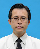 Minoru Ono (稔小野)合作论文数Department of Cardiovascular Surgery, Graduate School of Medicine, The University of Tokyo;Department of Cooperative Unit of Medicine and Engineering Research, The University of Tokyo Hospital26
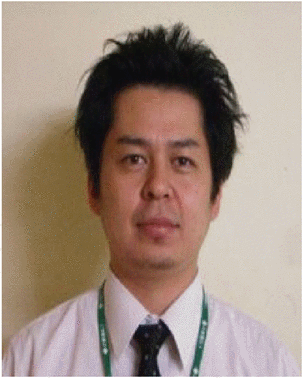 Masaki Tsuji合作论文数24Ishida Junichi合作论文数23
 Osamu Kinoshita合作论文数15Maki Hisataka合作论文数11Shimada Shogo合作论文数11
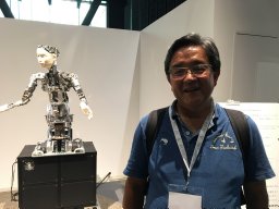 Masahiko Ando合作论文数Department of Preventative Services, Kyoto University School of Public Health11Kimura Mitsutoshi合作论文数10Nobutaka Kakuda合作论文数9Akihito Saito合作论文数9Minatsuki Shun合作论文数8Endo Miyoko合作论文数7Hosoya Yumiko合作论文数7Narita Koichi合作论文数7Yoshitaka Isotani合作论文数6Kagami Yukie合作论文数6
 Kan Nawata合作论文数6Hiroko Imai合作论文数5Hiroyuki Morita合作论文数5
 Satoshi Ishii合作论文数4Hiroki Yagi合作论文数4M. Nemoto合作论文数4Kanna Fujita合作论文数Univ Tokyo, Grad Sch Med, Dept Cardiovasc Med, Tokyo, Japan4Nitta Daisuke合作论文数4
 Norifumi Takeda合作论文数4Takahiro Kurihara合作论文数3Asako Shimada合作论文数3Genri Numata合作论文数Department of Cardiovascular Medicine (Y.H., The University of Tokyo3Takashi Hiruma合作论文数3
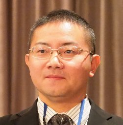 Seitaro Nomura合作论文数3Haruo Yamauchi合作论文数3Toru Hara合作论文数2Shunsuke Inoue合作论文数2Kensuke Tsushima合作论文数2Toshiyuki Ko合作论文数2Masamichi Ito合作论文数Univ Tokyo, Grad Sch Med, Dept Cardiovasc Med, Tokyo, Japan2Manami Katoh合作论文数Genome Science Division, Research Center for Advance Science and Technology, The University of Tokyo, Tokyo, Japan.2Yamada Takanobu合作论文数2
 Yasuyuki Shiraishi合作论文数2Koh Okamoto合作论文数2Norihiko Takeda (武田憲彦)合作论文数Division of Cardiology and Metabolism, Center for Molecular Medicine, Jichi Medical University;Department of Cardiovascular Medicine, Graduate School of Medicine, The University of Tokyo2Hiroyuki Abe合作论文数2The University of Tokyo Hospital合作论文数1Keita Murakami合作论文数1I Komuro合作论文数1Atsuko Ozeki合作论文数1




