OBJECTIVES The objective of this study is to test the wear of dual-cured bulkfill and flowable bulkfill composites. MATERIALS AND METHODS Six dual-cured bulkfill composites, Cention, Cention-Exp, Activa, Fill-up, Hyperfil Injectafill, and two flowable bulkfill composites Tetric Evoflow BulkFill and G-aenial Universal Flo were tested in this study (n = 8). Each composite was applied into an aluminum sample holder and cured with a Valo Grand (1230 mW/cm2 ) or self-cured according to manufacturer's recommendations, and stored in water for 3 weeks. The samples were subjected to 120 000 load cycles of 49 N (CS-4) against spherical steatite antagonists and simultaneously subjected to 4440 thermocycles (5°C-55°C). At intervals between load cycles, polyvinyl siloxane impressions were taken and scanned with a True Definition Laser Scanner. The volumetric wear was calculated using image software (Geomagic). Microscopic images of selected wear facets and their corresponding worn antagonists were obtained using SEM. All data were analyzed using analysis of variance (ANOVA) and Tukey post hoc test for multiple comparison (α = .05; β = .2). RESULTS The load cycles volumetric wear-increase was linear. Two-way ANOVA showed significant main effects (material and light-cured) and significant interactions. Self-cured materials (mean = 0.38 mm3 ) showed more wear than light-cured materials (mean = 0.35mm3 ). CONCLUSIONS The bioactive materials except Activa light-cured showed significantly more wear than the flowable composites. CLINICAL SIGNIFICANCE This study demonstrated that most of the dual-cured composites, if light-cured, showed the same wear as the flowable composites used as control. Therefore, to obtain adequate wear resistance the dual-cured composites should be routinely light-cured.
Objectives: To test wear of 10 universal composites and the antagonist.Null hypothesis: there are no differences in the composite and antagonist wear.Materials and Methods: Flat samples, light cured as of manufacturer's instructions and polished were made from Admira Fusion (AF), Filtek Supreme Ultra (FS), G-aenial Sculpt (GS), Harmonize (HR), Herculite Ultra (HU), Tetric Evoceram (TE), TPH Spectra (SP) and three Ultradent experimental materials (UPI Exp 1-3) (n = 8), and stored in water for 3 weeks.They were subjected to wear in a chewing simulator (1.2 x 10⁵ cycles, 49 N, 0.7 mm lateral movement, 1 Hz, steatite antagonists (Ø 6 mm), simultaneously thermocycled (5/55°C) every 90 s).The volumetric wear of the composite was measured with a 3D laser scanner) after 5,000, 10,000 then every 10,000, up to 120,000 cycles.The wear of antagonists was measured after 120,000 cycles.Results: From 5,000 -120,000 load cycles wear was linear.The total volumetric wear of composites was: GS 0.428 ± 0.083 mm 3 , UPI Exp 3 0.51 ± 0.042 mm 3 , HU 0.576 ± 0.072 mm 3 , SP 0.609 ± 0.088 mm 3 , FS 0.635 ± 0.077 mm 3 , HR 0.658 ± 0.116 mm 3 , TE 0.714 ± 0.097 mm 3 , Ultradent UPI Exp2 0.725 ± 0.132 mm 3 , UPI Exp 1 0.894 ± 0.278 mm 3 and AF 1.578 ± 0.37 mm 3 .The wear of AF was significantly the largrest (p < 0.0001).GS showed the lowest wear, but shared this position with UPI Exp 3, HU, SP, FS and HR.The total wear of UPI Exp3 was the lowest. Conclusion:The null hypothesis was rejected.
Materials and methods: The smart composites Activa (Pulpdent) and Cention N (Ivoclar Vivadent) and the glass ionomer cement Fuji IX (GC) were applied into aluminum sample holders, pressed against a glass plate and stored in water for 3 weeks after curing. The samples were subjected to 400,000 load cycles of 49 N in the CS4 chewing simulator (Mechatronik) against steatite antagonists and subjected to 4,440 thermocycles from 5 °C to 55 °C. Samples were evaluated with replicas after 5,000, 10,000, 20,000, 40,000, 60,000, 80,000, 100,000, 120,000, 160,000, 200,000, 240,000, 280,000, 320,000, 360,000 and 400,000 cycles with a laser scanner (LAS-20, Mechatronik) and the Geomagic software (wear volume). The data was analyzed with ANOVA and Tukey test. Selected wear facets were analyzed with a scanning electron microscope (SEM).
Aim:The study aimed to measure light scattering of a broad spectrum light curing unit (LCU) as influenced by ceramic type, shade and thickness as well as exposure distance and LCU' s position.Methodology: A broad spectrum LED LCU (ASCENT OL5) was mounted above a spectrometer (MARC Resin Calibrator, Blue, Light Analytics) at exposure distances of 1.0, 1.5 or 2.5 mm.The position of the center of the head was aligned with the spectrophotometer's sensor and then moved in 1 mm increments in the X-Y plane, while concomitantly recording the irradiance.The process was repeated with lithium disilicate and leucite glass ceramic slabs of similar thicknesses.The loss in irradiance related to the value measured at center position was analyzed by means of linear regressions and multiple ANOVA analysis.Results: The regressions showed a good fit (90% -99%).Moving away from the center showed decreased irradiance.Values of slope obtained were divided by their respective intercept to eliminate the influence of the irradiance measured at the center.Two three-way ANOVA's were performed.One examined the influence of ceramic slab, direction and translucency/shade.It shows only the direction of measurement exhibited significant influence (p < 0.0001) on the mean normalized slope values.The other one examined the influence of ceramic slab, direction and slab thickness.It shows the mean normalized slope values are significantly influenced by the direction of measurement and the slab thickness (p < 0.0001).Values of the slopes indicated the ceramic scattering effect of the light.Thicker samples showed more scattering. Conclusion:The ceramic types, translucency/ shade had no significant effect on the light scattering.The thicker the ceramic the less irradiance changes were found indicating that the ceramics were scattering the light and thus slightly alleviating the effect of the inhomogeneous beam profile.
Purpose of the Study: Compare occlusal wear of PMMA DCL denture teeth under two different loads in vitro.Materials & Methods: Sixteen mandibular second premolars (SR Orthoplane DCL) with a flat occlusal surface (specimens) were worn by sixteen maxillary second premolar-antagonists (Ortholingual DCL).These teeth were subjected in a chewing simulator (CS-4, SD Mechatronik) up to 240,000 loading cylces at 19.6N (LL ≈ full denture) and 68.6N (HL ≈ implant-overdenture) and TC (2,222 x 5 °C -55 °C).Replicas of mandibular teeth were obtained at 0, 10,000; 20,000; 40,000; up to 240,000 cycles with polyvinyl-siloxane impressions and dental stone.Antagonist-replicas were made at baseline and at 240,000 cycles.The volumetric wear was determined with Geomagic after scanning replicas with a laser scanner.Linear regressions and ANOVA were used for statistical analysis. Results:The wear rate of the HL-specimens was significantly higher than that of the LL-group (p < 0.0001).The LL-wear rate became linear after 60,000 cycles and was calculated to be 0.182 x 10 -6 mm 3 /stroke.The HL-wear rate was linear from 20,000 to 140,000 cycles and was 1.056 x 10 -6 mm 3 / stroke, then up to 240,000 cycles 0.656 x 10 -6 mm 3 /stroke.At 240,000 cycles the HL-group showed significantly higher antagonist-wear (p < 0.0001).The antagonists in both groups demonstrated higher wear than their opposing specimens (p < 0.08).Conclusions: HL generated significantly higher wear of both the specimens and the antagonists.The antagonists showed higher wear than the specimens.As a clinical consequence one may expect more wear of denture teeth in implant supported overdentures than in full dentures.
Aim: The study aimed to characterize a broad spectrum light curing unit (LCU) by measuring the light beam profile output of the LCU using a spectrometer-based method and correlate it with a standard camera-based beam profile method. Materials and Methods: A broad spectrum LED LCU (Ascent OL5, CAO Group) was mounted above a spectrometer (MARC® Resin Calibrator, BlueLight Analytics) at exposure distances of 1.0, 1.5 or 2.5 mm. The position of the center of the LCU was aligned with the spectrometer’s cosine corrector sensor, and then moved in 1-mm increments in the x-y plane, while concomitantly recording the irradiance. The recorded irradiance was systematically organized and reported in function of the distance from the center of the LCU exiting window. Using a standard camera-based beam profiler, a beam profile of the LCU was obtained and the above approach was emulated to the beam profile. For both methods, the irradiance decreases related to the value measured at the center position was analyzed by calculating the slope, using a linear correlation. Results: Both methods showed that moving away from the center showed decreased irradiation. The beam profile of the LCU is asymmetric. The inhomogeneity of the beam was slightly lower with farther distance from the LCU’s light exciting window. Conclusion: The spectrometer-based method was able to characterize the beam profile of the LCU and can be used in the evaluation of LCUs.
Purpose: Evaluate the microshear bond strength (µSBS) of orthodontic resin cement to monolithic zirconium oxide ceramic (MZ) under orthodontic load (OL) and thermocycling (TC) effect.Materials and Methods: Glazed MZ blocks (Zenostar, Ivoclar Vivadent) were tested after air abrasion with 30-µm silica coated aluminum oxide (Al 2 O 3 ) particles (CoJet, 3M ESPE).The specimens were randomly divided into 4 groups (n = 15): G1, OL with TC; G2, OL without TC; G3 no OL with TC; and G4, no OL, no TC (control).Orthodontic cement cylinders (Heliosit Orthodontic, Ivoclar Vivadent) were bonded to the primed samples (Monobond Plus, Ivoclar Vivadent) using the Ultradent SBS system and light cured (SmartLite Max, Dentsply Sirona, 1400 mW/cm 2 , 40 s).G1 and G2 were subjected to 70 ± 15 N load perpendicular to the cylinder axis, G1 and G3 were thermo-cycled (5000 cycles 5-55°C, 90 s/cycle).G2 and G4 were stored in distilled water at 37 ± 1°C.The specimens were subjected to µSBS test (crosshead speed 0.5 mm/min).Data were analyzed using two-way ANOVA, and one-way ANOVA and Tukey test (HSD).Results: Two-way ANOVA for µSBS values (MPa) showed significant (p = 0.0004) load effects, but not thermal effect (p = 0.2455) with significant load/thermocycling interactions (p < 0.0001).The ranking of the single groups by Tukey test (α = 0.05) showed that G1 exhibited the highest µSBS (8.4 ± 2.8 MPa), G4 (6.3 ± 1.1 MPa) and G2 (5.8 ± 1.1 MPa) as a group was second, and G2 and G3 (4.7 ± 1.1 MPa) as a group was the lowest.Conclusion: G1, which is the closest to clinical reality, yielded the best results.
The effect of restoration depth on the curing time of a conventional and two bulk-fill composite resins by measuring microhardness and the respective radiosity of the bottom surface of the specimen was investigated. 1-, 3- and 5-mm thick washers were filled with Surefil SDR Flow-U (SDR), Tetric EvoCeram Bulk Fill-IVA (TEC) or Esthet-X HD-B1 (EHD), and cured with Bluephase® G2 for 40s. Additional 1-mm washers were filled with SDR, TEC or EHD, placed above the light sensor of MARC®, stacked with pre-cured 1-, 3- or 5-mm washer of respective material, and cured for 2.5~60s to mimic 2-, 4- and 6-mm thick composite curing. The sensor measured the radiosity (EB) at the bottom of specimen stacks. Vickers hardness (VH) was measured immediately at 5 locations with triplicate specimens. Nonlinear regression of VH vs EB by VH=α[1-exp(-EB/β)] with all thickness shows that the values of α, maximum hardness, are 21.6±1.0 kg/mm2 for SDR, 38.3±0.6 kg/mm2 for TEC and 45.3±2.6 kg/mm2 for EHD, and the values of β, rate parameter, are 0.40±0.06 J/cm2 for SDR, 0.77±0.04 J/cm2 for TEC and 0.58±0.09 J/cm2 for EHD. The radiosity of the bottom surface was calculated when the bottom surface of each material attained 80% of α of each material. The curing times for each material are in agreement with manufacturer recommendation for thickness. It is possible to estimate time needed to cure composite resin of known depth adequately by the radiosity and microhardness of the bottom surface.
Introduction: This in vitro study aimed at testing the hypotheses that (1) there is no difference in wear in vitro among 3 bulk-fill composites investigated and their respective antagonists, and (2) the tested bulk-fill wear is not different from enamel.Methodology: X-tra fil (Voco; [X]), Tetric-N-Ceram Bulkfill (Ivoclar Vivadent; [T]), QuiXX (Dentsply; [Q]), and enamel [E] specimens (Ø=8 mm, depth=1.5 mm, n=8/material) were subjected to wear in a chewing simulator (CS 4.8, SD Mechatronik) with steatite antagonists (Ø=6mm).1.2x10 5 cycles (0-49 N, 0.7 mm lateral movement, 1 Hz) were performed while simultaneously thermocycling (5/55°C) every 90 s.The volumetric wear of the materials was measured with a 3D laser scanner.Results: The total wear of bulk-fills was: [X]: 0.64±0.07mm 3 ; [T]: 0.66±0.08mm 3 ; [Q]: 1.58±0.14mm 3 .The total wear of enamel (0.24±0.03 mm 3 ) was significantly lower than that of the bulk-fills (p<0.0001).The total wear of the antagonists was: [X]: 0.32±0.02mm 3 ; [T]: 0.24±0.04mm 3 ; [Q]: 0.27±0.02mm 3 ; [E]: 0.12 ±0.01 mm 3 .The wear of the antagonists by [X] was significantly higher than by [T] and [Q] (p<0.001).Enamel produced the lowest wear of the antagonists (p<0.0001).The wear was linear between 5x10 3 and 1.2x10 5 wear-cycles.A negative correlation between the wear of the composite materials and that of the antagonists was found. Conclusion:In vitro wear of Tetric-N-Ceram Bulkfill was in the expected range and equal to X-tra fil.QuiXX wear was 2.7 times higher.The antagonist wear was significantly lower, less than 50% of the wear of the composites and the enamel.Both hypotheses were rejected.
Objectives: To test the hypotheses: (1) there is no difference in the volumetric wear among composites tested, and (2) there is no difference in the wear rates calculated from the linear relationship of wear increase over cycling.Methods: Two composites comprising pre-polymerized particles (Herculite-Précis [H], Tetric-N-Ceram [T]), one composite with very fine glass fillers (Charisma Opal [C]), and one composite with a mixture of agglomerated and nonagglomerated silica, and zirconia fillers (Filtek Z 350 XT [F]) were tested in a chewing simulator (CS 4.8, SD Mechatronik) with spherical Steatite antagonists (Ø 6 mm).Eight specimens of each composite were made by applying two increments in aluminum specimen-holders with a cylindrical cavity (Ø 8 mm, depth 1.5 mm), light cured (Bluephase G2; 1383 mW/cm2) for 20 s, polished to high gloss, and subjected to mastication cycles (59 N, 1.2 Hz, lateral movement 0.7 mm) and thermocycles (5/55°C; 116 s per cycle) simultaneously.After each 100, 500, 1,000, 2,000, 5,000, 10,000, 20,000, 30,000, 40,000, 50,000, 60,000, 70,000, 80,000, 90,000, 100,000, 110,000, and 120,000 mastication cycles, 3D images of worn surfaces were captured with Laserscanner LAS-20 (SD Mechatronik), and volumetric wear in mm3 was calculated by Geomagic software.Results: Five samples were lost due to separation at interface between increments.The means of total volumetric wear (mean±SD) after 120,000 cycles are 0.78±0.26mm3, 0.91±0.15mm3, 0.99±0.29 mm3, and 1.15±0.36mm3for F, H, T and C. Wear rate of each surviving sample between 2,000 and 120,000 cycles was calculated by linear regression (R2>0.99 for all specimens).The wear rates (mean±SD ; μm3/ cycle) are, 5.97±2.29x103,6.85±1.06x103,8.91±2.81x10³and 6.43±0.58x10³for F, H, T and C. GLM shows statistically significant differences in the wear rate among the four materials (p=0.0488).Looking at the total volumetric wear of the four composites ) and wear of antagonists no differences were found (p=0.1183(p=0.3027)respectively.Conclusions: The first hypothesis was accepted and the second hypothesis was rejected.To prevent separation between increments, future specimen preparation should consider bulk fill.
To evaluate the effect of Dentaurum Grid Strengthener on flexural strength and modulus of two types of PMMA, 24 beams (75 mm x 10 mm x 2.5 mm) containing bonded and flattened Grid Strengthener and 24 beams without Grid Strengthener were subjected to a 3-point bending test at 5 mm/min. Flexural strength and modulus were calculated. Data were analyzed with a 2-way ANOVA. The highest mean strength and flexural modulus was measured for reinforced high impact resin (96.2±14.3 MPa and 3425±779 MPa, respectively). For flexural strength and modulus, resin and reinforcement had a significant effect.CLINICAL RELEVANCE:Independent of the resin used, the incorporation of a grid strengthener increases the strength and modulus, thus the fracture probability of dentures is reduced.
This in vitro study investigated the interfacial flexural strength (FS) of amalgam repairs and the optimal combination of repair materials and mechanical retention required for a consistent and durable repair bond. Amalgam bricks were created, each with 1 end roughened to expose a fresh surface before repair. Four groups followed separate repair protocols: group 1, bonding agent with amalgam; group 2, bonding agent with composite resin; group 3, mechanical retention (slot) with amalgam; and group 4, slot with bonding agent and amalgam. Repaired specimens were stored in artificial saliva for 1, 10, 30, 120, or 360 days before being loaded to failure in a 3-point bending test. Statistical analysis showed significant changes in median FS over time in groups 2 and 4. The effect of the repair method on the FS values after each storage period was significant for most groups except the 30-day storage groups. Amalgam-amalgam repair with adequate condensation yielded the most consistent and durable bond. An amalgam bonding agent could be beneficial when firm condensation on the repair surface cannot be achieved or when tooth structure is involved. Composite resin can be a viable option for amalgam repair in an esthetically demanding region, but proper mechanical modification of the amalgam surface and selection of the proper bonding system are essential.
Background: Full dentures show a high rate of fractures under clinical use.Therefore strengthening of the denture base is expected to be beneficial with respect to reducing the fracture risk.Preformed perforated thin metal plates seem to be a simple tool for reinforcement.Objective: To test the strengthening effect of perforated metal plates incorporated into a PMMA based resin as a function of the bonding strategy.Materials and Methods: Preformed gold plated stainless steel grid strengtheners were purchased, pressed flat and embedded in wax and processed as dentures using the Ivobase hybrid injection moldable material (Ivoclar Vivadent, Schaan, Liechtenstein).Prior to processing the surfaces were either not treated or treated with a primer (Monobond Plus, Ivoclar Vivadent, Schaan, Liechtenstein) and grit blasted and primed.They were then sectioned into beams for three point bending test.Resin beams of identical dimensions without reinforcement were used as control.Flexural strength and modulus were calculated based on the load to fracture determined by an Instron universal testing machine.The data were analyzed with ANOVA and Tukey's test.Results: Flexural strength: control beams fractured at 78.8 ± 5.9 MPa, while, with the exception of the grit blasted samples, the experimental groups showed significantly higher strength (97.2 respect.95.4 MPa).Flexural Modulus: The control group yielded 2261.7 ± 261.4 MPa, while all experimental groups had a significantly higher modulus (3239.1 -3952.4MPa) The surface treatments did not show significant differences.Conclusions: The grid strengtheners tested had a significant effect on strengthening.Under the conditions of this study, surface priming did not increase the mechanical properties of the reinforced bars.
Purpose: To test the hypothesis that stress distribution is more complex than generally assumed during microtensile testing by determining the stress level in the adhesive region of a virtual dentin-adhesive-composite stick using FEA (finite element analysis).Materials and Methods: A 3D FEA model simulating a dentin-adhesive-composite stick was analyzed. The length of the composite and the dentin was 5.0 mm each and the thickness of the adhesive layer was 0.02 mm. For the stress analysis, either only one lateral side of the stick or both end surfaces were attached. A 20-N load was then exerted on the stick with its 1.0 mm(2) cross-sectional area, and von Mises stresses were calculated.Results: Large variations in stress levels existed. The highest stresses were located in the dentin and composite sections, near the adhesive interface. The stress level in these regions in the stick attached to one lateral side was more than 5 times higher than the 20 MPa stress calculated by dividing the force with the cross-sectional area. For the specimen glued to the ends of the sticks, the stress level differences at the bonded interfaces were around 22 MPa, which decreased to 12 to 14 MPa in the center of the adhesive. Thus, this load condition yielded von Mises stress levels at the interface that were closer to the expected stress level than were the lateral-side attached specimens.Conclusion: The calculated stress levels were higher and more complex than the strength values obtained by dividing the load at failure by the cross-sectional area. Reported strength values from microtensile tests therefore do not represent the true bond strength values at either the dentin/adhesive or adhesive/composite interface. However, the high stress levels in dentin and composite may explain the cohesive failures reported in the literature.
Resin-based materials that release either fluoride or chlorhexidine have been formulated for inhibiting caries activity. It is not known if the two agents, when incorporated into one material, would interact and affect their release potential. We hypothesized that the ratio of fluoride to chlorhexidine incorporated into a resin, and the pH of the storage medium, will affect their releases from the material. The material investigated contained 23 wt% of filler, and the ratios of calcium fluoride to chlorhexidine diacetate were 8/2, 5/5, and 2/8. The release was conducted in pH 4, 5, and 6 acetate buffers. The results showed that release of either agent increased as the pH of the medium decreased. The presence of fluoride salt substantially reduced the chlorhexidine release, while the presence of a specific quantity of chlorhexidine significantly increased fluoride release. This interaction can be utilized to optimize the release of either agent for therapeutic purposes.
This study tested the hypothesis that short-term exposure to acidic drinks can reduce enamel surface hardness, and that subsequent brushing with toothpaste can cause detectable loss of enamel. Enamel specimens (three groups per storage medium) were immersed in one of four acidic drinks for five seconds, one minute, or 10 minutes. The pH values of all four drinks were measured. Six microhardness indentations were made on each specimen before immersion (baseline), after immersion, and after brushing with toothpaste. Changes in surface microhardness were expressed as the ratio of hardness values after each treatment to the baseline value of each specimen. Two-way ANOVA showed that both storage medium and duration of immersion influenced the ratio significantly. One-way ANOVA showed that the ratio was reduced significantly with increased time of immersion in drinks with a pH =2.8, but the ratio reduction was not significant in drinks with a pH =3.3. Subsequent brushing removed the softened layer and exposed intact surface underneath. Although short-term exposure (~5 seconds) did not significantly affect the surface hardness values, it left a lightly etched enamel surface among specimens immersed in acidic drinks with a low pH. Subsequent brushing could still cause detectable loss of enamel surface after multiple exposures.
STATEMENT OF PROBLEM:Titanium is the most biocompatible metal used for dental casting; however, there is concern about its marginal accuracy after porcelain application since this aspect has direct influence on marginal fit. PURPOSE:The purpose of this study was to determine the effect that metal selection and the porcelain firing procedure have on the marginal accuracy of metal ceramic prostheses. MATERIAL AND METHODS:Cast CP Ti, milled CP Ti, cast Ti-6Al-7Nb, and cast Ni-Cr copings (n=5) were fired with compatible porcelains (Triceram for titanium-based metals and VITA VMK 95 for Ni-Cr alloy). The Ni-Cr alloy fired with its porcelain served as the control. Photographs of metal copings placed on a master die were made. Marginal discrepancy was determined on the photographs using an image processing program at 8 predetermined locations before airborne-particle abrasion for porcelain application, after firing of the opaque layer, and after firing of the dentin layer. Repeated-measures 2-way ANOVA was used to investigate the effect of metal selection and firing stage, and paired t tests were used to determine the effect of each firing stage within each material group (alpha=.05). RESULTS:ANOVA showed that both metal selection and firing stage significantly influenced the measured marginal discrepancy (P<.001), and there was interaction between the 2 variables (P<.001). Student-Newman-Keuls multiple comparison tests showed that there were significant differences between any 2 metals compared, at each stage of measurement. Paired t tests showed that significant changes in marginal discrepancy occurred with opaque firing on milled CP Ti (P=.017) and cast Ti-6Al-7Nb alloy (P=.003). CONCLUSIONS:Titanium copings fabricated by CAD/CAM demonstrated the least marginal discrepancy among all groups, while the base metal (Ni-Cr) groups exhibited the most discrepancy of all groups tested.
 Jean-Francois Roulet合作论文数Center For Dental Biomaterials, College of Dentistry, University of Florida;Department of Restorative and Dental Sciences, College of Dentistry, University of Florida12
 Kenneth Anusavice合作论文数Department of Materials Science & Engineering, University of Florida10
 Valeria V. Gordan合作论文数10
 Ivar A Mjor合作论文数College of Dentistry, University of Florida;Department of Operative Dentistry, University of Florida9
 Nader Abdulhameed合作论文数8Eduardo Mondragon合作论文数6Hind S. Hussein合作论文数4AA Barrett合作论文数4Nz Zhang合作论文数3Marwah Majid Khudhair合作论文数2
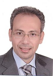 Tamer Shokry合作论文数Faculty of Dental Medicine, Al-Azhar University2
 Alvaro Della Bona合作论文数Dental School, Universidade de Passo Fundo2
 John J Mecholsky合作论文数Department of Materials Science and Engineering, Herbert Wertheim College of Engineering, University of Florida2
 Jason A. Griggs合作论文数Department of Biomedical Materials Science, The University of Mississippi Medical Center;School of Dentistry, The University of Mississippi Medical Center2EM Song合作论文数1P.V. Rao合作论文数1Ihab E Mosleh合作论文数1Snigdha Gummadi合作论文数1Raed Aloul合作论文数1John Cliff Jones合作论文数1
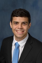 Mateus Garcia Rocha合作论文数College of Dentistry, University of Florida1
 Tamer Hamza合作论文数Al-Azhar University1
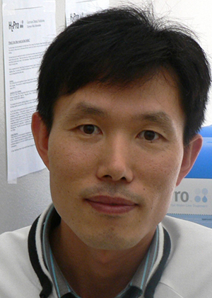 Won-Suck Oh合作论文数Department of Biologic and Materials Sciences & Prosthodontics, School of Dentistry, University of Michigan-Ann Arbor1
 Andre Reis合作论文数School of Dentistry, University of Guarulhos1Haisch Larry D合作论文数1Berry Steven E合作论文数1Setcos James C合作论文数1
 Dayane Oliveira合作论文数Piracicaba Dental School, UNICAMP - University of Campinas1Wilson Nairn Hf合作论文数1
 Seiichiro Tsutsumi合作论文数Department of Marine System Engineering, Kyushu University1Jun Yan合作论文数1G.M. Furnish合作论文数1
 Rasoul Khosravi合作论文数Faculty of Medical Sciences, Tarbiat Modares University1Venita J. Sposetti合作论文数1Mohamed M. El-Hosary合作论文数1Alan C. Levin合作论文数1Jacquelyn Moorhead合作论文数1Chris Batich合作论文数Department of Materials Science and Engineering, University of Florida1
 Mazen Attia合作论文数Faculty of Oral and Dental Medicine, Cairo University1Karl Johan M. Söderholm合作论文数Department of Materials Science and Engineering, College of Engineering, University of Florida;Division of Operative Dentistry, Department of Restorative Dental Sciences, College of Dentistry, University of Florida;Center for Dental Biomaterials, College of Dentistry, University of Florida1
 Kasthurirangan Gopalakrishnan合作论文数Iowa State University1
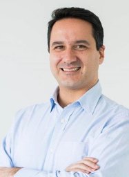 Paulo Francisco Cesar合作论文数School of Dentistry, University of São Paulo1
 Joseph Riley合作论文数Department of Community Dentistry and Behavioral Science, College of Dentistry, University of Florida1
 José Augusto Rodrigues合作论文数Department of Restorative Dentistry;Faculty of Dentistry of Piracicaba;UNICAMP, University of Campinas;Faculty of Dentistry of Piracicaba, University of Campinas1
 Uli Lohbauer合作论文数Department Materials Science and Engineering, Faculty of Engineering, Friedrich-Alexander-Universität;Fracto Forum International1
 Susanne Scherrer合作论文数Division of Fixed Prosthodontics-Biomaterials, University of Geneva1
 Saulo Geraldeli合作论文数University of Iowa College of Dentistry1




