BACKGROUND:Pathogenic variants in COL1A1 and COL1A2 account for 85-90% of osteogenesis imperfecta (OI). Glycine substitutions within the triple-helical domain are traditionally considered highly pathogenic. The COL1A2 c.2827G>A p.(Gly943Arg) variant was previously classified as pathogenic under ACGS 2019/2020 criteria. METHODS:One proband with this heterozygous variant was identified through national newborn genomic screening prompting further evaluation from the UK National OI Reference Laboratory. Four additional patients with the same variant were identified through referral to tertiary metabolic bone services/ Genetics clinic and phenotypic data were gathered to understand better clinical presentations and inheritance patterns. The variant was reassessed following the release of gnomAD v4.1, incorporating updated ACGS 2024 criteria. RESULTS:The expanded gnomAD v4.1 dataset incorporating UK Biobank (UKBB) demonstrated the presence of p.(Gly943Arg) in an unselected population at a frequency inconsistent with the pathogenicity of COL1A2, resulting in reclassification from pathogenic to variant of uncertain significance (VUS). Clinical presentations of 5 patients ranged from no fracture history to moderately severe OI. Cascade testing revealed multiple asymptomatic adult heterozygotes. UKBB data demonstrated no recorded OI diagnoses among 44 adult heterozygotes with evidence of a shared common haplotype in 40/45 enriched in Northwest England. CONCLUSION:The COL1A2 p.Gly943Arg variant demonstrates marked clinical heterogeneity and population frequency inconsistent with a fully penetrant dominant OI allele. This study highlights the dynamic nature of variant classification, the importance of large population datasets, and the ethical and clinical challenges of variant reclassification in the era of genomic screening.
Osteogenesis imperfecta (OI) is a rare genetic disorder characterised by bone fragility and frequent fractures. While current treatments aim to preserve bone mass, there is no cure. Boost Brittle Bones Before Birth (BOOSTB4) is an international clinical trial investigating postnatal (n = 15) and prenatal plus postnatal (n = 3) infusions of fetal mesenchymal stem cells as a potential treatment for severe OI. Each BOOSTB4 participant received four stem cell doses at four-month intervals. Here we explore the experiences of parents and of healthcare professionals involved in BOOSTB4. In this qualitative study, one or both parents of children participating in BOOSTB4 took part after the second dose (T1: 16 families, 22 participants) and at least 6 months after the final dose (T2: 12 families, 16 participants). Professionals delivering BOOSTB4 participated in one interview (n = 13). Data were analysed using codebook thematic analysis. Decisions to participate in the trial were influenced by the potential for improved prognosis and limited alternative therapies. Parents felt reassurance because they were taking action and gratitude for access to expertise in OI. Balancing hope, expectations and uncertainty around treatment efficacy was challenging. Parents also expressed disappointment and uncertainty about treatment ending. Practical challenges included travel for treatment with a child with OI or late in pregnancy. Professionals noted the difficulty in managing parental expectations and the regulatory and logistical barriers of an international trial. Findings emphasised the need for clear communication before, during and after the trial, transparent information and trial designs that accommodate the needs of individual families.
INTRODUCTION:Current tools to evaluate fracture risk in children with Osteogenesis Imperfecta (OI) are limited and bone mineral density (BMD) has limitations and is not widely available for children under 5 years. We hypothesized that the Bone Health Index (BHI), evaluating the average cortical thickness of the three middle metacarpal bones, adjusted for bone width and length, could be a useful tool in the assessment of fracture risk, particularly in infants. METHODS:The service at Great Ormond Street Hospital (London, UK) follows a cohort of over 277 children with classical OI and keeps a detailed and accurate record of confirmed fractures. We reviewed their BHI (analysed using the latest version of BoneXpert® software) at presentation (n = 123), the lumbar BMD (n = 47) taken on the same day where available, the subsequent two years fracture history and annual lateral spine X-rays. RESULTS:In multivariable logistic regression, each 1-SD decrease in BHI SDS was independently associated with a 5.3-fold higher odds of incident fracture within two years (OR 5.26, 95% CI 2.4-11.1, p < 0.001), while DXA BMD and BMAD Z-scores were not independent predictors. ROC analysis showed good discriminatory performance for BHI SDS (AUC 0.84), superior to DXA BMD Z-score (AUC 0.60) and BMAD Z-score (AUC 0.51). CONCLUSIONS:Our study indicates that lower BHI SDS values are associated with increased fracture risk in children with classical OI. In the available DXA subgroup, BHI demonstrated stronger discriminatory performance than DXA-derived measures. BHI may represent a clinically useful complementary tool for fracture risk stratification in young children with OI, particularly where DXA is unavailable or technically limited, including infancy.
IntroductionSevere osteogenesis imperfecta (OI) is a debilitating disease with no cure or sufficiently effective treatment. Mesenchymal stem cells (MSCs) have good safety profile, show promising effects and can form bone. The Boost Brittle Bones Before Birth (BOOSTB4) trial evaluates administration of allogeneic expanded human first trimester fetal liver MSCs (BOOST cells) for OI type 3 or severe type 4.Methods and analysisBOOSTB4 is an exploratory, open-label, multiple dose, phase I/II clinical trial evaluating safety and efficacy of postnatal (n=15) or prenatal and postnatal (n=3, originally n=15) administration of BOOST cells for the treatment of severe OI compared with a combination of historical (1–5/subject) and untreated prospective controls (≤30). Infants<18 months of age (originally<12 months) and singleton pregnant women whose fetus has severe OI with confirmed glycine substitution inCOL1A1orCOL1A2can be included in the trial.Each subject receives four intravenous doses of 3×10⁶/kg BOOST cells at 4 month intervals, with 48 (doses 1–2) or 24 (doses 3–4) hours in-patient follow-up, primary follow-up at 6 and 12 months after the last dose and long-term follow-up yearly until 10 years after the first dose. Prenatal subjects receive the first dose via ultrasound-guided injection into the umbilical vein within the fetal liver (16+0 to 35+6 weeks), and three doses postnatally.The primary outcome measures are safety and tolerability of repeated BOOST cell administration. The secondary outcome measures are number of fractures from baseline to primary and long-term follow-up, growth, change in bone mineral density, clinical OI status and biochemical bone turnover.Ethics and disseminationThe trial is approved by Competent Authorities in Sweden, the UK and the Netherlands (postnatal only). Results from the trial will be disseminated via CTIS, ClinicalTrials.gov and in scientific open-access scientific journals.Trial registration numbersEudraCT 2015-003699-60, EUCT: 2023-504593-38-00,NCT03706482.
DEPDC5 (DEP Domain-Containing Protein 5) encodes an inhibitory component of the mammalian target of rapamycin (mTOR) pathway and is commonly implicated in sporadic and familial focal epilepsies, both non-lesional and in association with focal cortical dysplasia. Germline pathogenic variants are typically heterozygous and inactivating. We describe a novel phenotype caused by germline biallelic missense variants in DEPDC5. Cases were identified clinically. Available records, including magnetic resonance imaging and electroencephalography, were reviewed. Genetic testing was performed by whole exome and whole-genome sequencing and cascade screening. In addition, immunohistochemistry was performed on skin biopsy. The phenotype was identified in nine children, eight of which are described in detail herein. Six of the children were of Irish Traveller, two of Tunisian and one of Lebanese origin. The Irish Traveller children shared the same DEPDC5 germline homozygous missense variant (p.Thr337Arg), whereas the Lebanese and Tunisian children shared a different germline homozygous variant (p.Arg806Cys). Consistent phenotypic features included extensive bilateral polymicrogyria, congenital macrocephaly and early-onset refractory epilepsy, in keeping with other mTOR-opathies. Eye and cardiac involvement and severe neutropenia were also observed in one or more patients. Five of the children died in infancy or childhood; the other four are currently aged between 5 months and 6 years. Skin biopsy immunohistochemistry was supportive of hyperactivation of the mTOR pathway. The clinical, histopathological and genetic evidence supports a causal role for the homozygous DEPDC5 variants, expanding our understanding of the biology of this gene.
Background The World Health Organization’s International Classification of Functioning, Health and Disabilities (ICF) is a biopsychosocial health model focusing on six domains: Health Condition, Body Structure and Function, Environmental Factors, Personal Factors, Activity, and Participation. In 2011 CanChild developed the ‘F-words’ – function, family, fitness, fun, friends, and future, mapped to ICF domains, as a tool to aid goal setting, personalise interventions and achieve child and family centred outcomes. We aimed to apply the ‘F-words’ to children with osteogenesis imperfecta (OI). Methods Children aged 6 years and over attending our multidisciplinary OI clinic were invited to complete the F-words Goal Sheet in the waiting area between March 2020 and March 2022, identifying personalised goals. This was reviewed during the appointment, to aid discussion and goal setting with professionals. Following the consultation participants completed a feedback questionnaire. Results 52 children (56% male, 44% female) aged 6-16 years (mean 11 years) completed the F-words Goal Sheet and feedback questionnaire. 87% of children found it helpful to complete the F-words Goal Sheet and 61% talked more in the appointment than previously. 90% reported they would be happy to use this tool again in the future and 90% would recommend the ‘F-words’ to other children with OI. Children ‘felt more involved’, ‘understood more’ and ‘got more information’ during the appointment which was ‘more personal’. Health professionals gained additional information, helping identify personalised goals and children requiring referral to the OI team psychologist. Conclusion The ‘F-words’ is a valuable tool in OI to set meaningful goals, whilst acting as a useful identification aid for children requiring psychosocial support. When using the ‘F-words’, communication in healthcare settings improves and children and families participate more actively. We promote application of this tool across the spectrum of OI and encourage its adoption in clinical practice, research, and advocacy.
By reading this article, you should be able to: •Recall the epidemiology and classification of osteogenesis imperfecta. •Identify the common clinical presentations. •Describe the common types of surgical procedures that these children undergo. •Discuss the important considerations for the anaesthetist before, during and after surgery. Edmund Chan MA FRCA MAcadMed is a specialty registrar in London. He has undertaken training in paediatric anaesthesia at Evelina London Children's Hospital and at Great Ormond Street Hospital for Children. Catherine DeVile MA MD FRCPCH FRCP is a consultant paediatric neurologist at Great Ormond Street Hospital for Children and is lead for their highly specialised osteogenesis imperfecta service. Vineetha Ratnamma DA MD FRCA is a consultant in paediatric anaesthesia at Great Ormond Street Hospital for Children. Her subspecialty interests are renal, renovascular, orthopaedic and general paediatric surgery.
Background Horizontal gaze palsy with progressive scoliosis (HGPPS) is a rare autosomal recessive neurological disorder characterised by absence of conjugate horizontal eye movements and early-onset rapidly progressive scoliosis. It is caused by variants in ROBO3, essential for axon decussation and contralateral neuronal migration during nervous system development. We describe a unique case of HGPPS and bone fragility. Presenting Problem A 19-month-old girl with genetically confirmed osteogenesis imperfecta (OI) type I presented to our OI Service, following intramedullary rod insertion for a femur fracture aged 16 months. Her Romanian family have mild OI, secondary to COL1A1 heterozygosity. Parents are non-consanguineous. Our assessment identified abnormal visual behaviour, horizontal gaze palsy, left torticollis and a flexible thoracic dextroconvex scoliosis. Ophthalmological assessment confirmed horizontal gaze palsy, finding significant myopia with astigmatism, anisometropia and reduced left ocular vision. ROBO3 gene testing was initiated. Neuroimaging revealed brainstem malformation with pontine hypoplasia, absent facial colliculi, butterfly configuration of the medulla and a deep midline pontine cleft in keeping with HGPPS. Clinical Management To promote vision glasses were prescribed. Aged 26 months scoliosis had progressed, with a levoconvex thoracolumbar component; T9-L5 Cobb angle 38°. By 32 months this progressed further; dextroconvex T2-T11 Cobb angle 45°, levoconvex T10-L4 Cobb angle 66°. This rapidly progressive scoliosis was atypical for mild OI – she had no vertebral compression fractures. Given concerns regarding rigid spinal bracing in OI a Lycra suit was provided. Multidisciplinary input including physiotherapy, spinal orthopaedic and ophthalmological monitoring is ongoing. Aged 35 months genetic results confirmed compound heterozygosity for two ROBO3 variants. Discussion Thorough clinical assessment and genotype-phenotype correlation is essential in children with known familial gene variants. When atypical clinical features present additional diagnoses should be sought. For children with dual diagnoses specialist multidisciplinary input is required to optimally manage comorbidities and achieve best possible developmental outcomes.
Background: Osteogenesis imperfecta (OI) is a rare genetic condition characterised by increased bone fragility. Recurrent fractures, pain and fatigue have a considerable impact on many aspects of the life of a person affected with OI and their families. Objective: To improve our understanding of the impact of OI on the daily lives of individuals and families and consider how the condition is managed so that support needs can be better addressed. Methods: Semi-structured qualitative interviews (n = 56) were conducted with adults affected with OI, with (n = 9) and without children (n = 8), parents of children affected with OI (n = 8), health professionals (n = 29) and patient advocates (n = 2). Interviews were digitally recorded, transcribed verbatim and analysed using thematic analysis. Results: Three overarching themes are described: OI is not just a physical condition, parenting and family functioning and managing the condition. Fractures, chronic pain and tiredness impact on daily life and emotional well-being. For parents with OI, pain, tiredness and mobility issues can limit interactions and activities with their children. Specialist paediatric health services for OI were highly valued. The need for more emotional support and improved coordination of adult health services was highlighted. Conclusions: Our findings allow a better understanding of the day-to-day experiences of individuals and families affected with OI. Supporting emotional well-being needs greater attention from policy makers and researchers. Improvements to the coordination of health services for adults with OI are needed and an in-depth exploration of young people's support needs is warranted with research focused on support through the teenage years. (c) 2021 The Authors. Published by Elsevier Inc. This is an open access article under the CC BY-NC-ND license (http://creativecommons.org/licenses/by-nc-nd/4.0/).
Background/Objectives In England, children (0-18 years) with severe, complex and atypical osteogenesis imperfecta (OI) are managed by four centres (Birmingham, Bristol, London, Sheffield) in a 'Highly Specialised Service' (HSS OI); affected children with a genetic origin for their disease that is not in COL1A1 or COL1A2 form the majority of the 'atypical' group, which has set criteria for entry into the service. We have used the data from the service to assess the range and frequency of non-collagen pathogenic variants resulting in OI in a single country. Methods Children with atypical OI were identified through the HSS OI service database. All genetic testing for children with OI in the service were undertaken at the Sheffield Diagnostic Genetics Service. Variant data were extracted and matched to individual patients. This study was done as part of a service evaluation project registered with the Sheffield Children's Hospital Clinical Governance Department. Results One hundred of 337 children in the HSS met the 'atypical' criteria. Eighty have had genetic testing undertaken; 72 had genetic changes detected, 67 in 13 genes known to be causative for OI. The most frequently affected genes were IFITM5 (22), P3H1 (12), SERPINF1 (8) and BMP1 (6). Conclusion Among children with more severe forms of OI (approximately one-third of all children with OI), around 20% have pathogenic variants in non-collagen genes. IFITM5 was the most commonly affected gene, followed by genes within the P3H1 complex. These data provide additional information regarding the likelihood of different genetic origins of the disease in children with OI, which may influence clinical care.
The hereditary ataxias are a heterogenous group of disorders with an increasing number of causative genes being described. Due to the clinical and genetic heterogeneity seen in these conditions, the majority of such individuals endure a diagnostic odyssey or remain undiagnosed. Defining the molecular etiology can bring insights into the responsible molecular pathways and eventually the identification of therapeutic targets. Here, we describe the identification of biallelic variants in the GEMIN5 gene among seven unrelated families with nine affected individuals presenting with spastic ataxia and cerebellar atrophy. GEMIN5, an RNA-binding protein, has been shown to regulate transcription and translation machinery. GEMIN5 is a component of small nuclear ribonucleoprotein (snRNP) complexes and helps in the assembly of the spliceosome complexes. We found that biallelic GEMIN5 variants cause structural abnormalities in the encoded protein and reduce expression of snRNP complex proteins in patient cells compared with unaffected controls. Finally, knocking out endogenous Gemin5 in mice caused early embryonic lethality, suggesting that Gemin5 expression is crucial for normal development. Our work further expands on the phenotypic spectrum associated with GEMIN5-related disease and implicates the role of GEMIN5 among patients with spastic ataxia, cerebellar atrophy, and motor predominant developmental delay.
Diagnostic whole genome sequencing (WGS) is increasingly used in rare diseases. However, standard, semi-automated WGS analysis may overlook diagnoses in complex disorders. Here, we show that specialist multidisciplinary analysis of WGS, following an initial ‘no primary findings’ (NPF) report, improves diagnostic rates and alters management. We undertook WGS in 102 adults with diagnostically challenging primary mitochondrial disease phenotypes. NPF cases were reviewed by a genomic medicine team, thus enabling bespoke informatic approaches, co-ordinated phenotypic validation, and functional work. We enhanced the diagnostic rate from 16.7% to 31.4%, with management implications for all new diagnoses, and detected strong candidate disease-causing variants in a further 3.9% of patients. This approach presents a standardised model of care that supports mainstream clinicians and enhances diagnostic equity for complex disorders, thereby facilitating access to the potential benefits of genomic healthcare. This research was made possible through access to the data and findings generated by the 100,000 Genomes Project: http://www.genomicsengland.co.uk .
Background Secreted protein, acidic, cysteine rich (SPARC)-related osteogenesis imperfecta (OI), also referred to as OI type XVII, was first described in 2015, since then there has been only one further report of this form of OI. SPARC is located on chromosome 5 between bands q31 and q33. The encoded protein is necessary for calcification of the collagen in bone, synthesis of extracellular matrix and the promotion of changes to cell shape. Methods We describe a further two patients with previously unreported homozygous SPARC variants with OI: one splice site; one nonsense pathogenic variant. We present detailed information on the clinical and radiological phenotype and correlate this with their genotype. There are only two previous reports by Mendozo-Londono et al and Hayat et al with clinical descriptions of patients with SPARC variants. Results From the data we have obtained, common clinical features in individuals with OI type XVII caused by SPARC variants include scoliosis (5/5), vertebral compression fractures (5/5), multiple long bone fractures (5/5) and delayed motor development (3/3). Interestingly, 2/4 patients also had abnormal brain MRI, including high subcortical white matter changes, abnormal fluid-attenuated inversion in the para-atrial white matter and a large spinal canal from T10 to L1. Of significance, both patients reported here presented with significant neuromuscular weakness prompting early workup. Conclusion Common phenotypic expressions include delayed motor development with neuromuscular weakness, scoliosis and multiple fractures. The data presented here broaden the phenotypic spectrum establishing similar patterns of neuromuscular presentation with a presumed diagnosis of 'myopathy'.
Our aim was to identify clinical outcomes, serological features and possible prognostic indicators of paediatric myasthenia gravis (MG). We collected 74 MG patients with disease onset before the age of 16 years (73% pre-pubertal onset defined as <= 10 years), seen regularly at two UK specialist centres, over a period of 11 years. The cohort was multi-ethnic, with a high number of non-Caucasians (52%). Ocular presentation was seen in 38 (51%) and only 8 (21%) of these generalised. Fifty-two (70%) patients had antibodies to the acetylcholine receptor (AChR) measured by radioimmunoprecipitation, 10 (14%) had antibodies only to clustered AChRs detected by a cell based assay, 3 (4%) had muscle-specific kinase and one (1%) low-density lipoprotein receptor-related protein 4 antibody. Only 8 (11%) had no detectable antibodies. Seventeen patients attained drug free remission (Kaplan Meyer survival curve estimates 25% by 7 years). Several factors were associated with a higher likelihood of free remission: onset age <= 10 years, Asian and Caucasian races, lack of AChR antibodies on RIA, and normal repetitive nerve stimulation at diagnosis. However, in a multifactorial regression analysis, the antibody status was the only significant predictor for drug free remission, with 60% of patients with antibodies only to clustered AChR achieving this outcome. Complete drug free remission is not uncommon in paediatric MG and several factors appear to influence this outcome with antibody status being the most important. These factors can be easily evaluated at diagnosis, and may help to determine whose patients are likely to require more intensive treatments. (C) 2019 Elsevier B.V. All rights reserved.
KIF1A-related disorders (KRD) were first described in 2011 and the phenotypic spectrum has subsequently expanded to encompass a range of central and peripheral nervous system involvement. Here we present a case series demonstrating the range of clinical, neurophysiological, and radiological features which may occur in childhood-onset KRD. We report on all the children and young people seen at a single large tertiary centre. Data were collected through a retrospective case-notes review. Twelve individuals from 10 families were identified. Eight different mutations were present, including four novel mutations. Two patients displayed a very severe phenotype including congenital contractures, severe spasticity and/or dystonia, dysautonomia, severe sensorimotor polyneuropathy and optic atrophy, significant white matter changes on brain MRI, respiratory insufficiency, and complete lack of neurodevelopmental progress. The remaining 10 patients represented a spectrum of severity with common features including a movement disorder with spasticity and/or dystonia, subtle features of dysautonomia, sensory axonal neuropathy, varying degrees of optic atrophy and of learning and/or behavioural difficulties, and subtle or absent-but sometimes progressive-changes in white matter on MRI. Epilepsy was common among the more severely affected children. This case series demonstrates that KRD comprise a range of neurological disorders, with both the milder and the more severe forms combining central and peripheral (including autonomic) nervous system deficits.
Glycosylphosphatidylinositol (GPI)-anchored proteins are critical for embryogenesis, neurogenesis, and cell signaling. Variants in several genes participating in GPI biosynthesis and processing lead to decreased cell surface presence of GPI-anchored proteins (GPI-APs) and cause inherited GPI deficiency disorders (IGDs). In this report, we describe 12 individuals from nine unrelated families with 10 different bi-allelic PIGK variants. PIGK encodes a component of the GPI transamidase complex, which attaches the GPI anchor to proteins. Clinical features found in most individuals include global developmental delay and/or intellectual disability, hypotonia, cerebellar ataxia, cerebellar atrophy, and facial dysmorphisms. The majority of the individuals have epilepsy. Two individuals have slightly decreased levels of serum alkaline phosphatase, while eight do not. Flow cytometric analysis of blood and fibroblasts from affected individuals showed decreased cell surface presence of GPI-APs. The overexpression of wild-type (WT) PIGK in fibroblasts rescued the levels of cell surface GPI-APs. In a knockout cell line, transfection with WT PIGK also rescued the GPI-AP levels, but transfection with the two tested mutant variants did not. Our study not only expands the clinical and known genetic spectrum of IGDs, but it also expands the genetic differential diagnosis for cerebellar atrophy. Given the fact that cerebellar atrophy is seen in other IGDs, flow cytometry for GPI-APs should be considered in the work-ups of individuals presenting this feature.
Coatomer complexes function in the sorting and trafficking of proteins between subcellular organelles. Pathogenic variants in coatomer subunits or associated factors have been reported in multi-systemic disorders, i.e., coatopathies, that can affect the skeletal and central nervous systems. We have identified loss-of-function variants in COPB2 , a component of the coatomer complex I (COPI), in individuals presenting with osteoporosis, fractures and developmental delay of variable severity. Because the role of COPB2 in bone has not been characterized, we studied the effect of COPB2 deficiency on skeletal development in mice and zebrafish. Copb2 +/− mice showed low bone mass and decreased bone strength. In zebrafish, larvae carrying a copb2 heterozygous frameshift variant showed delayed mineralization. copb2 -null embryos showed endoplasmic reticulum (ER) and Golgi disorganization, and embryonic lethality. COPB2 siRNA-treated fibroblasts showed delayed collagen trafficking with retention of type I collagen in the ER and Golgi, and altered distribution of Golgi markers. Our data suggest that COPB2 haploinsufficiency leads to disruption of intracellular collagen trafficking and osteoporosis, which may improve with ascorbic acid supplementation. This work highlights the role of COPI complex as a critical regulator of bone mass and identifies a new form of coatopathy due to COPB2 deficiency.### Competing Interest StatementThe authors have declared no competing interest.
Objective To characterize a cohort of children with epilepsia partialis continua(EPC) and develop a diagnostic algorithm incorporating key differential diagnoses. Methods Children presenting with EPC to a tertiary pediatric neurology center between 2002 and 2019 were characterized. Results Fifty-four children fulfilled EPC criteria. Median age at onset was 7 years (range 0.6-15), with median follow-up of 4.3 years (range 0.2-16). The diagnosis was Rasmussen encephalitis (RE) in 30 of 54 (56%), a mitochondrial disorder in 12 of 54 (22.2%), and magnetic resonance imaging (MRI) lesion-positive focal epilepsy in 6 of 54 (11.1%). No diagnosis was made in 5 of 54 (9%). Children with mitochondrial disorders developed EPC earlier; each additional year at presentation reduced the odds of a mitochondrial diagnosis by 26% (P = .02). Preceding developmental concerns (odds ratio [OR] 22,P < .001), no seizures prior to EPC (OR 22,P < .001), bilateral slowing on electroencephalogram (EEG) (OR 26,P < .001), and increased cerebrospinal fluid (CSF) protein level (OR 16) predicted a mitochondrial disorder. Asymmetry or hemiatrophy was evident on MRI at presentation with EPC in 18 of 30 (60%) children with RE, and in the remainder at a median of 6 months (range 3-15) after EPC onset. The first diagnostic test is brain MRI. Hemiatrophy may permit a diagnosis of RE with unilateral clinical and EEG findings. For children in whom a diagnosis of RE cannot be made on first scan but the clinical and radiological presentation resembles RE, repeat imaging every 6 months is recommended to detect progressive unicortical hemiatrophy, and brain biopsy should be considered. Evidence of intrathecal inflammation (oligoclonal bands and raised neopterin) can be supportive. In children with bihemispheric EPC, rapid polymerase gamma testing is recommended and if negative, sequencing mtDNA and whole-exome sequencing on blood-derived DNA should be performed. Significance Children presenting with EPC due to a mitochondrial disorder show clinical features distinguishing them from RE and structural epilepsies. A diagnostic algorithm for children with EPC will allow targeted investigation and timely diagnosis.
Purpose Unexpected fetal abnormalities occur in 2–5% of pregnancies. While traditional cytogenetic and microarray approaches achieve diagnosis in around 40% of cases, lack of diagnosis in others impedes parental counseling, informed decision making, and pregnancy management. Postnatally exome sequencing yields high diagnostic rates, but relies on careful phenotyping to interpret genotype results. Here we used a multidisciplinary approach to explore the utility of rapid fetal exome sequencing for prenatal diagnosis using skeletal dysplasias as an exemplar. Methods Parents in pregnancies undergoing invasive testing because of sonographic fetal abnormalities, where multidisciplinary review considered skeletal dysplasia a likely etiology, were consented for exome trio sequencing (both parents and fetus). Variant interpretation focused on a virtual panel of 240 genes known to cause skeletal dysplasias. Results Definitive molecular diagnosis was made in 13/16 (81%) cases. In some cases, fetal ultrasound findings alone were of sufficient severity for parents to opt for termination. In others, molecular diagnosis informed accurate prediction of outcome, improved parental counseling, and enabled parents to terminate or continue the pregnancy with certainty. Conclusion Trio sequencing with expert multidisciplinary review for case selection and data interpretation yields timely, high diagnostic rates in fetuses presenting with unexpected skeletal abnormalities. This improves parental counseling and pregnancy management.
 Belinda Crowe合作论文数11Emilie Hupin合作论文数8Jeremy Allgrove合作论文数8Karen Edwards合作论文数6Philip Zack合作论文数6Robert A Surtees合作论文数5Lucinda J Carr合作论文数5Lyn Chitty合作论文数UCL Great Ormond Street Institute of Child Health, Faculty of Pop Health Sciences, University College London5Christine Clark合作论文数5Isabel Sarmiento合作论文数4Karin Henriksson合作论文数4Alex Bultitude合作论文数4
 Maria-Eugenia Sanchez合作论文数4Neil A. Holtzman合作论文数4Arnaud Sandrine合作论文数4Melissa Hill合作论文数Clinical and Molecular Genetics, Institute of Child Health and Great Ormond Street Hospital for Children NHS Trust4Leo P. Ten Kate合作论文数4Ulf Kristoffersson合作论文数4
 Alexa McCray合作论文数Department of Biomedical Informatics, Harvard Medical School;Department of Medicine, Beth Israel Deaconess Medical Center;Blavatnik Institute Biomedical Informatics, Harvard Medical School4
 Kirsty Challen合作论文数GenEd Coordinating Centre, University of Manchester4
 Matthew Pitt合作论文数4Irmgard Nippert合作论文数4Frits A. Beemer合作论文数4
 Caroline M Benjamin合作论文数Liverpool Women’s NHS Foundation Trust4Cathy Fomous合作论文数4
 Anne Marie Plass合作论文数4Marieke J. H. Baars合作论文数4
 Claire Julian-Reynier合作论文数4
 Cecilia Gotherstrom合作论文数the Karolinska Institute, Karolinska University Hospital4Stephanie S. Weinreich合作论文数4Mark Heathfield合作论文数3Megan Riddington合作论文数3M. van den Berg合作论文数3Muin J. Khoury合作论文数3Alison Male合作论文数3Bishop Nick合作论文数3
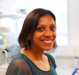 Manju Kurian合作论文数Great Ormond Street Institute of Child Health, University College London;Developmental Neurosciences Department, University College London3Arundel P合作论文数3
 Shamima Rahman合作论文数Department of Genetics & Genomic Medicine, GOS Institute of Child Health, Faculty of Pop Health Sciences, University College London3Calder Alistair D合作论文数3Kaliakatsos Marios合作论文数3
 Francesco Muntoni合作论文数Department of Developmental Neurosciences, Great Ormond Street Institute of Child Health, Faculty of Population Health Sciences, University College London;Dubowitz Neuromuscular Centre, Great Ormond Street Hospital for Children NHS Foundation Trust3Jörg Schmidtke合作论文数3
 Joyce A. Mitchell合作论文数3Stephanie A. Robb合作论文数3
 Athina Ververi合作论文数1st Department of Pediatrics, Aristotle University of Thessaloniki3Hilary Harris合作论文数Department of General Medicine, Manchester University NHS Foundation Trust3
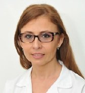 Paola E. Leone合作论文数Facultad de Medicina, Pontificia Universidad Católica del Ecuador3
 Meena Balasubramanian合作论文数Department of Oncology and Metabolism, The Medical School, Faculty of Medicine, Dentistry & Health, The University of Sheffield3




