Reticuloendothelial cells such as tissue-resident macrophages have an important function of clearing unwanted material from our bloodstream. Kupffer cells residing in liver sinusoids comprise the largest contingent of tissue-resident macrophages in our body. Different radiological techniques used to diagnose and treat patients with liver diseases employ the scavenging function of Kupffer cells to clear the contrast agents administered into peripheral vein or hepatic artery. It is useful for the clinician to understand the utility of these “unsung heroes” in liver microcirculation: The Kupffer cells act as scavengers removing waste material from bloodstream and work silently to maintain homeostasis.
Acute fatty liver of pregnancy (AFLP) is a rare cause of catastrophic liver dysfunction and failure in late pregnancy. Defective mitochondrial fatty acid oxidation (FAO) seems to be the underlying pathogenic mechanism. Stressors of both late pregnancy (increased maternal dependence on fats as energy source) and fetus with homozygous defect in mitochondrial FAO, precipitate AFLP in a woman who was previously asymptomatic. Mitochondrial disorders exhibit significant clinical heterogeneity, but many of these primary and secondary disorders have liver as the primary organ affected. The liver injury pattern in these patients also has a varied spectrum and course. Unlike other primary mitochondrial disorders affecting the liver, AFLP represents a potentially completely reversible form of liver injury. Thus, AFLP behaves as a secondary mitochondrial disorder precipitated by inciting factors from fetus and placenta. In this review, we attempt to highlight the varied aspects of mitochondrial dysfunction in AFLP. Mitochondrial injury is the mainstay of pathogenesis and can be recognized in typical clinical features and histopathological findings in patients with AFLP. We also present the consequent impact of recognizing underlying mitochondrial injury on clinical diagnosis and management strategies employed in AFLP.
Background: To assess the prevalence and impact of Clostridium difficile infection (CDI) in hospitalized patients with cirrhosis in India.Methods: In this prospective observational study from June 2015 to March 2016, all hospitalized patients with cirrhosis and acute diarrhea at the time of admission or during hospitalization were included.We studied hospitalized patients with cirrhosis without diarrhea during the same period to detect asymptomatic colonizers.Stool samples were tested for CDI, bacterial cultures, and parasite microscopy in patients with diarrhea.CDI was detected using a stool PCR test that detects the pathogenicity locus of toxigenic Clostridium difficile gene.We analysed the impact of CDI on hospital outcomes and also assessed the risk factors for acquiring CDI.Result: Among 92 hospitalized cirrhotic patients with acute diarrhea [male: 74; median age: 50 (range 19 to 80) years; Child's class A: B: C: 8:41:43; median MELD score: 18 (range 6 to 44)], 6 (6.5%) had CDI by positive stool PCR.Use of antibiotics (100% CDI Vs 55.8% non-CDI, p= 0.04) and steroids (50% CDI vs 10.5% non-CDI, p =0.028) emerged as risk factors for CDI among cirrhosis patients.Two of the 6 patients (33.3%) with CDI as compared to 6/86 patients (7%) with no CDI died (p-value: 0.08).There were no asymptomatic colonizers amongst 35 hospitalized cirrhosis patients without diarrhea.Conclusions: C. difficile, although uncommon, was an important cause of mortality in cirrhosis patients hospitalized with diarrhea in India.Prior use of antibiotics or steroids were identified as risk factors for CDI.
High-volume plasma exchange (PLEX) to treat acute liver failure is now categorized as a Category I indication (i.e., first-line treatment as a stand-alone treatment or with other treatment modalities) by the American Society for Apheresis after a randomized controlled trial of 183 patients demonstrated survival benefit by this treatment. In this review, we provide an introduction to PLEX to treat acute liver failure syndromes for hepatologists and gastroenterologists. From our recent experience of treating 188 patients with acute liver failure syndromes with low-volume PLEX and low-dose steroid, we present five illustrative case histories of patients who benefitted from this management protocol. We discuss some postulated mechanisms how PLEX benefits patients with acute liver failure syndromes. PLEX appears to provide survival benefit in patients with acute liver failure syndromes (severe acute liver injury, acute liver failure, and acute-on chronic liver failure) and may be a nontransplant treatment option for some of these patients, especially in resource-constrained settings.
Background: Liver Transplant recipients have an increased susceptibility to SARS CoV2 infection with a possible more severe disease course. There is paucity of data of SARSCoV2 infection in this cohort from Asia. We report on the data of an on-going APCOLIS-1 (APASL Liver Injury Spectrum, APCOLIS) registry with the aim to define the profile, risk factors for severity and predictors of survival among the liver transplant recipients. Methods: In a multinational study, data was recorded between April 2020 to May 2021 across 13 countries in Asia. The data was compiled on a survey monkey under the APASL COVID-19 study task force [NCT04345640]. We performed multivariate logistic regression to identify independent predictors of severity and all cause mortality among the liver transplant (LT) recipients suffering from COVID-19. Results: Altogether, 130 LT recipients were enrolled [mean age 53 ± 12 years, median post-transplant period, 54 months (range 2-77 months)]. Majority (92, 71% ) of the patients had undergone live-donor transplant. Severe COVID was seen in 21/130 (16%) and 17/130 (13%) required ICU care. COVID related organ failures (OF) were seen in 17 patients (13%), predominantly as respiratory (16/21, 76.2%) followed by renal (9/21, 42.8%) and circulatory (5/21, 23.8%) with ventilatory requirement in 12/21 (57.1%) of the severe cases. Among the baseline parameters age [HR=1.08, 95CI 1.01-1.16, p=0.03], presentation with dyspnea [HR=6.34, 95CI 1.78-22.9, p=0.004] and Neutrophil to Lymphocyte ratio (NLR) [HR=1.08, 95CI 1.01-1.17, p=0.04] independently predicted a severe course of the COVID-19 among LT recipients. The baseline NLR of 8.47 ± 1.45 peaked to 17.94 ± 3.68 in median of 15 days (range 1-37) among severe cases [p<0.001] indicating rapid progression of disease. Age above 55 years increased the disease severity with AUROC of 0.78, sensitivity of 72.7% and specificity of 74.8%. Time from LT, immunosuppression dosage or presence of co-morbidities did not influence the outcome. Graft dysfunction was seen in 21/130 (16%);predominantly as acute cellular rejection in 13/130 (10%) and graft failure 7/130 (5%). The all cause mortality was 8% (11/130). Among non-survivors, the baseline NLR of 4.88 ± 1.63 increased to a peak value of 25.14 ± 5.49 [p<0.001] i.e 5 folds. The baseline NLR [HR=1.17, 95CI 1.03-1.34, p=0.02], development of graft injury [HR=27.21, 95CI 2.55-290., p=0.006] and COVID related OF [ HR=21.87, 95CI 2.39-203.85, p=0.007] independently predicted mortality due to SARSCoV2 infection. Conclusion: COVID-19 infection precipitates a severe disease course in one fifth of the liver transplant recipients, leading to graft dysfunction and early mortality. LT recipients above 55 years of age, presenting with dyspnea and high NLR need to be specifically watched for a progressive disease course. Dynamic NLR determination can help in early stratification and referral to a specialized liver unit to improve outcomes.
Background: COVID-19 among liver transplant (LT) recipients varies with symptoms, severity, time from LT and with treatment from region to region. Here-with we report the clinical presentation, spectrum of disease and outcome from the Asia Pacific region. Methods: In this multinational study, data was recorded between April 2020 to May 2021 across 13 countries in Asia. The data was compiled on survey monkey under the APASL COVID-19 study task force [NCT04345640]. Severity of COVID infection was defined as per WHO guidelines. We analysed symptoms, demography, treatment, clinical course and treatment among LT patients with COVID-19. Results: Among a total 130 LT patients, males were 117 with mean age of 53±12 years and majority were live donor transplant (92,71%). The most common etiology of liver disease was ethanol (37, 28%) followed by NASH (28,22%) and cryptogenic(21,16%). Median post LT period was 54 months. Co-morbidity was present in 101 patients (78%), the most common being obesity (61,47%),followed by Diabetes (56,43%) and hypertension(30,23%). The presenting complaints were fever(82%), cough(61%), dyspnoea(29%) and diarrhoea(7%);8% were asymptomatic. Respiratory distress was seen in 29 (22%) cases;17 received only oxygen and 12 needed ventilator support. Prior to illness, only CNI was used as immunosuppressant in 30% (39), CNI & MMF in 31% (40) and in another 31% (40) mTOR inhibitors alone or with a CNI and triple regimen in 9%. During COVID, only low dose steroid was used in 50% (64), low dose CNI in mild to moderate ( 32%,41), complete avoidance of MMF in 4% cases where as low dose CNI and MMF in 16% (20) patients. Immunosuppression reintroduction done in 68% to pre-COVID dose and 28% were maintained on low dose CNI with steroids with close monitoring of LFT and pre-COVID dose was restarted after 14 days of recovery .4% had graft dysfunction leading to early augmentation of immunosuppression. Any antiviral was received by 36%, convalescent plasma by 26% and immunomodulators like tocilizumab or bevacizumab in 13%. Home care offered to 46% cases while those admitted, 41% were managed in ward only and 13% needed ICU admission. Graft function was unaffected in majority (109, 84%) but Acute Cellular Rejection was noted in 10% cases and DILI or other causes in 6%. Liver injury in the form of raised transaminases or bilirubin was noted in 19% at hospital stay and in 40% at presentation. Severe COVID was in 16% (21) cases with mortality in 8% (11). Conclusion: Compared to general population post-LT patients have an increased mortality due to COVID. Home based care could be feasible in only half of the patients.
Objectives: The aim of this study is to assess the utility of plasma von-Willebrand factor (VWF) levels in predicting in-hospital survival for patients with severe alcoholic hepatitis. Methods: From a prospectively collected database of acute-on-chronic liver failure (ACLF) patients, we retrospectively selected all severe alcoholic hepatitis (discriminant function [DF] ≥32) patients and correlated baseline plasma VWF (antigen, Ag and collagen-binding activity [CBA]) levels with disease severity. In-hospital survival was classified as discharged alive (or) died/discharged in a terminal condition (poor outcome). Results: Of 34 consecutive severe alcoholic hepatitis patients (age: 40.5, 30–63 years; median, range, hospital stay: 6, 2–15 days) studied, 15 had a poor outcome. Plasma VWF-CBA was higher in patients with poor outcome (736 [396–1157] IU/dL) as compared to patients discharged in stable state (492 [97–986] IU/dL; P = 0.03). VWF-CBA correlated well with VWF-Ag, model for end-stage liver disease (MELD) score, sequential organ failure assessment score and ACLF grades. AUROC to predict composite poor outcome was similar for plasma VWF-CBA (0.72, 0.54–0.89) and MELD score (0.71, 0.53–0.89). Plasma VWF-CBA was higher in 23 “very severe” alcoholic hepatitis (DF >60 or MELD >30) patients. Combining plasma VWF-CBA (>750 IU/dL) and criteria for “very severe” alcoholic hepatitis had a sensitivity of 100% (81%–100%) and negative predictive value of 100% (68%–100%) for predicting poor outcome. Conclusions: Plasma VWF levels are markedly elevated, correlate with organ failure, and predict in-hospital survival in severe alcoholic hepatitis, and also in “very severe” alcoholic hepatitis, patients.
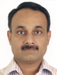 Goel Apul合作论文数Dept Urol, King George Med Univ8
 Shiv Kumar Sarin合作论文数Department of Hepatology, Institute of Liver and Biliary Sciences;Jawaharlal Nehru Center for Advanced Scientific Research8
 Uday Zachariah合作论文数7
 Kalal Chetan Ramesh合作论文数Department of Hepatology, Institute of Liver and Biliary Sciences7
 Balakrishnan Ramakrishna合作论文数SRM Institutes for Medical Science7j ramachandran合作论文数6
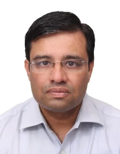 U Zachariah合作论文数Christian Medical College Vellore6
 Ashok Kumar Choudhury合作论文数Institute of Liver & Biliary Sciences6
 Sunil Taneja合作论文数Madhuraj Nursing Home6
 Radha Krishan Dhiman合作论文数Department of Hepatology, Postgraduate Institute of Medical Education and Research, Chandigarh6G Kurian合作论文数5Samir Shah合作论文数5
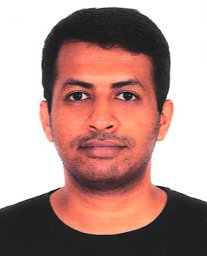 Vijay Alexander合作论文数Christian Medical College5
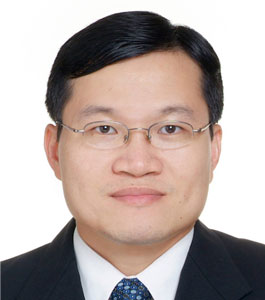 Guan Huei Lee合作论文数Yong Loo Lin School of Medicine, National University of Singapore5Devarbhavi Harshad合作论文数5
 Gagandeep (Cherry) Kang合作论文数Department of Public Health and Community Medicine, Tufts University;Wellcome Trust Research Laboratory, Division of Gastrointestinal Sciences, Christian Medical College, Vellore5Sajith Kattiparambil Gangadharan合作论文数5
 Madhumita Premkumar合作论文数National Centre for Antarctic and Ocean Research5
 Sombat Treeprasertsuk合作论文数Department of Medicine, Faculty of Medicine, Chulalongkorn University5
 Qin Ning (宁琴)合作论文数Tongji Hospital, Tongji Medical College of Huazhong University of Science & Technology5George K.K. Lau合作论文数5
 S. M. Wasim Jafri合作论文数Section of Gastroenterology, Department of Medicine, Medical College, Pakistan, The Aga Khan University;South City Hospital5
 Saeed Sadiq Hamid合作论文数Section of Gastroenterology, Department of Medicine, Aga Khan University5M Chandy合作论文数4Zaigham Abbas合作论文数4
 Mamun Mahtab合作论文数Interventional Hepatology Division, Bangabandhu Sheikh Mujib Medical University;Department of Hepatology, Bangabandhu Sheikh Mujib Medical University4
 Nagaraja P Rao合作论文数4
 Soek Siam Tan合作论文数4
 Ajay Duseja合作论文数Department of Hepatology, Postgraduate Institute of Medical Education and Research4Ramachandran Jeyamani合作论文数4
 Rakhi Maiwall合作论文数4
 Anand Kulkarni合作论文数4
 Ashish Goel合作论文数Department of Management Science and Engineering, Stanford University;Coinbase, Inc.4
 Osamu Yokosuka合作论文数Chiba University4
 Virendra Singh合作论文数Department of Electrical Engineering, Indian Institute of Technology Bombay;Department of Computer Science & Engineering, Indian Institute of Technology Bombay4George Chandy合作论文数4K. A. Balasubramanian合作论文数4
 Akash Shukla合作论文数3Gian Carpio合作论文数3Irene Paulson合作论文数3
 Shahinul Alam合作论文数3
 Jimil Shah合作论文数Dept Gastroenterol, Postgrad Inst Med Educ & Res PGIMER3V. G. Mohan Prasad合作论文数3Diana A. Payawal合作论文数Cardinal Santos Med Ctr3Laurentius A. Lesmana合作论文数3
 Shyamkumar Nidugala Keshava合作论文数Department of Radiology, Christian Medical College3
 Omesh Goyal合作论文数Dayanand Medical College and Hospital3
 Seng Gee Lim合作论文数Division of Gastroenterology and Hepatology, Department of Medicine, National University Hospital;National University Centre for Organ Transplantation, National University Hospital;Department of Medicine, Yong Loo Lin School of Medicine, National University of Singapore3




