TEAD (transcriptional enhanced associate domain) transcription factors (TEAD1-4) serve as the primary effectors of the Hippo signaling pathway in various cancers. Targeted therapy leads to the emergence of resistance and the underlying mechanism of resistance to TEAD inhibition in cancers is less characterized. We uncover that upregulation of the AP-1 (activator protein-1) transcription factors, along with restored YAP (yes-associated protein) and TEAD activity, drives resistance to GNE-7883, a pan-TEAD inhibitor. Acute GNE-7883 treatment abrogates YAP-TEAD binding and attenuates FOSL1 (FOS like 1) activity. TEAD inhibitor resistant cells restore YAP and TEAD chromatin occupancy, acquire additional FOSL1 binding and exhibit increased MAPK (mitogen-activated protein kinase) pathway activity. FOSL1 is required for the chromatin binding of YAP and TEAD. This study describes a clinically relevant interplay between the Hippo and MAPK pathway and highlights the key role of MAPK pathway inhibitors in mitigating resistance to TEAD inhibition in Hippo pathway dependent cancers.
Lipid synthesis increases during the cell cycle to ensure sufficient membrane mass, but how insufficient synthesis restricts cell-cycle entry is not understood. Here, we identify a lipid checkpoint in G1 phase of the mammalian cell cycle by using live single-cell imaging, lipidome, and transcriptome analysis of a non-transformed cell. We show that synthesis of fatty acids in G1 not only increases lipid mass but extensively shifts the lipid composition to unsaturated phospholipids and neutral lipids. Strikingly, acute lowering of lipid synthesis rapidly activates the PERK/ATF4 endoplasmic reticulum (ER) stress pathway that blocks cell-cycle entry by increasing p21 levels, decreasing Cyclin D levels, and suppressing Retinoblastoma protein phosphorylation. Together, our study identifies a rapid anticipatory ER lipid checkpoint in G1 that prevents cells from starting the cell cycle as long as lipid synthesis is low, thereby preventing mitotic defects, which are triggered by low lipid synthesis much later in mitosis.
Transcriptional enhanced associate domain family members 1 to 4 (TEADs) are a family of four transcription factors and the major transcriptional effectors of the Hippo pathway. In order to activate transcription, TEADs rely on interactions with other proteins, such as the transcriptional effectors Yes-associated protein and transcriptional co-activator with PDZ-binding motif. Nuclear protein interactions involving TEADs influence the transcriptional regulation of genes involved in cell growth, tissue homeostasis, and tumorigenesis. Clearly, protein interactions for TEADs are functionally important, but the full repertoire of TEAD interaction partners remains unknown. Here, we employed an affinity purification mass spectrometry approach to identify nuclear interacting partners of TEADs. We performed affinity purification mass spectrometry experiment in parallel in two different cell types and compared a wildtype TEAD bait protein to a nuclear localization sequence mutant that does not localize to the nucleus. We quantified the results using SAINT analysis and found a significant enrichment of proteins linked to DNA damage including X-ray repair cross-complementing protein 5 (XRCC5), X-ray repair cross-complementing protein 6 (XRCC6), poly(ADP-ribose) polymerase 1 (PARP1), and Rap1-interacting factor 1 (RIF1). In cellular assays, we found that TEADs co-localize with DNA damage-induced nuclear foci marked by histone H2AX phosphorylated on S139 (γH2AX) and Rap1-interacting factor 1. We also found that depletion of TEAD proteins makes cells more susceptible to DNA damage by various agents and that depletion of TEADs promotes genomic instability. Additionally, depleting TEADs dampens the efficiency of DNA double-stranded break repair in reporter assays. Our results connect TEADs to DNA damage response processes, positioning DNA damage as an important avenue for further research of TEAD proteins.
Cell-to-cell heterogeneity is vital for tumor evolution and survival. How cancer cells achieve and exploit this heterogeneity remains an active area of research. Here, we identify c-Myc as a highly heterogeneously expressed transcription factor and an orchestrator of transcriptional and phenotypic diversity in cancer cells. By monitoring endogenous c-Myc protein in individual living cells, we report the surprising pulsatile nature of c-Myc expression and the extensive cell-to-cell variability in its dynamics. We further show that heterogeneity in c-Myc dynamics leads to variable target gene transcription and that timing of c-Myc expression predicts cell-cycle progression rates and drug sensitivities. Together, our data advocate for a model in which cancer cells increase the heterogeneity of functionally diverse transcription factors such as c-Myc to rapidly survey transcriptional landscapes and survive stress.
Abstract The TEA domain family members 1-4 (TEADs) are major transcription factors for YAP/TAZ transcription activators in the Hippo pathway, regulating many biological processes, including development, tissue homeostasis, and tumorigenesis through target genes. Their amplification/upregulation correlates with poor prognosis in cancer patients. Although the Hippo pathway continues to be elucidated, it is clear that TEAD largely exerts its actions via transcriptional regulation. Here, we uncover an unexpected role for TEADs in the DNA damage response. Using comparative mass spectrometry, we demonstrate that TEADs interact with several DNA repair proteins. We further show that TEADs form DNA damage-induced nuclear foci that co-localize with DNA damage markers, γH2AX, and RIF1. We also found that TEADs are required for resistance to DNA damage, maintaining genome stability, and resolution of double strand break repair that is independent from the Hippo pathway and its transcriptional role. Our results establish a new role for TEADs in DNA repair and therefore, highlight a critical consideration in therapeutically targeting the Hippo pathway.
Myc is the most commonly amplified oncogene, and is known to play critical roles in cancer establishment and maintenance. Myc expression heterogeneity has also been linked to cancer drug resistance; however, the source of this heterogeneity remains poorly characterized. Even in isogenic cancer cell lines, we found that Myc is one of the most heterogeneously expressed transcription factors at the single-cell level. This heterogeneity is independent of cell cycle progression, local cell density, and p53/p21 signaling. To elucidate the source and duration of Myc heterogeneity, we endogenously tagged Myc with a fluorescent protein and used long-term live-cell imaging techniques to monitor Myc expression over time at single-cell resolution. Surprisingly, we found that Myc expression is pulsatile, explaining why Myc expression appears heterogeneous at a given point in time. Furthermore, we found that Myc dynamics are heterogeneous, and the dynamics are not always heritable. Additionally, Myc pulse amplitude is correlated with the duration of G1 and the overall cell cycle, suggesting that the pulses may be a mechanism for escaping many chemotherapy agents. Finally, we show that Myc pulses are primarily transcription-driven and controlled by at least one negative feedback loop. Our findings demonstrate for the first time that Myc expression is pulsatile, and provide insights into Myc regulation in cancer. Citation Format: Chad Liu, Karen Gascoigne. Pulsatile Myc expression as a source of Myc heterogeneity in cancer cells [abstract]. In: Proceedings of the American Association for Cancer Research Annual Meeting 2022; 2022 Apr 8-13. Philadelphia (PA): AACR; Cancer Res 2022;82(12_Suppl):Abstract nr 1592.
After two converging DNA replication forks meet, active replisomes are disassembled and unloaded from chromatin. A key process in replisome disassembly is the unloading of CMG helicases (CDC45-MCM-GINS), which is initiated in Caenorhabditis elegans and Xenopus laevis by the E3 ubiquitin ligase CRL2(LRR1). Here, we show that human cells lacking LRR1 fail to unload CMG helicases and accumulate increasing amounts of chromatin-bound replisome components as cells progress through S phase. Markedly, we demonstrate that the failure to disassemble replisomes reduces the rate of DNA replication increasingly throughout S phase by sequestering rate-limiting replisome components on chromatin and blocking their recycling. Continued binding of CMG helicases to chromatin during G2 phase blocks mitosis by activating an ATR-mediated G2/M checkpoint. Finally, we provide evidence that LRR1 is an essential gene for human cell division, suggesting that CRL2(LRR1) enzyme activity is required for the proliferation of cancer cells and is thus a potential target for cancer therapy.
During muscle regeneration, extracellular signal-regulated kinase (ERK) promotes both proliferation and migration. However, the relationship between proliferation and migration is poorly understood in this context. To elucidate this complex relationship on a physiological level, we established an intravital imaging system for measuring ERK activity, migration speed, and cell-cycle phases in mouse muscle satellite cell-derived myogenic cells. We found that in vivo, ERK is maximally activated in myogenic cells two days after injury, and this is then followed by increases in cell number and motility. With limited effects of ERK activity on migration on an acute timescale, we hypothesized that ERK increases migration speed in the later phase by promoting cell-cycle progression. Our cell-cycle analysis further revealed that in myogenic cells, ERK activity is critical for G1/S transition, and cells migrate more rapidly in S/G2 phase 3 days after injury. Finally, migration speed of myogenic cells was suppressed after CDK1/2-but not CDK1-inhibitor treatment, demonstrating a critical role of CDK2 in myogenic cell migration. Overall, our study demonstrates that in myogenic cells, the ERK-CDK2 axis promotes not only G1/S transition but also migration, thus providing a novel mechanism for efficient muscle regeneration.
Cell-cycle entry relies on an orderly progression of signaling events. To start, cells first activate the kinase cyclin D-CDK4/6, which leads to eventual inactivation of the retinoblastoma protein Rb. Hours later, cells inactivate APC/CCDH1 and cross the final commitment point. However, many cells with genetically deleted cyclin Ds, which activate and confer specificity to CDK4/6, can compensate and proliferate. Despite its importance in cancer, how this entry mechanism operates remains poorly characterized, and whether cells use this path under normal conditions remains unknown. Here, using single-cell microscopy, we demonstrate that cells with acutely inhibited CDK4/6 enter the cell cycle with a slowed and fluctuating cyclin E-CDK2 activity increase. Surprisingly, with low CDK4/6 activity, the order of APC/CCDH1 and Rb inactivation is reversed in both cell lines and wild-type mice. Finally, we show that as a consequence of this signaling inversion, Rb inactivation replaces APC/CCDH1 inactivation as the point of no return. Together, we elucidate the molecular steps that enable cell-cycle entry without CDK4/6 activity. Our findings not only have implications in cancer resistance, but also reveal temporal plasticity underlying the G1 regulatory circuit.
Mammalian cells typically start the cell-cycle entry program by activating cyclin-dependent protein kinase 4/6 (CDK4/6). CDK4/6 activity is clinically relevant as mutations, deletions, and amplifications that increase CDK4/6 activity contribute to the progression of many cancers. However, when CDK4/6 is activated relative to CDK2 remained incompletely understood. Here, we developed a reporter system to simultaneously monitor CDK4/6 and CDK2 activities in single cells and found that CDK4/6 activity increases rapidly before CDK2 activity gradually increases, and that CDK4/6 activity can be active after mitosis or inactive for variable time periods. Markedly, stress signals in G1 can rapidly inactivate CDK4/6 to return cells to quiescence but with reduced probability as cells approach S phase. Together, our study reveals a regulation of G1 length by temporary inactivation of CDK4/6 activity after mitosis, and a progressively increasing persistence in CDK4/6 activity that restricts cells from returning to quiescence as cells approach S phase.
Cell-cycle entry relies on an orderly progression of signaling events. To start, cells first activate the kinase cyclin D-CDK4/6, which leads to eventual inactivation of the retinoblastoma protein Rb. Hours later, cells inactivate APC/C CDH1 and cross the final commitment point. However, many cells with genetically deleted cyclin Ds, which activate and confer specificity to CDK4/6, can compensate and proliferate. Despite its importance in cancer, how this alternate pathway operates and whether wild-type cells use this pathway remain unknown. Here, using single-cell microscopy, we demonstrate that cells with acutely inhibited CDK4/6 enter the cell cycle with slowed and fluctuating cyclin E-CDK2 activity. Surprisingly, in this alternate pathway, the order of APC/C CDH1 and Rb inactivation is inverted in both cell lines and wild-type mice. Finally, we show that as a consequence of the signaling inversion, Rb inactivation replaces APC/C CDH1 inactivation as the point of no return. Together, we provide molecular characterization of a parallel cell-cycle entry pathway, and reveal temporal plasticity that underlies the G1 regulatory circuit.
Cells escape the need for mitogens at a restriction point several hours before entering S phase. The restriction point has been proposed to result from CDK4/6 initiating partial Rb phosphorylation to trigger a bistable switch whereby cyclin E-CDK2 and Rb mutually reinforce each other to induce Rb hyperphosphorylation. Here, using single-cell analysis, we unexpectedly found that cyclin E/A-CDK activity can only maintain Rb hyperphosphorylation starting at the onset of S phase and that CDK4/6 activity, but not cyclin E/A-CDK activity, is required to hyperphosphorylate Rb throughout G1 phase. Mitogen removal in G1 results in a gradual loss of CDK4/6 activity with a high likelihood of cells sustaining Rb hyperphosphorylation until S phase, at which point cyclin E/A-CDK activity takes over. Thus, it is short-term memory, or transient hysteresis, in CDK4/6 activity following mitogen removal that sustains Rb hyperphosphorylation, demonstrating a probabilistic rather than an irreversible molecular mechanism underlying the restriction point.
Faithful DNA replication is challenged by stalling of replication forks during S phase. Replication stress is further increased in cancer cells or in response to genotoxic insults. Using live single-cell image analysis, we found that CDK2 activity fluctuates throughout an unperturbed S phase. We show that CDK2 fluctuations result from transient ATR signals triggered by stochastic replication stress events. In turn, fluctuating endogenous CDK2 activity causes corresponding decreases and increases in DNA synthesis rates, linking changes in stochastic replication stress to fluctuating global DNA replication rates throughout S phase. Moreover, cells that reenter the cell cycle after mitogen stimulation have increased CDK2 fluctuations and prolonged S phase resulting from increased replication stress induced CDK2 suppression. Thus, our study reveals a dynamic control principle for DNA replication whereby CDK2 activity is suppressed and fluctuates throughout S phase to continually adjust global DNA synthesis rates in response to recurring stochastic replication stress events.
Apoptosis is a tightly controlled process in mammalian cells. It is important for embryogenesis, tissue homoeostasis, and cancer treatment. Apoptosis not only induces cell death, but also leads to the release of signals that promote rapid proliferation of surrounding cells through the Phoenix Rising (PR) pathway. To quantitatively understand the kinetics of interactions of different molecules in this pathway, we developed a mathematical model to simulate the effects of various changes in the PR pathway on the secretion of prostaglandin E2 (PGE2), a key factor for promoting cell proliferation. These changes include activation of caspase 3 (C3), caspase 7 (C7), and nuclear factor κB (NFκB). In addition, we simulated the effects of cyclooxygenase-2 (COX2) inhibition and C3 knockout on the level of secreted PGE2. The model predictions on PGE2 in MEF and 4T1 cells at 48 hours after 10-Gray radiation were quantitatively consistent with the experimental data in the literature. Compared to C7, the model predicted that C3 activation was more critical for PGE2 production. The model also predicted that PGE2 production could be significantly reduced when COX2 expression was blocked via either NFκB inactivation or treatment of cells with exogenous COX2 inhibitors, which led to a decrease in the rate of conversion from arachidonic acid to prostaglandin H2 in the PR pathway. In conclusion, the mathematical model developed in this study yielded new insights into the process of tissue regrowth stimulated by signals from apoptotic cells. In future studies, the model can be used for experimental data analysis and assisting development of novel strategies/drugs for improving cancer treatment or normal tissue regeneration.
 Tobias Meyer合作论文数Weill Cornell Medical College, Cornell University8
 Mingyu Chung合作论文数Stanford Univ, Sch Med, Dept Chem & Syst Biol, Stanford, CA 94305 USA6
 Yilin Fan合作论文数Tsinghua Univ, Sch Life Sci, MOE Key Lab Prot Sci, Beijing 100084, Peoples R China5
 Daigh Leighton H合作论文数Department of Microbiology and Immunology, Georgetown University Medical Center4
 Hee Won Yang合作论文数Dept. of Biological Sciences, Korea Advanced Institute of Science and Technology4
 Marielle S Koberlin合作论文数Dept Chem & Syst Biol, Stanford Univ3
 Yumi Konagaya合作论文数Laboratory of Bioimaging and Cell Signaling, Graduate School of Biostudies, Kyoto University, Kyoto 606-8501, Japan3Anwesha Dey合作论文数3
 Kenta Terai (寺井健太)合作论文数Department of Histology, Graduate School of Biomedical Sciences, Tokushima University3
 Michiyuki Matsuda合作论文数Department of Tumor Virology;Research Institute for Microbial Diseases;Osaka University;Research Institute for Microbial Diseases, Osaka University3Noelyn M. Kljavin合作论文数3
 Philamer Calses合作论文数2Rebecca K Rowntree合作论文数2Trent Hinkle合作论文数MPL, Genentech2Trang Pham合作论文数2Alissa D. Guarnaccia合作论文数2
 Lill Jennie R合作论文数Dept Prot Chem, Genentech Inc2Haley Benjamin合作论文数Dept Mol Biol, Genentech Inc2
 Matthew Chang合作论文数Department of Bioengineering and Therapeutic Sciences,2Bakker Sietske T合作论文数Division of Molecular Biology, Netherlands Cancer Institute2
 Erik Verschueren合作论文数2
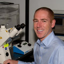 Steven Cappell合作论文数Department of Pharmacology, University of North Carolina at Chapel Hill2
 Alan Ashworth合作论文数Division of Hematology and Oncology, Department of Medicine, UCSF;Helen Diller Family Comprehensive Cancer Center, UCSF2Victoria Pham合作论文数2
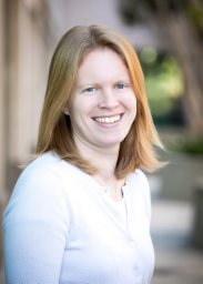 Karen E. Gascoigne合作论文数Whitehead Institute for Biomedical Research, and Department of Biology, Massachusetts Institute of Technology2Antônio F M Pinto合作论文数1Kanako Takakura合作论文数1Christy Ong合作论文数1
 Thijs J Hagenbeek合作论文数1Lindsey R Pack合作论文数1Sayantanee Paul合作论文数Department of Biology, University of Massachusetts Boston1
 Maina Sogabe合作论文数1
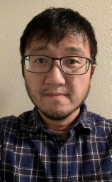 Peter L Hsu合作论文数Department of Pharmacology, University of Washington;Howard Hughes Medical Institute, University of Washington;University of Washington, University of Washington1Madhura Deshpande合作论文数Icahn Sch Med Mt Sinai, Precis Immunol Inst, New York, NY 10029 USA1
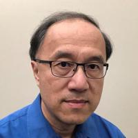 Fan Yuan合作论文数Department of Biomedical Engineering, Pratt School of Engineering, Duke University1
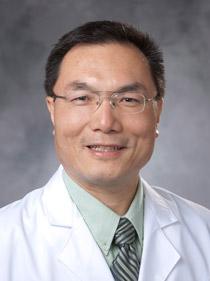 Chuan-Yuan Li合作论文数Department of Dermatology, Duke University School of Medicine;Duke Cancer Institute;Duke Regeneration Center1
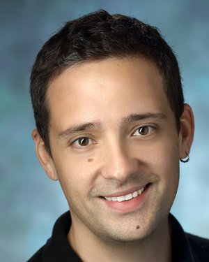 Sergi Regot合作论文数Department of Otolaryngology-Head and Neck Surgery, School of Medicine, Johns Hopkins University1
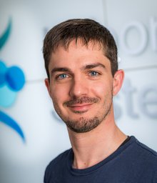 Luca Gerosa合作论文数Institute of Molecular System Biology, ETH Zurich;Institute of Molecular System Biology, ETH Zurich1
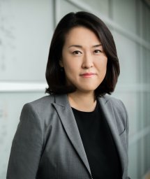 Meena Choi合作论文数1Blake Robert A合作论文数1
 Takamasa Kudo合作论文数Genenetech, Inc;AWAKENS, Inc.1
 Ratnayeke Nalin合作论文数Center for Nonlinear Dynamics and Department of Physics, University of Texas1Bisaria Anjali合作论文数Lewis-Sigler Institute for Integrative Genomics, Princeton University1
 Jeannine Gerhardt合作论文数Department of Cell Biology, Albert Einstein College of Medicine1
 Xiaosai Yao合作论文数Koch Institute for Integrative Cancer Research, Massachusetts Institute of Technology;Department of Biological Engineering, Massachusetts Institute of Technology;Massachusetts Institute of Technology, Massachusetts Institute of Technology1Lacap Jennifer A合作论文数1
 Xie Shiqi合作论文数Shanghai Institutes for Biological Sciences, Chinese Academy of Sciences1Corey E. Bakalarski合作论文数1
 Markus Covert合作论文数Department of Bioengineering, Stanford University;Department of Chemical and Systems Biology, Stanford University;hriram Center for Bioengineering and Chemical Engineering, Stanford University1




