This Innovative Practice Full Paper presents a new digitized Draw-A-Scientist Test (DAST) tool and discusses the results of deploying it to minority students in at-risk schools. Pervasive science stereotypes, particularly stereotypes of scientists, continue to negatively affect minoritized students' perceptions of science and commitment to pursuing STEM-related careers. One helpful tool for better understanding students' perceptions of scientists through projective tests is Chambers's DAST, initially created in 1983. The original protocol for deploying the DAST in a research setting asked students to “draw a scientist” on paper. Then, researchers could assess the seven indicators of stereotypical scientists (i.e., eyeglasses, facial hair, white lab coats, symbols of knowledge, symbols of research, technology, and captions). However, to improve inter-rater reliability and capture a more accurate reflection of students' perceptions of scientists, Finson, Beaver, and Cramond reenvisioned DAST scoring and assessment in 1995 to include eight additional indicators (i.e., indicators of danger, indicators of secrecy, mythic figures, presence of light bulbs, sex indicator for male, Caucasian, middle-aged or elderly, and scientists working indoors). Finson and team called the new DAST scoring rubric the DAST-C, which remains the most widely used one. However, methodological issues still impact DAST assessments, and updated tools for technology have made paper/pencil tasks obsolete, even for young students. With support from the National Science Foundation's Innovative Technology Experiences for Students and Teachers (NSF ITEST) program, our collaborative interdisciplinary team infused technology-rich STEM learning modules that incorporated a proof-of-concept digitized version of the DAST into at-risk 3 rd –5 th -grade classrooms serving minoritized students. We compared findings from the digitized DAST with a priori paper/pencil data to highlight critical differences in DAST implementation and scoring. The current study had two primary objectives related to updating the DAST as a tool and in terms of streamlining DAST scoring: 1) digitizing the DAST to bring the paper/pencil version into the 21st century and 2) streamlining DAST assessment by integrating digital assessment tools into the new digitized DAST tool. Here, we discuss digitizing the DAST tool, assessing indicators of stereotypical scientists, and deploying the digitized DAST to a population of minoritized students studying in at-risk schools in America.
Constantly evolving, high-intensity functional training (HIFT) exercise consists of various modalities, orders, weights, and repetition schemes. High-intensity functional training has gained popularity among the general population, but lacks empirical evidence regarding acute adaptive responses. The purpose of this study was to describe the acute effects of 2 representative bouts of HIFT on physiologic markers of growth. For convenience, the bouts are designated "short" (<5 minutes) and "long" (similar to 15 minutes), although duration was not the only difference between bouts. Ten apparently healthy men (28.1 +/- 5 years) performed 2 HIFT bouts in a randomized crossover design. Blood was collected at 5 time points (Pre, Post, 1, 3, and 6 hours) to examine growth hormone (GH), insulin-like growth factor (IGF-1), insulin-like growth factor binding proteins 1 and 2 (IGFBP-1 and IGFBP-2), and vascular endothelial growth factor (VEGF). Blood lactate concentration ([La]) was analyzed at the Pre and Post time points. A repeated-measures analysis of variance (ANOVA) revealed no trial differences among the markers (IGF-1, IGFBP-1, IGFBP-2, and VEGF) except GH at Post, where the long bout produced a greater effect (p = 0.005). Mean GH levels (pgml-1) in the short bout increased from 68.4 to 106.5, and in the long bout, mean GH levels increased from 38.5 to 286.4. The repeated-measures ANOVA revealed a main time effect in GH (p = 0.037), while a post hoc t-test demonstrated elevated GH at 1 hour (p = 0.018) when compared with Pre. No time-dependent change (p > 0.05) was observed in IGF-1, IGFBP-1, IGFBP-2, or VEGF. Mean blood [lactate] increased more than tenfold in both bouts. The findings of this descriptive study suggest that, other than GH, there are no acute differences in markers of skeletal muscle or vascular growth between these 2 specific HIFT bouts.
Effects of varying types of short duration workouts in high-intensity functional training (HIFT) on inflammatory biomarkers have not been adequately characterized. Objectives: The purpose of this descriptive study was to examine the acute effects of HIFT workouts on biomarkers of inflammation, over time, in two HIFT bouts. Materials and Methods: Ten apparently healthy males (28.1 ± 5 yrs) completed two HIFT sessions (“short bout:” sub-5-minute vs. “long bout:” 15-minute) in a randomized crossover design. Blood was drawn pre and post-exercise, and 1 hour, 3 hours, and 6 hours post-exercise, centrifuged, and plasma frozen for analysis. Inflammation was assessed through plasma interleukin-6 (IL-6), interleukin-10 (IL-10), and tumour necrosis factor alpha (TNF-α). Results: Repeated measures ANOVA revealed a single trial-dependent difference (IL-6, p≤ 0.05), and while statistically significant, this difference may not be biologically significant. The biomarkers IL-6, IL-10, and TNF-α all follow a similar pattern of peaking post-exercise and returning to baseline within 6 hours in both trials. Conclusions: Both temporal responses and concentrations were similar in the short and long bout. A practical implication is that both bouts of a HIFT elicit certain specific physiologic inflammatory responses.
Plasma biomarkers of skeletal muscle growth are influenced by the modality, duration, and intensity of an exercise bout. High-Intensity Training Programs (HITP) are prescribed using various modalities, orders, weights, and repetition schemes. The duration of these bouts varies greatly, from shorter bouts of less than 5 minutes to longer bouts of 15 minutes or more. PURPOSE: To examine the effects of short- (<5min) and long- (15min) duration bouts of HITP on markers of skeletal muscle growth. METHODS: Ten apparently healthy males (28.1 ± 5yrs) participated in this study. Two HITP sessions (SHORT and LONG) were performed in a randomized crossover fashion. Blood plasma was collected at five time points: PRE, POST, 1HR, 3HR, and 6HR in order to examine growth hormone (GH), insulin-like growth factor (IGF-1), and insulin- like growth factor binding proteins 1 & 2 (IGFBP-1, IGFBP-2). RESULTS: The repeated measures ANOVA revealed no trial differences among any of the markers (IGF-1, IGFBP-1 & IGFBP-2) except GH at POST, where the LONG bout produced a greater effect (p = 0.005). A repeated measures ANOVA revealed a main time effect in GH (p = 0.037), while a posthoc t-test demonstrated elevated GH at 1HR (p = 0.018) when compared to PRE, while no time-dependent change (p > 0.05) was observed in IGF-1, IGFBP-1, or IGFBP-2. CONCLUSIONS: The findings suggest that there are no differences in markers of skeletal muscle growth other than GH between the SHORT and LONG bouts of HITP.
Background: Duchenne muscular dystrophy (DMD) is a common and lethal genetic disease. Cardiomyopathy frequently causes mortality in DMD patients. However, its cardiac pathophysiology is unclear. Wh...
Effects of varying types of high-intensity training (HIT) on the appearance and time course of circulating biomarkers have not been adequately characterized. PURPOSE: The purpose of this study was to examine the effects of HIT on biomarkers of inflammation and angiogenesis, over time, in HIT bouts of varying duration (short bout, 5 min, and long bout, 15 min). METHODS: Ten males, 18-45 years old, who had participated in CrossFit for at least 6 months, were recruited from local affiliates. The participants completed a total of 3 lab visits [Visit 1: collection of descriptive data; the next 2 visits were randomized between the short bout and long bout]. All subjects completed the same exercises. Blood was drawn pre and post-exercise, and 1 hour, 3 hours, and 6 hours post-exercise, centrifuged, and plasma frozen for analysis. A multiplex assay (Millipore MagPix) was used to determine concentrations of the biomarkers of interest [interleukin 6 (IL-6), interleukin 10 (IL-10), tumor necrosis factor alpha (TNF-alpha), and vascular endothelial growth factor (VEGF)]. RESULTS: The short bout produced results similar to those of the long bout. Repeated measures ANOVA revealed no trial-dependent differences (p≤ 0.05) in any of the biomarkers. Both temporal responses and concentrations were similar in the short and long bout. CONCLUSIONS: The biomarkers IL-6, IL-10, TNF-alpha, and VEGF all follow a similar pattern of peaking post-exercise, and returning to baseline within 6 hours, regardless of the duration of the HIT. A practical implication is that a 5 min bout of HIT may be just as effective as a 15 min bout in terms of eliciting certain specific physiologic responses. Supported by WSSU Office of Student Research, Honors Student Research Grant (K.N.C.).
Regenerative medicine is a novel discipline that both excites undergraduates and may be used as a vehicle to expose students to scientific concepts and opportunities. The goal of this article is to describe the implementation of a National Science Foundation-funded Targeted Infusion Project in which underrepresented minority undergraduates are exposed to laboratory-bench skills and summer research opportunities that they may not have encountered otherwise. A 3-wk infusion of laboratory-bench and data presentation skills, in the context of a regenerative medicine/bioengineering project, aimed to engage students and expose them to opportunities as summer researchers and teaching assistants. The infusion aimed to assess the extent to which students improved 1) attitudes toward laboratory-bench-based techniques, using attitudes toward science as a proxy; 2) perceptions of scientific inquiry; 3) intentions to engage in undergraduate research; and 4) intentions to persist in science, technology, engineering, and mathematics (STEM)-related fields. Results indicate that the 3-wk infusion had no effect on science attitudes, but transcribed responses to structured interviews administered after the summer research experience indicated that students who completed summer research projects had positive experiences. Differences in intentions to engage in research were detected between groups of students in different STEM majors, in addition to differences in intentions to pursue a career in science. We describe the implementation of the infusion and briefly discuss quantitative outcomes. We conclude that infusion of laboratory-bench modules in the context of a regenerative medicine/ bioengineering project may play a small but important role in increasing (minority) participation and persistence in the STEM pipeline.
PURPOSE: The purpose of these pilot studies is to validate PCR primer sets intended to describe mitochondrial damage-associated molecular patterns (mtDAMPs, which influence the immune system) in human plasma samples, after the muscle injury evoked by extreme exercise, such as a marathon. We hypothesize that circulating concentrations of mtDAMPs—specifically, mtDNA—are elevated post-marathon relative to pre-marathon. With quantitative PCR, evaluation of the change in mtDAMP levels pre- vs. post-marathon is enabled via comparing these levels to an internal reference (“housekeeping”) gene that is unaffected by exercise. METHODS: All procedures were IRB approved and all subjects (n=11) provided informed consent. Blood was obtained by antecubital venipuncture at baseline and within 48 hours post-race. Blood was centrifuged, plasma aliquotted, and stored at -80°C for further analyses. Plasma DNA was isolated using a commercially available mini kit (Qiagen). Primers for human mtDNA (cytochrome B, cytochrome C oxidase subunit III, NADH dehydrogenase), and bacterial 16S rRNA were synthesized by Eurofins Genomics using published sequences. The human sequences have no significant homology with DNA found in any bacterial species published on BLAST. To determine whether levels of 3 candidate housekeeping genes are consistent pre- vs. post-marathon, the housekeeping primer sequences were added to pre- and post-marathon DNA samples, amplified via thermocycling in routine endpoint PCR, and visualized on 1.5% agarose-EtBr gels. Amplicons of the mtDNA primer sets were similarly assayed. RESULTS: Pilot endpoint PCR studies of 3 candidate housekeeping genes (one primer set for beta-actin, and two distinct GAPDH primer sets) indicate that a specific GAPDH primer set provides data which are unaffected by marathon participation, and also indicate that the pattern of these data is consistent among individuals. Additionally, endpoint PCR validated the mtDNA primer sets. CONCLUSION: These pilot studies validated the following human primer sets: Housekeeping gene—GAPDH; cytochrome B; cytochrome C oxidase subunit III; NADH dehydrogenase. Supported by the WSSU Research Initiation Program.
[PDF] [Full Text] [Abstract] , March 1, 2010; 95 (3): 411-421. Exp Physiol Robert F. Chapman, Jonathon L. Stickford and Benjamin D. Levine Altitude training considerations for the winter sport athlete [PDF] [Full Text] [Abstract] , October , 2010; 109 (4): 1140-1147. J Appl Physiol Santiago Lorenzo, John R. Halliwill, Michael N. Sawka and Christopher T. Minson Heat acclimation improves exercise performance [PDF] [Full Text] , January , 2011; 110 (1): 278-293. J Appl Physiol Taylor, Bruce D. Johnson, Jeeser Alves de Almeida, Herbert Gustavo Simões and Thomas Seifert Stephen M. Roth, Jos J. de Koning, Hein A. Daanen, Joanna Majerczak, Bruno Grassi, Bryan J. Boussuges, Pascal Rossi, Hidetaka Okada, Yasunori Morioka, Zhichao Zhou, Nathan T. Jenkins, Davinia Vicente-Campos, Kenneth W. Rundell, Ryan M. Broxterman, Thomas J. Barstow, Alain Jonas R. Mureika, Denis E. O'Donnell, Abigail Laymon, Carsten Lundby, Benjamin D. Levine, Andrew G. Edwards, Guido Ferretti, Andres E. Carrillo, Lawrence I. Sinoway, Michael J. White, Benjamin T. Corona, Fabiola C. Nunes, Shane P. Esau, R. John Holash, Brian R. MacIntosh, Chin Leong Lim, Asker E. Jeukendrup, Olivier Girard, Andrew M. Jones, Scott J. Montain, Carl Foster, Jerzy A. Zoladz, Thomas P. Olson, Daniel Alexandre Boullosa, Henning Bay Nielsen, Delliaux, Alun G. Williams, Wissam H. Joumaa, Yutaka Kano, Daphne Merkus, Erik D. Hanson, Frank E. Marino, Jose L. Chicharro, Charles L. Dumke, James M. Smoliga, Carl J. Ade, Stephane Andreas D. Flouris, Conrad P. Earnest, Rachel C. Drew, Jordan A. Guenette, Robert F. Chapman, Braga, Jared R. Fletcher, Sandra K. Hunter, Martin Buchheit, Anthony J. Donato, Carlo Capelli, Philip F. Skiba, Claudio Marabotti, Matthew R. Ely, Beth A. Parker, Chad D. Markert, Valdir A. Trent Stellingwerff, Stephane Perrey, Roy J. Shephard, Matthew M. Schubert, Gregoire P. Millet, Commentaries on Viewpoint: The two-hour marathon: Who and when? [PDF] [Full Text] [Abstract] , October , 2011; 301 (4): R1078-R1087. Am J Physiol Regul Integr Comp Physiol Kobel, Walther Parson, Martin Burtscher, Michael Schocke and Erich Gnaiger Dominik Pesta, Florian Hoppel, Christian Macek, Hubert Messner, Martin Faulhaber, Conrad and endurance training in normoxia and hypoxia in sedentary humans Similar qualitative and quantitative changes of mitochondrial respiration following strength
Our objective was to characterize the elasticity of hydrogel formulations intended to mimic physical properties that cells and tissues experience in vivo. Using atomic force microscopy (AFM), we tested a variety of concentrations in a variety of biomaterials, including agarose, alginate, the collagens, fibrin, hyaluronic acid, kerateine, laminin, Matrigel, polyacrylamide, polyethylene glycol diacrylate (PEGDA) and silicone elastomer (polydimethylsiloxane). Manipulations of the concentration of biomaterials were detectable in AFM measurements of elasticity (Young′s modulus, E), and E tended to increase with increased concentration. Depending on the biomaterials chosen, and their concentrations, generation of tunable biocompatible hydrogels in the physiologic range is possible.
Previous research has indicated that the material properties of the cellular microenvironment modulate the developmental fate of stem cells: stem cells grown on soft surfaces tended to adopt a neuronal fate, while cells grown on hard surfaces tended towards an osteoblast phenotype. Our objective was to characterize the elasticity of hydrogel formulations intended to mimic physical properties that cells and tissues experience in vivo. We approached this objective with atomic force microscopy (AFM) indentation experiments, using a 5.3 micron sphere attached to the probe. We tested a variety of concentrations in a variety of biomaterials, including agarose, alginate, the collagens, fibrin, hyaluronic acid, keratin, Matrigel, and polyethylene glycol diacrylate (PEGDA). Manipulations of the concentration of biomaterials were detectable in AFM measurements as a function of elasticity E, and E tended to increase with increased concentration. Depending on the biomaterials chosen, and their concentrations, generation of tunable biocompatible hydrogels in the physiologic range is possible.
The ability to extract somatic cells from a patient and reprogram them to pluripotency opens up new possibilities for personalized medicine. Induced pluripotent stem cells (iPSCs) have been employed to generate beating cardiomyocytes from a patient's skin or blood cells. Here, iPSC methods were used to generate cardiomyocytes starting from the urine of a patient with Duchenne muscular dystrophy (DMD). Urine was chosen as a starting material because it contains adult stem cells called urine-derived stem cells (USCs). USCs express the canonical reprogramming factors c-myc and klf4, and possess high telomerase activity. Pluripotency of urine-derived iPSC clones was confirmed by immunocytochemistry, RT-PCR and teratoma formation. Urine-derived iPSC clones generated from healthy volunteers and a DMD patient were differentiated into beating cardiomyocytes using a series of small molecules in monolayer culture. Results indicate that cardiomyocytes retain the DMD patient's dystrophin mutation. Physiological assays suggest that dystrophin-deficient cardiomyocytes possess phenotypic differences from normal cardiomyocytes. These results demonstrate the feasibility of generating cardiomyocytes from a urine sample and that urine-derived cardiomyocytes retain characteristic features that might be further exploited for mechanistic studies and drug discovery.
Muscle & NerveVolume 45, Issue 5 p. 746-751 Issues and Opinions Exercise and duchenne muscular dystrophy: Where we have been and where we need to go† Chad D. Markert PhD, Corresponding Author Chad D. Markert PhD [email protected] Wake Forest Institute for Regenerative Medicine, Wake Forest University, Winston-Salem, North Carolina 27157, USAWake Forest Institute for Regenerative Medicine, Wake Forest University, Winston-Salem, North Carolina 27157, USASearch for more papers by this authorLaura E. Case DPT, PCS, Laura E. Case DPT, PCS Division of Physical Therapy, Department of Community and Family Medicine, Duke University Medical Center, Durham, North Carolina, USASearch for more papers by this authorGregory T. Carter MD, MS, Gregory T. Carter MD, MS Department of Clinical Neurosciences, Providence Medical Group, Olympia, Washington, USASearch for more papers by this authorPatricia A. Furlong RN, BSN, MS, Patricia A. Furlong RN, BSN, MS Parent Project Muscular Dystrophy, Middletown, Ohio, USASearch for more papers by this authorRobert W. Grange PhD, Robert W. Grange PhD Department of Human Nutrition, Foods and Exercise, Virginia Tech, Blacksburg, Virginia, USASearch for more papers by this author Chad D. Markert PhD, Corresponding Author Chad D. Markert PhD [email protected] Wake Forest Institute for Regenerative Medicine, Wake Forest University, Winston-Salem, North Carolina 27157, USAWake Forest Institute for Regenerative Medicine, Wake Forest University, Winston-Salem, North Carolina 27157, USASearch for more papers by this authorLaura E. Case DPT, PCS, Laura E. Case DPT, PCS Division of Physical Therapy, Department of Community and Family Medicine, Duke University Medical Center, Durham, North Carolina, USASearch for more papers by this authorGregory T. Carter MD, MS, Gregory T. Carter MD, MS Department of Clinical Neurosciences, Providence Medical Group, Olympia, Washington, USASearch for more papers by this authorPatricia A. Furlong RN, BSN, MS, Patricia A. Furlong RN, BSN, MS Parent Project Muscular Dystrophy, Middletown, Ohio, USASearch for more papers by this authorRobert W. Grange PhD, Robert W. Grange PhD Department of Human Nutrition, Foods and Exercise, Virginia Tech, Blacksburg, Virginia, USASearch for more papers by this author First published: 18 November 2011 https://doi.org/10.1002/mus.23244Citations: 35 † This study is an overview of the recommendations that emerged from an Exercise and Duchenne Muscular Dystrophy Roundtable held on May 1, 2008 as part of the New Directions in Biology and Disease of Skeletal Muscle Conference, New Orleans, Louisiana, April 27 to May 1, 2008. Contributors to the roundtable included physicians, therapists, basic scientists, and societal stakeholders. Read the full textAboutPDF ToolsRequest permissionExport citationAdd to favoritesTrack citation ShareShare Give accessShare full text accessShare full-text accessPlease review our Terms and Conditions of Use and check box below to share full-text version of article.I have read and accept the Wiley Online Library Terms and Conditions of UseShareable LinkUse the link below to share a full-text version of this article with your friends and colleagues. Learn more.Copy URL Citing Literature Volume45, Issue5May 2012Pages 746-751 RelatedInformation
To develop a rational framework for answering questions about the role of exercise in Duchenne muscular dystrophy (DMD), we focused on five pathophysiological mechanisms and offer brief hypotheses regarding how exercise may beneficially modulate pertinent cellular and molecular pathways. We aimed to provide an integrative overview of mechanisms of DMD pathology that may improve or worsen as a result of exercise. We also sought to stimulate discussion of what outcomes/dependent variables most appropriately measure these mechanisms, with the purpose of defining criteria for well‐designed, controlled studies of exercise in DMD. The five mechanisms include pathways that are both intrinsic and extrinsic to the diseased muscle cells. Muscle Nerve 43: 464–478, 2011
 Martin K. Childers合作论文数Neurology and Regenerative Medicine, Wake Forest University Health Sciences19
 Yuanyuan Zhang合作论文数Wake Forest Institute for Regenerative Medicine, Wake Forest University;Bowman Gray Center for Medical Education, School of Medicine, Wake Forest University7
 Michael J Mckenzie合作论文数5
 Cassie Williamson-Reisdorph合作论文数4
 Emily Bechke合作论文数4
 Khala N. Clemons合作论文数4YingAi Shi (石英爱)合作论文数4
 Brian Matthew Kliszczewicz合作论文数4
 Robert Grange合作论文数Department of Physiology, University of Texas Southwestern Medical Center at Dallas4
 Emily Moorefield合作论文数3
 Kornegay Joe N合作论文数Dept Pathol & Lab Med, Univ N Carolina3Corona Benjamin T合作论文数Extrem Trauma & Regenerat Med Task Area, US Army Inst Surg Res3
 Xuan Guan (官璇)合作论文数3
 Mark Furth合作论文数3Guihua Liu合作论文数3
 Shantaram Bharadwaj合作论文数3Y Kano合作论文数2Tara N. Jones合作论文数2
 Carl J. Ade合作论文数2chin leong lim合作论文数2
 John Holash合作论文数Human Performance Lab, Univ Calgary2
 Kerry S. McDonald合作论文数Dept Med Pharmacol/Physiol, Univ Missouri2
 Hein A M Daanen合作论文数Faculty of Behavioural and Movement Sciences, Vrije Universiteit Amsterdam2
 Andrew M Jones合作论文数Department of Sport and Health Sciences, University of Exeter2
 Herbert Gustavo Simões合作论文数Universidade Católica de Brasília2
 Andreas Flouris合作论文数Department of Physical Education & Sport Science, University of Thessaly;FAME Lab2
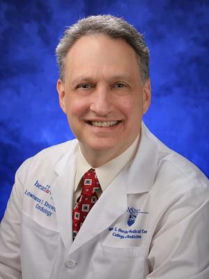 Lawrence I. Sinoway合作论文数Penn State Health;Division of Cardiology, Department of Medicine, Penn State College of Medicine, The Pennsylvania State University2
 Jerzy A. Zoladz合作论文数Faculty of Rehabilitation, University School of Physical Education Kraków2
 Bruno Grassi合作论文数Department of Medicine, University of Udine2
 Zhou Zhichao合作论文数Erasmus University Rotterdam, University Medical Center Rotterdam2Hunter Sandra K合作论文数2
 Almeida Jeeser A合作论文数Centro de Análises Proteômicas e Bioquímicas, Universidade Católica de Brasília;UDF - Centro Universitário, Brasília, DF, Brazil;Programa de Pós Graduação em Ciências e Tecnologias em Saúde, Universidade de Brasília - UnB;UDF - Centro Universitário, Universidade de Brasília - UnB2
 Daniel A. Boullosa合作论文数Graduação em Fisioterapia, Universidade Católica de Brasília2
 James M Smoliga合作论文数Dept Phys Therapy, High Point Univ2
 Carl Foster合作论文数Department of Exercise and Sport Science, University of Wisconsin-La Crosse2
 Broxterman Ryan M合作论文数2
 Ronald Snarr合作论文数School of Nutrition and Health Promotion, Arizona State University2Davinia Vicente-Campos合作论文数Universidad Francisco de Vitoria2Guo Xinyi合作论文数Department of Physics, Wake Forest University2
 Thomas Jackson Barstow合作论文数Department of Kinesiology, Kansas State University2
 Conrad P. Earnest合作论文数Exercise & Sport Nutrition Lab, Department of Health and Kinesiology, Texas A&M University2
 Asker Jeukendrup合作论文数Mysportscience;Core Nutrition Planning LLC;Loughborough University2
 Anthony Atala合作论文数Wake Forest Institute for Regenerative Medicine;Department of Urology, Wake Forest School of Medicine;Brenner Children's Hospital, Atrium Health Levine Children's2Olivier Girard合作论文数2
 Michael W Lawlor合作论文数Department of Medicine, Children’s Hospital Boston2Henning Bay Nielsen合作论文数Department of Anaesthesia, Rigshopitalet, University of Copenhagen2Scott J. Montain合作论文数2
 Jose Lopez Chicharro合作论文数2Guido Ferretti合作论文数2




