Understanding the biological mechanisms responsible for resilience to stress—the ability to overcome adverse conditions—remains a major challenge. While sleep is a known regulator of resilience, the localized cortical dynamics that facilitate this process remain unclear. We hypothesized that resilience to social stress is determined by local sleep changes within the prelimbic cortex (PrL), a region critical for top-down control of stress-responsive circuits. To test this, we conducted longitudinal single-unit and local field potential (LFP) recordings in the prelimbic (PrL) cortex of male mice before and after a five-day social defeat stress paradigm. Our results demonstrate that population-wide neuronal silences, or "OFF-periods," primarily occur during NREM sleep and strongly correlate with the local slow-waves. Notably, resilient mice exhibit significantly higher baseline coupling between OFF-period density and the number of slow-waves compared to susceptible animals. Following stress, resilient mice showed a unique reorganization of cortical silence patterns, characterized by an increase in shorter OFF-periods and a more uniform temporal distribution across NREM sleep. Furthermore, social stress was associated with a widespread, stochastic-like redistribution of cortical firing rates that was most pronounced in the resilient phenotype. These findings suggest that behavioral resilience is predicted by the capacity for flexible network redistribution and heightened synchronization during local NREM sleep. Furthermore, this study identifies pre-existing, localized sleep-dependent signatures— specifically a stronger coupling between OFF-period density and slow-wave activity —that serve as potential predictors of resilience prior to stress. Significance Statement The biological mechanisms driving resilience—the ability to overcome stressful conditions—remain poorly understood. While non-rapid eye movement sleep promotes resilience, the cortical mechanisms underlying sleep’s actions are unclear. Here, we demonstrate that local sleep within the prelimbic cortex—specifically neuronal silence—predicts stress resilience. Using single-unit recordings, we show that neuronal silence is coupled with slow-waves, the hallmark of non-rapid eye movement sleep, and this relationship is strengthened in resilient mice. Furthermore, resilience is associated with a post-stress reorganization of neuronal silence and a redistribution of cortical firing rates. These findings suggest that behavioral resilience is predicted by the prefrontal cortex’s capacity for reorganization during sleep and provide a framework for understanding how sleep-dependent circuit plasticity protects against stress.
Chronic exposure to repeated social stressors contributes to the development of anxiety and depressive-related behavioral phenotypes. Identifying molecular pathways that can be targeted during stress exposure to bias behavioral outcomes toward resilience is therefore a major goal of neuropsychiatric research. This study investigated the behavioral impact of inhibiting Acetyl-CoA Synthetase Short Chain Family Member 2 (ACSS2), an enzyme that links cellular metabolism to epigenetic signaling, in a mouse model of chronic social defeat stress. To evaluate the therapeutic potential of ACSS2 inhibition, mice were treated with either an ACSS2 inhibitor or vehicle starting at the midpoint of a 10-day defeat protocol. Behavioral assays revealed that ACSS2 inhibition exerted a potent, context-dependent pro-resilient effect. Specifically, ACSS2i-treated mice exhibited significantly higher social interaction scores and greater exploratory activity compared to vehicle-treated stressed controls following social defeat stress. Notably, these effects were observed exclusively in the stressed cohort, with no behavioral alterations detected in gentle-handled or novel-cage-exposed control groups. Longitudinal tracking showed that these pro-resilient improvements were not only present immediately following the stressor but persisted after seven days. These findings demonstrate that mid-stress ACSS2 inhibition can rescue behavioral deficits and promote a durable resilient state, positioning ACSS2 as a promising target for interventions aimed at mitigating the long-term psychiatric consequences of chronic stress.
Sleep disturbances and stress have a well-established link with neuropsychiatric illness; however, the nature of this relationship remains unclear. Recently, studies using the mouse social-defeat stress model revealed a causal role for non-rapid eye movement (NREM) sleep in the maladaptive behavioral responses to stress. These results suggest a novel function for NREM sleep; as a response by cortical neurons to mitigate the maladaptive effects of stress. A major limitation in many social defeat studies has been the exclusion of females. Women exhibit a greater prevalence of both affective disorders and sleep disturbances compared to men, thus there is a clear need to understand sleep–stress interactions in females. The present study adapts recently developed female social-defeat stress models to allow social-defeat and EEG in male–female pairs. Our findings duplicate the behavioral responses that occur in other female, nondiscriminatory, and male models of social-defeat stress. Analysis of electroencephalographic (EEG) recordings, before exposure to stress, reveal that resilience is associated with differences in both NREM and REM sleep that are dependent on sex. After social defeat stress, NREM sleep was increased only in resilient males. In females, susceptibility to stress was associated with increased durations in NREM-sleep bouts. A potential cause of these sleep differences was also identified prior to stress exposure, sex differences in recovery from NREM-sleep loss; thus, suggesting an underlying sex-difference in the homeostatic process regulating sleep interactions with social-defeat stress. These findings suggest that NREM sleep quality is lower in resilient females, whereas the amount of REM sleep is decreased in susceptible females—when compared to males of the same behavioral phenotype. Overall, our findings reveal sexual dimorphism in both the sleep characteristics predicting resilience and sleep changes induced by social-defeat stress.
Circadian gene expression is fundamental to the establishment and functions of the circadian clock, a cell-autonomous and evolutionary-conserved timing system. Yet, how it is affected by environmental-circadian disruption (ECD) such as shiftwork and jetlag are ill-defined. Here, we provided a comprehensive and comparative description of male liver circadian gene expression, encompassing transcriptomes, whole-cell proteomes and nuclear proteomes, under normal and after ECD conditions. Under both conditions, post-translation, rather than transcription, is the dominant contributor to circadian functional outputs. After ECD, post-transcriptional and post-translational processes are the major contributors to whole-cell or nuclear circadian proteome, respectively. Furthermore, ECD re-writes the rhythmicity of 64% transcriptome, 98% whole-cell proteome and 95% nuclear proteome. The re-writing, which is associated with changes of circadian regulatory cis-elements, RNA-processing and protein localization, diminishes circadian regulation of fat and carbohydrate metabolism and persists after one week of ECD-recovery.
There are well-documented gender differences in the risk and severity of sleep disorders and associated comorbidities. While fundamental sex differences in sleep regulatory mechanisms may contribute to gender disparities, biological responses to sleep loss and stress may underlie many of the risks for sleep disorders in women and men. Some of these sex differences appear to be dependent on sex chromosome complement (XX or XY) and the organizational effects of reproductive hormones. Reproductive development plays a critical role in the ability of sex chromosomes and reproductive hormones to produce sex differences in sleep and wakefulness. Rodent models reveal that reproductive hormones drive many but not all sex differences in sleep–wake architecture. The ability of reproductive hormones to alter sleep are often dependent on responses to sleep loss and stress. However, in the absence of reproductive hormones (in gonadectomized rodents) sex differences in sleep amount and the ability to recover from sleep loss persist. The suprachiasmatic nucleus (SCN) and the ventrolateral preoptic nucleus (VLPO) of the hypothalamus play crucial regulatory roles in mediating the effects of reproductive hormones on the sleep–wake cycle. Taken together, the work reviewed here reveals that the reproductive hormone environment and sex chromosome complement may underlie gender disparities in sleep patterns and the risk for sleep disorders.
The suprachiasmatic nucleus (SCN) is composed of functionally distinct subpopulations of GABAergic neurons which form a neural network responsible for synchronizing most physiological and behavioral circadian rhythms in mammals. To date, little is known regarding which aspects of SCN rhythmicity are generated by individual SCN neurons, and which aspects result from neuronal interaction within a network. Here, we utilize in vivo miniaturized microscopy to measure fluorescent GCaMP-reported calcium dynamics in arginine vasopressin (AVP)-expressing neurons in the intact SCN of awake, behaving mice. We report that SCN AVP neurons exhibit periodic, slow calcium waves which we demonstrate, using in vivo electrical recordings, likely reflect burst firing. Further, we observe substantial heterogeneity of function in that AVP neurons exhibit unstable rhythms, and relatively weak rhythmicity at the population level. Network analysis reveals that correlated cellular behavior, or coherence, among neuron pairs also exhibited stochastic rhythms with about 33% of pairs rhythmic at any time. Unlike single-cell variables, coherence exhibited a strong rhythm at the population level with time of maximal coherence among AVP neuronal pairs at CT/ZT 6 and 9, coinciding with the timing of maximal neuronal activity for the SCN as a whole. These results demonstrate robust circadian variation in the coordination between stochastically rhythmic neurons and that interactions between AVP neurons in the SCN may be more influential than single-cell activity in the regulation of circadian rhythms. Furthermore, they demonstrate that cells in this circuit, like those in many other circuits, exhibit profound heterogenicity of function over time and space.
Circadian gene expression is fundamental to the establishment and functions of the circadian clock, a cell-autonomous and evolutionary-conserved timing system. Yet, how it is affected by environmental-circadian disruption (ECD) such as shiftwork and jetlag, which impact millions of people worldwide, are ill-defined. Here, we provided the first comprehensive description of liver circadian gene expression under normal and after ECD conditions. We found that post-transcription and post-translation processes are dominant contributors to whole-cell or nuclear circadian proteome, respectively. Furthermore, rhythmicity of 64% transcriptome, 98% whole-cell proteome and 95% nuclear proteome is re-written by ECD. The re-writing, which is associated with changes of circadian cis-regulatory elements, RNA-processing and protein trafficking, diminishes circadian regulation of fat and carbohydrate metabolism and persists after one week of ECD-recovery. One-Sentence Summary Environmental-circadian disruption re-writes the circadian gene expression process which persists after one week of recovery.
Circadian clocks enable organisms to predict and align their behaviors and physiologies to constant daily day-night environmental cycle. Because the ubiquitin ligase Siah2 has been identified as a potential regulator of circadian clock function in cultured cells, we have used SIAH2-deficient mice to examine its function in vivo. Our experiments demonstrate a striking and unexpected sexually dimorphic effect of SIAH2-deficiency on the regulation of rhythmically expressed genes in the liver. The absence of SIAH2 in females, but not in males, altered the expression of core circadian clock genes and drastically remodeled the rhythmic transcriptome in the liver by increasing the number of day-time expressed genes, and flipping the rhythmic expression from nighttime expressed genes to the daytime. These effects are not readily explained by effects on known sexually dimorphic pathways in females. Moreover, loss of SIAH2 in females, not males, preferentially altered the expression of transcription factors and genes involved in regulating lipid and lipoprotein metabolism. Consequently, SIAH2-deficient females, but not males, displayed disrupted daily lipid and lipoprotein patterns, increased adiposity and impaired metabolic homeostasis. Overall, these data suggest that SIAH2 may be a key component of a female-specific circadian transcriptional output circuit that directs the circadian timing of gene expression to regulate physiological rhythms, at least in the liver. In turn, our findings imply that sex-specific transcriptional mechanisms may closely interact with the circadian clock to tailor overt rhythms for sex-specific needs.
Resilience, the ability to overcome stressful conditions, is found in most mammals and varies significantly among individuals. A lack of resilience can lead to the development of neuropsychiatric and sleep disorders, often within the same individual. Despite extensive research into the brain mechanisms causing maladaptive behavioral-responses to stress, it is not clear why some individuals exhibit resilience. To examine if sleep has a determinative role in maladaptive behavioral-response to social stress, we investigated individual variations in resilience using a social-defeat model for male mice. Our results reveal a direct, causal relationship between sleep amount and resilience-demonstrating that sleep increases after social-defeat stress only occur in resilient mice. Further, we found that within the prefrontal cortex, a regulator of maladaptive responses to stress, pre-existing differences in sleep regulation predict resilience. Overall, these results demonstrate that increased NREM sleep, mediated cortically, is an active response to social-defeat stress that plays a determinative role in promoting resilience. They also show that differences in resilience are strongly correlated with inter-individual variability in sleep regulation.
Introduction: Stress can have a variety of detrimental effects on humans. From depression and anxiety to schizophrenia, stress plays a factor in the development of these diseases through neurochemical changes in the brain and elevated levels of hormones. Among the geriatric population, decreased sleep levels are known to be a frequent issue; insomnia rates among the senior population are much higher in frequency compared to any other age group. Sleep deprivation also leads to major consequences in the brain and sleep disruption is linked to neuropsychological illness; however, the specific mechanisms involved in these effects are not understood. This study focuses on the resilient effects of ΔFosB, a protein known to mediate resilience to stress and the direct effect of sleep deprivation on ΔFosB expression in areas known to mediate resilience to social stress. We hypothesize that ΔFosB may be part of the mechanism through which sleep alters resilience to social stress. Methods: Mice were sleep-deprived for an eight-hour period for five days. After sleep deprivation they were subject to social defeat and underwent avoidance testing. The brains of these mice were removed, and immunohistochemistry analysis was conducted to determine ∆FosB expression in various sections of the brain. Results: The preliminary findings of this study indicates that sleep is altered in resilient animals and that sleep deprivation may lead to increased resilience to social defeat. The most significant decrease in ∆FosB expression was found in the prelimbic cortex, a change associated with resilience, and which was observed after chronic sleep deprivation. Contrarily, there was also an increase of ∆FosB expression in the nucleus accumbens. Conclusion: These findings indicate that changes in ∆FosB activation in the brain is a significant factor for promoting resilient behavior in situations of social stress. In particular, a decrease in ∆FosB activation in the PLC plays a role in explaining how sleep deprivation contributes to decreased social resilience in situations of social stress.
In 1965, Dr Harry Angelman reported a neurodevelopmental disorder affecting three unrelated children who had similar symptoms: brachycephaly, mental retardation, ataxia, seizures, protruding tongues, and remarkable paroxysms of laughter. Over the past 50 years, the disorder became Angelman's namesake and symptomology was expanded to include hyper-activity, stereotypies, and severe sleep disturbances. The sleep disorders in many Angelman syndrome (AS) patients are broadly characterized by difficulty falling and staying asleep at night. Some of these patients sleep less than 4 hours a night and, in most cases, do not make up this lost sleep during the day-leading to the speculation that AS patients may "need" less sleep. Most AS patients also have severely reduced levels of melatonin, a hormone produced by the pineal gland exclusively at night. This nightly pattern of melatonin production is thought to help synchronize internal circadian rhythms and promote nighttime sleep in humans and other diurnal species. It has been proposed that reduced melatonin levels contribute to the sleep problems in AS patients. Indeed, emerging evidence suggests melatonin replacement therapy can improve sleep in many AS patients. However, AS mice show sleep problems that are arguably similar to those in humans despite being on genetic backgrounds that do not make melatonin. This suggests the hypothesis that the change in nighttime melatonin may be a secondary factor rather than the root cause of the sleeping disorder. The goals of this review article are to revisit the sleep and melatonin findings in both AS patients and animal models of AS and discuss what AS may tell us about the underlying mechanisms of, and interplay between, melatonin and sleep.
Poor sleep is a hazard of daily life that oftentimes cannot be avoided. Gender differences in daily sleep and wake patterns are widely reported; however, it remains unclear how biological sex, which is comprised of genetic and endocrine components, directly influences sleep regulatory processes. In the majority of model systems studied thus far, sex differences in daily sleep amount are predominant during the active (wake) phase of the sleep-wake cycle. The pervasiveness of sex differences in sleep amount throughout phylogeny suggests a strong underlying genetic component. The goal of the current study is to determine if sex differences in active-phase sleep amount are dependent on sex chromosomes in mice.Sleep was examined in the four-core genotype (FCG) mouse model, whose sex chromosome complement (XY, XX) is independent of sex phenotype (male or female). In this line, sex phenotype is determined by the presence or absence of the Sry gene, which is dissociated from the Y chromosome. Polysomnographic sleep recordings were obtained from gonadectomized (GDX) FCG mice to examine spontaneous sleep states and the ability to recover from sleep loss. We report that during the active-phase, the presence of the Sry gene accounts for most sex differences during spontaneous sleep; however, during recovery from sleep loss, sex differences in sleep amount are partially driven by sex chromosome complement. These results suggest that genetic factors on the sex chromosomes encode the homeostatic response to sleep loss.
In 1965, Dr Harry Angelman reported a neurodevelopmental disorder affecting three unrelated children who had similar symptoms: brachycephaly, mental retardation, ataxia, seizures, protruding tongues, and remarkable paroxysms of laughter. Over the past 50 years, the disorder became Angelman's namesake and symptomology was expanded to include hyper‐activity, stereotypies, and severe sleep disturbances. The sleep disorders in many Angelman syndrome (AS) patients are broadly characterized by difficulty falling and staying asleep at night. Some of these patients sleep less than 4 hours a night and, in most cases, do not make up this lost sleep during the day—leading to the speculation that AS patients may “need” less sleep. Most AS patients also have severely reduced levels of melatonin, a hormone produced by the pineal gland exclusively at night. This nightly pattern of melatonin production is thought to help synchronize internal circadian rhythms and promote nighttime sleep in humans and other diurnal species. It has been proposed that reduced melatonin levels contribute to the sleep problems in AS patients. Indeed, emerging evidence suggests melatonin replacement therapy can improve sleep in many AS patients. However, AS mice show sleep problems that are arguably similar to those in humans despite being on genetic backgrounds that do not make melatonin. This suggests the hypothesis that the change in nighttime melatonin may be a secondary factor rather than the root cause of the sleeping disorder. The goals of this review article are to revisit the sleep and melatonin findings in both AS patients and animal models of AS and discuss what AS may tell us about the underlying mechanisms of, and interplay between, melatonin and sleep.
Circadian clocks enable organisms to predict and align their behaviors and physiologies to constant daily day-night environmental cycle. Because the ubiquitin ligase Siah2 has been identified as a potential regulator of circadian clock function in cultured cells, we have used Siah2-deficient mice to examine its function in vivo . Our experiments demonstrate a striking and unexpected sexually dimorphic effect of Siah2 deficiency on the regulation of rhythmically expressed genes. The absence of Siah2 in females, but not in males, altered the expression of core circadian clock genes and drastically remodeled the rhythmic hepatic transcriptome. Siah2 loss, only in females, increased the expression of 100’s of genes selectively at mid-day, resulting in a >50% increase in the number of rhythmically expressed genes, and shifted the expression of 100’s of other genes from a mid-night peak, to a mid-day peak. The combined result is a near inversion of overall rhythmicity in gene expression selectively in Siah2-deficient females. This dramatic reorganization created a substantial misalignment between rhythmic liver functions and feeding/behavioral rhythms, and consequently disrupted daily patterns of lipid/lipoprotein metabolism and metabolic responses to high-fat diet. Collectively, our results suggest that Siah2 is part of a female-specific circadian mechanism important for maintaining metabolic homeostasis and may play a key role in establishing sexual dimorphisms in metabolism, and broadly reveal that circadian clocks may drive rhythms using novel sex-specific transcriptional pathways.### Competing Interest StatementThe authors have declared no competing interest.
There are no Food and Drug Administration approved pharmacotherapies for methamphetamine (METH) overdose, thus identifying novel drug targets to prevent this devastating adverse event is a public-health imperative. Previous research suggests that serotonin and sigma receptors may contribute to the adverse effects of METH. The present study assessed whether pretreatment with the 5-HT2A receptor antagonist M100907 or the sigma 1 (sigma(1)) receptor antagonist BD 1047 attenuated METH-induced lethality, hyperthermia, convulsions, and seizures. Male, Swiss-Webster mice received intraperitoneal injections of M100907 (1 and 10 mg/kg), BD 1047 (10 mg/kg), or a combination of M100907 (1 mg/kg) and BD 1047 (10 mg/kg) prior to treatment with METH (78 mg/kg). Convulsions and lethality were assessed by observation, core body temperature was assessed by surgically implanted telemetric probes, and seizures were assessed by electroencephalography. M100907 reduced METH-elicited lethality from 67% to 33%, BD1047 reduced METH-elicited lethality from 67% to 50%, and combined administration of both agents eliminated lethality in all mice tested. Similarly, both agents and their combination reduced METH-elicited seizures and convulsions. None of the treatments decreased METH-induced hyperthermia. This research suggests that reducing METH-induced seizures is an important factor in reducing lethality associated with METH overdose. However, future studies should examine whether M100907 and BD 1047 modulate METH-induced hypertension and other adverse effects that may also contribute to METH overdose. Our data support the continued investigation of compounds that target 5-HT2A and sigma(1) receptors in METH-induced overdose, including their potential to yield emergency reversal agents.
Nitrergic neurons of the dorsal raphe nucleus (DRN) may play a role in physiological stress responses. The caudal lateral wings (CLW) are unique compared to other rostral-caudal DRN sub-regions because they contain distinct nitric oxide (NO) synthase (NOS) populations that are independent of tryptophan hydroxylase (TPH). NOS neurons in the CLW are also highly activated during acute restraint stress. However, the effects of acute stress duration on NOS activation in the CLW are unclear. Here NADPH-d, an index of NOS activity, is used to show that sub-regions of the DRN have differential NOS activation in response to 6 hours of restraint stress in rats. We report increased NOS activity through 6 hours of restraint in the caudal lateral wings and ventromedial sub-regions. These data suggest that, NOS neurons may play a dynamic role in the response to stress duration.
Sleep loss can severely impair the ability to perform, yet the ability to recover from sleep loss is not well understood. Sleep regulatory processes are assumed to lie exclusively within the brain mainly due to the strong behavioral manifestations of sleep. Whole-body knockout of the circadian clock gene Bmal1 in mice affects several aspects of sleep, however, the cells/tissues responsible are unknown. We found that restoring Bmal1 expression in the brains of Bmal1-knockout mice did not rescue Bmal1-dependent sleep phenotypes. Surprisingly, most sleep-amount, but not sleep-timing, phenotypes could be reproduced or rescued by knocking out or restoring BMAL1 exclusively in skeletal muscle, respectively. We also found that overexpression of skeletal-muscle Bmal1 reduced the recovery response to sleep loss. Together, these findings demonstrate that Bmal1 expression in skeletal muscle is both necessary and sufficient to regulate total sleep amount and reveal that critical components of normal sleep regulation occur in muscle.
Brain and muscle-ARNT-like factor (Bmal1/BMAL1) is an essential transcriptional/translational factor of circadian clocks. Loss of function of Bmal1/BMAL1 is highly disruptive to physiological and behavioral processes. In light of these previous findings, we examined if transgenic overexpression of Bmal1/BMAL1 in skeletal muscle could alter metabolic processes. First, we characterized in vivo and ex vivo metabolic phenotypes of muscle overexpressed mice (male and female) compared to wild-type littermates (WT). Second, we examined in vivo and ex vivo metabolic processes in the presence of positive and negative homeostatic challenges: high-intensity treadmill running (positive) and acute sleep deprivation (negative). In vivo measures of metabolic processes included body composition, respiratory exchange ratio (RER; VCO2/VO2), energy expenditure, total activity counts, and food intake collected from small animal indirect calorimetry. Ex vivo measure of insulin sensitivity in skeletal muscle was determined from radioassays. RER was lower for muscle overexpressed females compared to female WTs. There were no genotype-dependent differences in metabolic phenotypes for males. With homeostatic challenges, muscle overexpressed mice had lower energy expenditure after high-intensity treadmill running. Acute sleep deprivation reduced insulin sensitivity in skeletal muscle in overexpressed male mice, but not male WTs. The present study contributes to a body of evidence showing pleiotropic, non-circadian, and homeostatic effects of altered Bmal1/BMAL1 expression on metabolic processes, demonstrating a critical need to further investigate the broad and complex actions of Bmal1/BMAL1 on physiology and behavior.
STUDY OBJECTIVES:Episodes of brief limb ischemia (remote preconditioning) in mice induce tolerance to modeled ischemic stroke (focal brain ischemia). Since stroke outcomes are in part dependent on sleep-wake history, we sought to determine if sleep is critical for the neuroprotective effect of limb ischemia.METHODS:EEG/EMG recording electrodes were implanted in mice. After a 24 h baseline recording, limb ischemia was induced by tightening an elastic band around the left quadriceps for 10 minutes followed by 10 minutes of release for two cycles. Two days following remote preconditioning, a second 24 h EEG/EMG recording was completed and was immediately followed by a 60-minute suture occlusion of the middle cerebral artery (modeled ischemic stroke). This experiment was then repeated in a model of circadian and sleep abnormalities (Bmal1 knockout [KO] mice sleep 2 h more than wild-type littermates). Brain infarction was determined by vital dye staining, and sleep was assessed by trained identification of EEG/EMG recordings.RESULTS:Two days after limb ischemia, wild-type mice slept an additional 2.4 h. This additional sleep was primarily comprised of non-rapid eye movement (NREM) sleep during the middle of the light-phase (i.e., naps). Repeating the experiment but preventing increases in sleep after limb ischemia abolished tolerance to ischemic stroke. In Bmal1 knockout mice, remote preconditioning did not increase daily sleep nor provide tolerance to subsequent focal ischemia.CONCLUSIONS:These results suggest that sleep induced by remote preconditioning is both sufficient and necessary for its neuroprotective effects on stroke outcome.
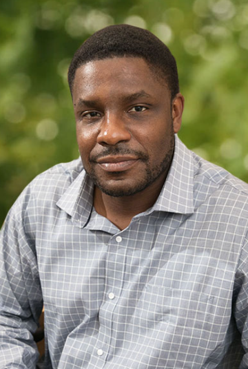 Ketema Paul合作论文数Department of Integrative Biology and Physiology, College of Life Sciences, University of California22
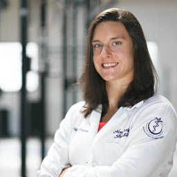 Allison Brager合作论文数Department of Neurobiology, Morehouse School of Medicine11
 H Elliott Albers合作论文数College of Arts & Sciences, Georgia State University;Neuroscience Institute, Georgia State University10
 Jason P. Debruyne合作论文数8Cloe L Gray合作论文数7Alec J. Davidson合作论文数Department of Psychology, Florida State University7
 Kenkichi Baba合作论文数Morehouse Sch Med, Inst Neurosci6
 Gianluca Tosini合作论文数Morehouse School of Medicine6
 Colleen M Novak合作论文数Department of Psychology and The Neuroscience Program, Michigan State University6Daniel L Hummer合作论文数5Ariel Armstrong合作论文数4B Bush合作论文数4Zhimei Qiao合作论文数4
 Karen L Gamble合作论文数Department of Psychiatry and Behavioral Neurobiology, University of Alabama at Birmingham4Benveniste Morris合作论文数4J. David Glass合作论文数Department of Biological Sciences, Kent State University4Lennisha Pinckney合作论文数3Darielle Lewis-Sanders合作论文数3Caroline Donnay合作论文数3Hadiya Johnson合作论文数3Ting-Chung Suen合作论文数3Hamadi Brewer合作论文数3K. Paul合作论文数3
 Gregory H. Grossman合作论文数3
 Daniella Do Carmo Buonfiglio合作论文数3
 Timothy J Bartness合作论文数Departments of Biology, Georgia State University3
 Oscar Castanon合作论文数3Tsedey Mekbib合作论文数2Kiandra Smith合作论文数2Aisha Rollins-Hairston合作论文数2Sahil Sood合作论文数2Talib Saafir合作论文数2Eva-Jeneé A. Andrews合作论文数2F. Jefferson合作论文数2
 Evans Jennifer A合作论文数Department of Psychology, University of California, San Diego2Hesse September合作论文数2
 Sharon Owino合作论文数2
 Michael D Powell合作论文数Purdue University2
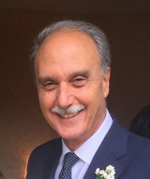 Saverio Cinti合作论文数Obesity Center, Department of Experimental and Clinical Medicine, School of Medicine, Università Politecnica delle Marche;The Adipose Organ Department of Biology, University of Copenhagen2
 Robert R Bowers合作论文数Molecular and Cellular Biology and Pathobiology, Medical University of South Carolina2C. Kay Song合作论文数2
 Andrea Frontini合作论文数Department of Molecular Pathology and Innovative Therapies, University of Ancona (Politecnica delle Marche),2
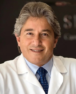 Antonio Giordano合作论文数Sbarro Institute for Cancer Research and Molecular Medicine, Temple University;Center for Biotechnology, College of Science & Technology, Temple University;Department of Pathology & Oncology, University of Siena2Hao A Duong合作论文数Department of Chemistry and Biochemistry, University of California, Los Angeles2Mary C. Karom合作论文数2
 Karyn Esser合作论文数School of Kinesiology, University of Illinois at Chicago2Julie E. Baggs合作论文数2Azizi Ray合作论文数1




