Mitochondrial dynamics relies on the function of dynamin family GTPase proteins including mitofusin 1 (MFN1), mitofusin 2 (MFN2) and dynamin-related protein 1 (DRP1; also known as DNM1L). The mitochondrial phosphatase phosphoglycerate mutase 5 (PGAM5) protein can regulate the phosphorylation levels and the function of both MFN2 and DRP1; however, the precise regulation of PGAM5 activity is unknown. Here, we show that PGAM5 oligomerization and localization controls its function. Under depolarization and/or metabolic stress PGAM5 changes its association and, instead of forming dodecamers, forms dimers. These PGAM5 oligomers have differential affinity towards MFN2 and DRP1. Simultaneously, PGAM5 is cleaved by the inner mitochondrial membrane-resident proteases PARL and OMA1 and a fraction of the cleaved PGAM5 translocates to the cytosol. These two events play an important role in regulating mitochondrial dynamics under depolarization and/or metabolic stress. Taken together, our results identify PGAM5 oligomerization and cleavage-induced relocalization as crucial regulators of its function.
Resilin is an elastomeric protein found in insects that provides flexibility and locomotive function in numerous biological contexts. Recombinant resilin and resilin-derived proteins share resilin's capacity for liquid-liquid phase separation (LLPS) and formation of materials with high elasticity and biocompatibility, making it a promising candidate for regenerative medicine and tissue engineering applications. While prior research has focused on domain 1 of resilin, the role of domain 3 (D3) in resilin self-assembly and material properties is not well understood. Here, we used nuclear magnetic resonance, electron paramagnetic resonance, and small-angle X-ray scattering to study the conformation, dynamics, and intermolecular interactions of D3 as a monomer, in the phase-separated state, and as a cross-linked gel. We show that D3 remains unusually dynamic and is primarily disordered in all three states. In elucidating the mechanism of D3 LLPS, we find a complex set of electrostatic and π-based interactions complemented by the hydrophobic effect that finely tune the solution sensitivity of D3 and its capacity for LLPS. Overall, these results highlight the complex mechanisms governing resilin LLPS with implications for utilizing resilin-derived sequence features in the rational design of self-assembling biomaterials.
Septin5 interacts with SNARE proteins to regulate exocytosis in neurons, but its role in pancreatic β-cells is unknown. Here, we report that Septin5 is abundant in rodent and human β-cells, deletion of which dramatically enhances biphasic glucose-stimulated insulin secretion, including in type 2 diabetes (T2D). Super-resolution imaging shows that Septin5 is preferentially assembled in microtubule-plasma membrane contact sites in a microtubule-dependent manner, which provides discrete harbor for secretory granule anchoring. By decreasing the stability of the cortical microtubule meshwork, Septin5 depletion increases insulin granule dynamics and access to the plasma membrane. Analysis of spatiotemporal coupling of fusion events and localized Ca2+ influx through L-type Ca2+ channels show that Septin5 depletion increases releasable granule pool clustering on Ca2+ channels, previously shown to be impaired in T2D, thus rectifying this T2D defect. Hence, inhibition of Septin5 can improve insulin secretion. Septins are the key component of the cytoskeleton. Here, the authors show that Septin5 is assembled in microtubule-plasma membrane contact sites and mediates insulin secretion in pancreatic islet β-cells through cortical microtubule remodeling.
Light sheet microscopy has become a popular imaging modality for multi-dimensional imaging. Its unconventional orthogonal setup between the excitation and emission axis allows for light to optically section the sample reducing the amount of background signal when compared to epifluorescence and reducing the laser power deposited into the sample when compared to confocal imaging. As the optics are different from a traditional inverted microscope, traditional mounting methods cannot be used. Instead, different mounting methods were created to work around the setup and adapt to the sample.
The field of nano-tracking has recently witnessed rapid growth, promising great potential for novel biomedical studies. Interferometric scattering microscopy (iSCAT) is a commonly used tool for the tracking of non-fluorescent nanoparticles tagged to viruses, DNA, and proteins. An iSCAT microscope can track particles as small as 5 nm in diameter, with sub-nanometer lateral resolution, since the destructive interference formed between the reference beam and the object beam is able to suppress background noise and enhance sample contrast.
The ability to sense and respond to osmotic fluctuations is critical for the maintenance of cellular integrity. We used gene co-essentiality analysis to identify an unappreciated relationship between TSC22D2, WNK1, and NRBP1 in regulating cell volume homeostasis. All of these genes have paralogs and are functionally buffered for osmo-sensing and cell volume control. Within seconds of hyperosmotic stress, TSC22D, WNK, and NRBP family members physically associate into biomolecular condensates, a process that is dependent on intrinsically disordered regions (IDRs). A close examination of these protein families across metazoans revealed that TSC22D genes evolved alongside a domain in NRBPs that specifically binds to TSC22D proteins, which we have termed NbrT (NRBP binding region with TSC22D), and this co-evolution is accompanied by rapid IDR length expansion in WNK-family kinases. Our study reveals that TSC22D, WNK, and NRBP genes evolved in metazoans to co-regulate rapid cell volume changes in response to osmolarity.
Current microscope stages position a given sample along linear XYZ axes. In order to capture further three-dimensional (3D) views of biological specimens, multi-view microscopy can be used. It images samples from multiple angles and reconstructs these views in a 3D image. However, it is costly and has a limited number of views due to the physical constraints of the microscope. Live organoid-based studies also face light penetration limitations in deeper microscopic planes due to light scattering.
SUMMARY The ability to sense and respond to osmotic fluctuations is critical for the maintenance of cellular integrity. Myriad redundancies have evolved across all facets of osmosensing in metazoans, including among water and ion transporters, regulators of cellular morphology, and macromolecular crowding sensors, hampering efforts to gain a clear understanding of how cells respond to rapid water loss. In this study, we harness the power of gene co-essentiality analysis and genome-scale CRISPR-Cas9 screening to identify an unappreciated relationship between TSC22D2 , WNK1 and NRBP1 in regulating cell volume homeostasis. Each of these genes have paralogs and are functionally buffered for macromolecular crowd sensing and cell volume control. Within seconds of hyperosmotic stress, TSC22D, WNK and NRBP family members physically associate into cytoplasmic biocondensates, a process that is dependent on intrinsically disordered regions (IDRs). A close examination of these protein families across metazoans reveals that TSC22D genes evolved alongside a domain in NRBPs that specifically binds to TSC22D proteins, which we have termed NbrT ( N RBP b inding region with T SC22D), and this co-evolution is concomitant with rapid IDR length expansion in WNK family kinases. Our study identifies functions for unrecognized components of the cell volume sensing machinery and reveals that TSC22D , WNK and NRBP genes evolved as cytoplasmic crowding sensors in metazoans to co-regulate rapid cell volume changes in response to osmolarity.
Astrocytes are a type of glial cell in the central nervous system responsible for modulating synaptic transmissions, tissue repair, maintaining homeostasis, and are therefore implicated in many neurological diseases. Human cortical astrocytes are more structurally complex, larger, and have unique subtypes in comparison to the commonly studied rodent cortical astrocytes. Morphological differences between species have been characterized, but there have been minimal functional experiments performed. As access to live human cortical tissue is sparse, emerging cerebral organoids (COs) derived from human stem cells have become a popular in vitro model for studying the human cortex. Here, we show that GFAP-positive astrocytes in COs are functional and undergo a range of calcium signaling modalities. Lentiviral transduction was used to express jGCaMP7c under the hGFAP promoter in the COs, and the spontaneous calcium signaling behaviour of astrocytes was characterized at varying spatio-temporal scales. COs were also exposed to neurotransmitters including glutamate, ATP, GABA, and acetylcholine and the astrocytic response was characterized. Establishing astrocyte functionality in COs is an important step in understanding the diverse functions of astrocytes in a human context, and further developing COs as a model system for studying human astrocytes and their roles in neurological diseases.
Preclinical in vitro and non-human animal models often inadequately replicate human physiology and disease processes. Human stem cell-derived organoids generated using microfluidic devices hold great promise for improved mimicry and monitoring. However, a critical challenge lies in long-term live organoid imaging using standard sample plates. In this study, we demonstrate the efficacy of fast, low-photobleaching fluorescence scanning and provide novel insights into calcium signaling within astrocytes in cerebral organoids. Our imaging pipeline is founded on single-objective selective plane illumination microscopy (SoSPIM). Achieving rapid and stable scanning involves integrating a high-speed 12 kHz galvanometer into the microscope's illumination train, precisely synchronized with the back focal plane of both the primary and secondary objectives. This configuration enables swift light sheet scanning within organoids, substantially reducing photobleaching by up to 80% in live cell imaging. A data acquisition (DAQ) card seamlessly bridges all hardware components and software, serving as the master controller. Pycro-manager supports the entire imaging workflow. In contrast to rigid systems, our detection arm, featuring the tertiary objective ("Mr. Snouty"), is mounted on a rotation stage, facilitating seamless adaptation of imaging angles based on sample thickness and opacity. Our imaging pipeline reveals significant potential in biophysical research, including visualization of vascularization within 3D printed organoids, shedding light on their intricate microarchitecture and perfusion dynamics. Additionally, our method unveils the morphology of GFAP-positive astrocytes, providing valuable insights into their spatial organization and interactions within cerebral organoids. Furthermore, we delve into the realm of cellular activity by capturing dynamic calcium signaling in live organoids cultivated in both six-well and single-well plates. This comprehensive approach not only enhances our understanding of organoid physiology but also extends opportunities for investigating the functional aspects of complex cellular networks within these in vitro models.
We present the design of a low-cost, portable telecentric digital holographic microscope (P-TDHM) that utilizes off-the-shelf components. We describe the system’s hardware and software elements and evaluate its performance by imaging samples ranging from nano-printed targets to live HeLa cells, HEK293 cells, and Dolichospermum via both in-line and off-axis modes. Our results demonstrate that the system can acquire high quality quantitative phase images with nanometer axial and sub-micron lateral resolution in a small form factor, making it a promising candidate for resource-limited settings and remote locations. Our design represents a significant step forward in making telecentric digital holographic microscopy accessible and affordable to the broader community.
Salmonella utilizes a type 3 secretion system to translocate virulence proteins (effectors) into host cells during infection1. The effectors modulate host cell machinery to drive uptake of the bacteria into vacuoles, where they can establish an intracellular replicative niche. A remarkable feature of Salmonella invasion is the formation of actin-rich protuberances (ruffles) on the host cell surface that contribute to bacterial uptake. However, the membrane source for ruffle formation and how these bacteria regulate membrane mobilization within host cells remains unclear. Here, we show that Salmonella exploits membrane reservoirs for the generation of invasion ruffles. The reservoirs are pre-existing tubular compartments associated with the plasma membrane (PM) and are formed through the activity of RAB10 GTPase. Under normal growth conditions, membrane reservoirs contribute to PM homeostasis and are preloaded with the exocyst subunit EXOC2. During Salmonella invasion, the bacterial effectors SipC, SopE2, and SopB recruit exocyst subunits from membrane reservoirs and other cellular compartments, thereby allowing exocyst complex assembly and membrane delivery required for bacterial uptake. Our findings reveal an important role for RAB10 in the establishment of membrane reservoirs and the mechanisms by which Salmonella can exploit these compartments during host cell invasion.
Nanoparticle tracking has received growing attention in molecular biology for its ability to understand cellular processes at the single molecule level. Digital holographic microscopy (DHM)'s ability to reconstruct 3D complex fields with deep sub-micron axial resolution with one shot lends to its credibility and increasing usage in tracking proteins and viruses. Given a certain distance from the image plane, DHM achieves numerical reconstruction by applying Angular Spectrum algorithm. A focused image can thus be discovered by iteratively computing multiple slices at various reconstruction distances and comparing the focusedness of the planes.
 Aaron Au合作论文数33James E. Shaw合作论文数Research Center, Phillips Petroleum Company20Amy Won合作论文数18
 Andrea L Slade合作论文数Institute of Biomaterials and Biomedical Engineering, University of Toronto17
 Peter K. Kim合作论文数Department of Biochemistry, Temerty Faculty of Medicine, University of Toronto15Adriano Vissa合作论文数14
 Scott Gray-Owen合作论文数Department of Molecular Genetics, Temerty Faculty of Medicine, University of Toronto14Kaitlin Szederkenyi合作论文数12
 Amine Driouchi合作论文数11
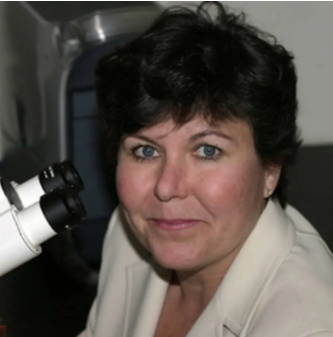 Joanne McLaurin合作论文数Department of Laboratory Medicine and Pathobiology, Temerty Faculty of Medicine, University of Toronto11
 Richard M. Epand合作论文数Department of Biochemistry and Biomedical Sciences, McMaster University11
 Lei Jin合作论文数University of Toronto10
 Paul Fraser合作论文数Department of Medical Biophysics, Temerty Faculty of Medicine, University of Toronto10
 John Oreopoulos合作论文数10
 Ernesto Abel-Santos合作论文数Department of Chemistry and Biochemistry, University of Nevada, Las Vegas10
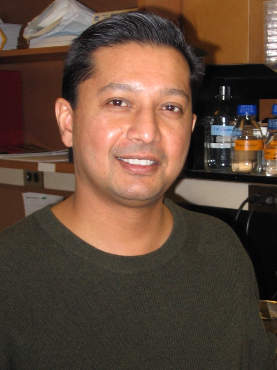 Avi Chakrabartty合作论文数Department of Biochemistry, Temerty Faculty of Medicine, University of Toronto9Ziyang Yu合作论文数8Giuliani Maximiliano合作论文数8
 G. Angus McQuibban合作论文数Department of Biochemistry, Temerty Faculty of Medicine, University of Toronto7
 William S. Trimble合作论文数SickKids Research Institute, Department of Biochemistry, Temerty Faculty of Medicine, University of Toronto;Hospital for Sick Children7
 Anthony Gramolini合作论文数Ted Rogers Centre for Heart Research, Department of Physiology, Temerty Faculty of Medicine, University of Toronto7
 Craig A. Simmons合作论文数Simmons Lab for Cellular Mechanobiology, Department of Mechanical & Industrial Engineering, Faculty of Applied Science and Engineering, University of Toronto;Boutiq Solutions Inc.;Ted Rogers Centre for Heart Research6Dun-Sheng Yang合作论文数Department of Psychiatry, Grossman School of Medicine, New York University6
 Neal I. Callaghan合作论文数6
 Ian Manners合作论文数Department of Chemistry, Faculty of Science, University of Victoria6
 Raquel F. Epand合作论文数Department of Biochemistry and Biomedical Sciences, McMaster University6
 Sergio Grinstein合作论文数Department of Biochemistry, Temerty Faculty of Medicine, University of Toronto;Research Institute, The Hospital for Sick Children;Keenan Research Centre, Li Ka Shing Knowledge Institute, St. Michael's Hospital6
 Martin Oheim合作论文数INSERM U603, Paris, France6Christopher McFaul合作论文数5Jenny C. Y. Hsu合作论文数5Dy Sasaki合作论文数5Scott B. Clendenning合作论文数5
 Jacqueline R. Phan合作论文数5
 Simon Sharpe合作论文数Department of Biochemistry, Temerty Faculty of Medicine, University of Toronto5
 Jonathan Rocheleau合作论文数Institute of Biomedical Engineering, Faculty of Applied Science and Engineering, University of Toronto5
 Brent Sleep合作论文数Department of Civil & Mineral Engineering, Faculty of Applied Science & Engineering, University of Toronto5Gary C. H. Mo合作论文数5
 Erica Pensini合作论文数†Department of Chemical and Materials Engineering, University of Alberta5
 Zhenghong Lu合作论文数Department of Materials Science & Engineering, Faculty of Applied Science and Engineering, University of Toronto5Vanderlee Gillian合作论文数5
 Michael Ward合作论文数College of Arts & Science, New York University;Department of Chemistry, New York University;Molecular Design Institute, New York University5
 Guocheng Yang (杨国程)合作论文数School of Chemical & Life Science, Changchun University of Technology5
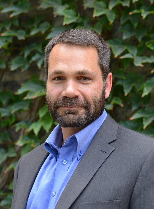 Peter Roy合作论文数Department of Molecular Genetics, Temerty Faculty of Medicine, University of Toronto5Jonathan V. Rocheleau合作论文数Banting & Best Diabetes Centre, Temerty Faculty of Medicine, University of Toronto5Michael T. Wong合作论文数5michael rosario defelippis合作论文数4Paul M Gorman合作论文数4Michelle A Edwards合作论文数4




