RATIONALE AND OBJECTIVES:To investigate the optimal time window of the postvascular phase of perfluorobutane-enhanced ultrasound in differentiating between benign and malignant lymph nodes (LNs) in patients with superficial lymphadenopathy. MATERIALS AND METHODS:This study retrospectively analyzed 150 suspicious LNs in the superficial area from 141 patients. All LNs were evaluated by the postvascular phase of perfluorobutane-enhanced ultrasound at a dose of 0.4-0.6 mL and a mechanical index range of 0.18-0.21. Quantitative parameters, including peak intensity (PI) and mean postvascular phase intensity (MPI), were obtained at 6, 8, and 10 min post-injection. Receiver operating characteristic (ROC) analysis was performed at each time point. Area under the curve (AUC) differences between earlier time points and the 10 min assessment were calculated with 95% confidence intervals. Diagnostic performance and classification stability were further assessed using the 10 min cutoff. RESULTS:PI and MPI were significantly lower in malignant LNs than in benign LNs at all three time points. For PI, the AUCs were 0.919, 0.942, and 0.949 at 6, 8, and 10 min, respectively. For MPI, the corresponding AUCs were 0.936, 0.933, and 0.940. In the exploratory AUC based comparison with 10 min, PI at 6 min was not within the exploratory margin, whereas PI at 8 min and MPI at both 6 and 8 min were within the margin. When the 10 min cutoff was applied to earlier measurements, the 6 min assessment showed reduced sensitivity and lower classification agreement, whereas the 8 min assessment showed more stable classification relative to 10 min CONCLUSIONS: In suspicious superficial LNs evaluated with perfluorobutane enhanced ultrasound, quantitative postvascular phase assessment showed good diagnostic performance. However, the 6 min assessment showed less stable classification, while the 8 min assessment may represent a more reliable earlier observation time point under this specific imaging protocol.
The heel fat pad plays a vital role in shock absorption during weight-bearing and may exhibit structural and textural differences associated with long-term mechanical loading. This study employed ultrasound radiomics to investigate the association between exercise volume and layer-specific radiomic features of the macrochamber and microchamber layers of the heel fat pad. Ultrasound images were obtained from 51 healthy young adults aged 18-35 years, who were classified into high-exercise-volume (HEV, n = 18), moderate-exercise-volume (MEV, n = 15), and low-exercise-volume (LEV, n = 18) groups according to weekly physical activity energy expenditure. A total of 101 radiomic features were extracted from each layer to characterize intensity, heterogeneity, and spatial texture patterns. Intergroup differences were analyzed using one-way analysis of variance or the Kruskal-Wallis test, followed by Bonferroni-adjusted pairwise comparisons. Significant radiomic differences were identified in the macrochamber layer, with 19 features differing among the three groups (P < 0.05), and six representative features retained after LASSO feature selection. The main pairwise differences occurred between the LEV group and the MEV or HEV groups, whereas no significant differences were detected between the MEV and HEV groups in the main distinguishing features. In contrast, the microchamber layer showed limited variation, with only Uniformity differing significantly between the LEV group and the MEV/HEV groups. These findings suggest that the macrochamber layer may be more sensitive to activity-associated radiomic texture differences than the microchamber layer. Overall, ultrasound radiomics shows potential as a non-invasive approach for quantifying subtle, layer-specific texture patterns in the heel fat pad.
Gout, a prevalent and treatable form of crystal-induced arthritis, results from monosodium urate (MSU) crystal deposition in articular and periarticular tissues. Early diagnosis is crucial to prevent progression to chronic gouty arthritis, tophus formation, and structural joint damage. Musculoskeletal ultrasound (MUS) has emerged as a sensitive, noninvasive, and cost-effective imaging modality that enables the visualization of urate crystal deposition, evaluation of treatment response, and prediction of disease flares. This narrative review summarizes recent advances in MUS for the diagnosis and management of gout, including its integration into the 2015 ACR/EULAR classification criteria and the development of OMERACT consensus-based definitions and semi-quantitative scoring systems. Compared with dual-energy computed tomography (DECT), MUS is more accessible and radiation-free, and offers superior performance in detecting early-stage gout. MUS also provides valuable insights into comorbidities such as cardiovascular disease and chronic kidney disease. Furthermore, emerging technologies-such as superb microvascular imaging (SMI) and artificial intelligence (AI)-based deep learning-show promise in enhancing diagnostic accuracy and automation. MUS is expected to play an increasingly pivotal role in the comprehensive management of gout.
Multidrug resistance in breast cancer is asignificant clinical challenge that often leads to treatment failure and tumor recurrence. Light‐activated therapies such as photothermal and photodynamic therapies offer spatially and temporally controlled treatments with minimal invasiveness. However, their effectiveness is limited by the instability of conventional agents, hostile tumor microenvironment, and shallow tissue light penetration. The combination of these light‐based approaches with nanotechnology has enabled the development of multifunctional nanoplatforms capable of overcoming multidrug resistance. Through rational design and surface modification, these platforms can improve drug targeting, biocompatibility, and therapeutic outcomes. Smart nanosystems for the codelivery of light‐responsive agents and drugs promote enhanced tumor accumulation, controlled drug release, and integrated diagnostics and therapies. This review examines resistance mechanisms in breast cancer and discusses light‐based strategies, emphasizing the design of advanced photoactivated nanoplatforms, their material properties, and synergistic antitumor effects. These multifunctional nanosystems demonstrate considerable potential for overcoming biological barriers and enabling precise drug release and real‐time monitoring, indicating their strong prospects for clinical applications. Future challenges and directions for clinical translation have also been addressed.
Objective: Microvascular alterations are early biomarkers of many diseases, but clinical translation is hindered by the lack of noninvasive deep-tissue imaging tools with capillary resolution and standardized analysis approaches. Impact Statement: ULM-based Vascular Biomarker Automated Analysis (U-VBA) provides a standardized framework that bridges microvascular imaging reconstruction and automated biomarker analysis, with feasibility demonstration in animals models in vivo and clinical patient data. Introduction: Ultrasound localization microscopy (ULM) can overcome the diffraction limit but lacks generalizable imaging reconstruction and biomarker quantification pipelines. Methods: We developed U-VBA, integrating motion correction, cascaded denoising (singular value decomposition, high-pass filtering, and background subtraction), and a panel of 6 vascular biomarkers (density, intervessel distance, diameter, velocity, perfusion, and tortuosity). Performance was validated in various settings, including flow phantoms, chicken chorioallantoic membranes (n = 5), rabbit optic nerve injury models (n = 4), and a clinical patient cohort with cervical lymph node tumors (n = 39). Results: U-VBA achieved robust performance in flow phantoms and resolved the finest capillaries down to 16.2 μm in chorioallantoic membrane models. In rabbit eye models, it precisely captures the dynamic vascular changes and establishes the vascular biomarkers during elevated intraocular pressure and recovery. In the clinical cohort, we demonstrated the potential value of U-VBA, which leverages 4 biomarkers (intervessel distance, diameter, perfusion, and tortuosity) to differentiate 3 lymph node conditions, with 85% accuracy in 5-fold cross-validation. Conclusion: U-VBA standardizes ULM-based microvascular phenotyping and integrates into routine ultrasound workflows, offering a noninvasive tool for preclinical and clinical vascular biomarker analysis.
Accurate evaluation of renal fibrosis and interstitial inflammation grades is necessary to improve the prognosis of patients with chronic kidney disease (CKD). However, non-invasive and accurate diagnostic methods are lacking. In this regard, shear wave elastography, which measures tissue viscosity and elasticity, may be a promising alternative. Therefore, this study aimed to evaluate the diagnostic performance of individual viscoelastic parameters, combined viscoelastic parameter models, and combined clinical parameter models in assessing the grades of renal fibrosis and interstitial inflammation. This prospective study recruited 22 and 47 patients with CKD in the reproducibility and validity trials, respectively, between October 2023 and June 2024. The mean elasticity (Emean), maximum elasticity (Emax), minimum elasticity (Emin), and mean viscosity (Vmean) were obtained using two-dimensional shear-wave elastography (2D-SWE). The biopsy specimens were assessed to determine the grades of renal fibrosis and interstitial inflammation. Additionally, clinical data, including protein-to-creatinine ratio, and estimated glomerular filtration rate (eGFR), were obtained from the patients’ medical records. Multiple logistic regression models were constructed, including viscoelastic models and clinical models, and their diagnostic performance was evaluated using receiver operating characteristic curves. The Emean, Emax, Emin and Vmean demonstrated good to excellent intra-observer consistency (ICCs = 0.839–0.929, p < 0.001). The area under the curve (AUC) of Emax combined with Vmean was not significantly superior to that of Emax alone in distinguishing fibrosis grades (0.740 [95
Ultrasound computed tomography is emerging as a promising safe and accessible modality for soft-tissue medical imaging, with full waveform inversion playing a key role in unlocking its full potential for high-resolution, quantitative reconstructions. Frequency domain full waveform inversion (FDFWI) for reconstructing spatial maps of acoustic properties in the musculoskeletal system is highly sensitive to the quality of low-frequency signals, making the final imaging outcome vulnerable to issues such as inappropriate initial models and strong scatterings related to bones. To address these challenges, we propose a hybrid full waveform inversion (HFWI) algorithm that incorporates a traveltime inversion algorithm based on the generalized Rytov approximation into the FDFWI framework. This hybrid strategy enhances early-stage inversion quality and substantially reduces sensitivity to the initial model, all while maintaining computational efficiency. Importantly, HFWI achieves results comparable to those obtained using well-constructed initial models, without incurring extra computational cost, thus enabling accurate imaging under realistic, bandwidth-limited conditions. In addition, we introduce a near real-time strategy to update first-arrival traveltimes based on forward-scattered phase variations without requiring extra wavefield simulations. Numerical simulations, as well as in vitro and in vivo experiments confirm the robustness and efficiency of the proposed approach. HFWI also shows promise to extend to more complex scenarios of musculoskeletal parametric reconstruction.
Full-waveform inversion (FWI) is a promising strategy for quantitative musculoskeletal ultrasound computed tomography (USCT), but bone-related scattering, attenuation, and signal degradation make it highly sensitive to the accuracy of the initial acoustic-property distributions and prone to cycle skipping. First-arrival traveltimes provide important kinematic information for initial-model construction, yet conventional trace-wise picking is unreliable when arrivals are weak, spatially heterogeneous, or buried in system noise. We propose a learning-assisted reconstruction pipeline that combines segmentation-based first-arrival extraction with hybrid full-waveform inversion (HFWI), which incorporates Rytov-approximation-based traveltime information together with waveform fitting during the early inversion stage. A lightweight 2D U-Net treats the first-arrival trajectory across receiver channels as a first-break segmentation target and exploits its spatial continuity rather than processing each trace independently. To address both limited manual annotations and the simulation-to-real gap, the network is pretrained on task-specific simulations augmented with real system-noise recordings, followed by stage-wise training with progressively increased signal degradation and decoder-only fine-tuning using limited weakly labeled experimental data. The method is evaluated on in vitro phantom, ex vivo bovine-limb, and in vivo human-thigh datasets. Compared with conventional STA/LTA picking, the proposed network yields more spatially coherent first-arrival trajectories, lower mean extraction errors, and processes a full-matrix-capture dataset within seconds. When integrated into HFWI, the extracted arrivals improve initial-model construction and lead to stable subsequent FWI reconstructions, including challenging cases with estimated local first-arrival SNRs below 3 dB.
PurposeTo assess the clinical efficacy of robot-assisted remote ultrasound (RARUS) for the evaluation of the carotid and vertebral arteries.Materials and methodsIn this prospective study, participants were consecutively enrolled between June and December 2025. Each participant underwent carotid and vertebral artery examinations using both robot-assisted remote ultrasound and conventional handheld probe ultrasound (CHPUS). Examination findings, scanning time, and image quality were compared between the two modalities. Participant questionnaires were also collected to evaluate the examination experience.ResultsA total of 91 participants were included (mean age, 46 ± 10 years [SD]; 20 men, 22.0%). No significant differences were observed between CHPUS and RARUS in the positive detection rates for carotid artery lesions (24.2% vs. 20.9%, P = 0.595) or vertebral artery lesions (9.9% vs. 9.9%, P = 1.000). However, the visualization rate of the vertebral artery origin was significantly lower with RARUS than with CHPUS on both the right side (65.9% vs. 92.3%, P < 0.001) and the left side (61.5% vs. 92.3%, P < 0.001). The overall mean image quality score was also lower for RARUS than for CHPUS (43.47 ± 4.410 vs. 48.79 ± 3.906, P < 0.001). In addition, RARUS required a longer examination time (13.52 ± 1.803 min vs. 6.21 ± 1.160 min, P < 0.001).ConclusionRARUS represents a feasible modality for the evaluation of the carotid and vertebral arteries. Nevertheless, it exhibits limitations in delineating the origin segment of the vertebral artery, necessitating further technical refinement.
Background: In clinical practice, decision-making for Breast Imaging Reporting and Data System (BI-RADS) category 4A breast nodules poses significant challenges. Although 2-10% of such nodules are malignant, the majority are benign or high-risk lesions. Conventional management strategies-ranging from short-term imaging follow-up to open surgical excision (SE)-are associated with limitations: the former increases psychological burden and risk of loss to follow-up, while the latter entails trauma, cost, and aesthetic concerns. Ultrasound-guided vacuum-assisted excision (VAE), as a minimally invasive technique, enables both diagnosis and treatment. Compared with core needle biopsy (CNB), VAE achieves more complete removal; compared with open surgery, it is less traumatic, allows faster recovery, and yields better cosmetic outcomes. Nevertheless, the precise clinical value of VAE in managing nodules initially assessed as BI-RADS 4A or higher but pathologically confirmed as non-malignant remains inadequately defined. In particular, robust evidence regarding its complete excision rate, long-term local recurrence rate, and risk of malignant transformation is lacking, contributing to variability in clinical practice. This study aimed to evaluate the efficacy of ultrasound-guided VAE in treating non-malignant breast nodules diagnosed as BI-RADS 4A or higher by ultrasound, and to assess the rates of recurrence and malignant transformation post-VAE. Methods: A retrospective analysis was conducted on 262 patients diagnosed with non-malignant breast nodules classified as BI-RADS 4A or higher by ultrasound who underwent VAE between January 2014 and December 2022. Post-VAE follow-up was performed to observe the rates of nodule recurrence and malignant transformation. Results: Among the 262 patients, 10 experienced recurrence post-VAE, resulting in a local recurrence rate of 3.8%. Of these, 3 cases were benign phyllodes tumors, and 7 were intraductal papillomas. One patient developed malignant transformation post-VAE, yielding a malignant transformation rate of 0.4%. The patient underwent VAE surgery and the pathological findings suggested breast adenosis. The overall rate of recurrence and malignant transformation was 4.2%. No statistically significant differences were observed between the recurrence/malignant transformation group and the non-recurrence/non-malignant transformation group in terms of age, distance of the nodule from the nipple or BI-RADS classification (P<0.05). Conclusions: VAE is an effective treatment for non-malignant breast nodules diagnosed as BI-RADS 4A or higher by ultrasound, with a low rate of recurrence and malignant transformation, indicating a certain level of safety. However, we recommend regular follow-up after VAE, with follow-up conducted every two years, and any suspicious lesions detected during follow-up should be actively diagnosed and treated.
Breast cancer is the most common malignancy in women. Ultrasound plays a critical role in dense breasts, and BI-RADS provides a standardized framework for lesion assessment. However, conventional reports may suffer from variability. Deep learning and large language models (LLMs) show promise in automated report generation. We propose a workflow integrating deep learning with GPT-4o for structured breast ultrasound reports. We retrospectively collected 2,243 ultrasound images from 362 patients (BI-RADS 4B, 4C, 5; 2019–2024). The proposed BreastViT model, a VisionEncoderDecoderModel (pretrained: nlpconnect/vit-gpt2-image-captioning), was compared against three baseline architectures: CNN-Transformer (R2Gen), CNN-Attention-LSTM, and CNN-RNN. Generated texts were refined by GPT-4o for language optimization and terminology standardization. An external validation set (49 cases, Oct–Dec 2024) compared three outputs: GPT-4o alone, BreastViT outputs, and BreastViT + GPT-4o. Internally, BreastViT achieved a best BLEU of 0.9187 and loss of 0.1277. GPT-4o refinement markedly improved fluency and structure. In external validation, GPT-4o alone produced natural language but occasional image inconsistencies; BreastViT outputs captured key findings but lacked structure; the combined approach yielded the best accuracy, completeness, and terminological consistency. In blinded radiologist evaluation, the BreastViT + GPT-4o reports were rated highest for structural integrity and terminology standardization. In the external validation, a blinded evaluation was conducted by three senior radiologists. The intraclass correlation coefficient (ICC) demonstrated excellent inter-rater reliability (ICC = 0.8808; 95
Assessment of sperm retrieval outcomes is important in assisted reproduction for nonobstructive azoospermia (NOA). Traditional assessment methods rely on microscopic observation or surgical sampling, which are invasive, highly operator-dependent, and subjective. In recent years, the integration of medical imaging and artificial intelligence has offered new approaches for noninvasive diagnosis. However, research on automated analysis of testicular ultrasound images remains scarce. Currently, most studies are confined to clinical indicators or microscopic imaging, with a lack of systematic exploration and modeling of potential structural features within ultrasound images. To realize preoperative noninvasive assessment of sperm retrieval outcomes with ultrasound images, a multilayer dense convolutional sparse coding (DCSC) network is proposed in this study. First, the testicular region is segmented using 3-D Slicer, and quantitative features are extracted from the segmented images via PyRadiomics. Subsequently, the features are input into the DCSC model, which employs an iterative soft thresholding algorithm (ISTA) to efficiently optimize sparse representations. This enables rapid extraction of key features while suppressing interfering information. Channel weighting is then performed using an enhanced squeeze-stimulate module. Finally, a dataset comprising 1014 testicular ultrasound images is used to demonstrate the effectiveness of the proposed model. After multiple rounds of testing, the DCSC model achieved an area under the curve (AUC) value ranging from 0.8285 to 0.8520 on the validation set and from 0.8093 to 0.8254 on the test set, with the accuracy of 0.7783-0.7980 and 0.7586- 0.7635, respectively. These results significantly outperform traditional methods such as convolutional neural networks (CNNs) and random forests (RFs).
OBJECTIVES:To evaluate the clinical feasibility of telerobotic ultrasound for assessing acute sport-related Achilles tendon ruptures and gastrocnemius muscle tears. DESIGN:A prospective controlled study. METHODS:Patients with suspected acute sport-related Achilles tendon rupture or gastrocnemius muscle tear and healthy volunteers were enrolled in this study. All participants underwent both conventional handheld ultrasound and telerobotic ultrasound examinations of the Achilles tendon and gastrocnemius muscle. The two examination methods were compared in terms of their scanning safety, examination duration, and image quality. Physicians and participants were also surveyed regarding their experience with the telerobotic ultrasound system. RESULTS:Of the 61 participants, 12 had acute sport-related injuries, and 49 were healthy controls. Both ultrasound modalities identified the same 12 positive cases. Magnetic resonance imaging served as the diagnostic reference standard. The telerobotic ultrasound took significantly longer than the handheld ultrasound (10.03 ± 2.05 min vs 4.59 ± 0.67 min; t = -19.724, p < 0.001). The inter-rater concordance rate for the image quality scores was 88.7% (κ = 0.384; 95% CI, 0.289-0.472; p < 0.001). The concordance rate between the two imaging modalities was 84.4% (κ = 0.148; 95% CI, 0.060-0.232; p < 0.001). Among the participants, 70.5% (43/61) reported high satisfaction with the telerobotic ultrasound examination. Furthermore, the tele-doctor reported a willingness to use the telerobotic ultrasound as a routine procedure in 91.8% (56/61) of the examinations. CONCLUSIONS:This study preliminarily demonstrates the clinical feasibility of telerobotic ultrasound for evaluating acute sport-related Achilles tendon ruptures and gastrocnemius muscle tears.
Preoperatively distinguishing follicular thyroid carcinoma (FTC) from follicular thyroid adenoma (FTA) remains a significant clinical challenge. Current ultrasound risk stratification systems show limited efficacy for follicular neoplasms, and existing artificial intelligence (AI) approaches lack sufficient validation. We developed and validated a deep learning model using ultrasound images to differentiate FTC from FTA and classify FTC into invasion subtypes. This multicenter retrospective study incorporated data from 31 hospitals, using 1531 patients for model development and 900 across three external test sets for validation. The model demonstrated high diagnostic performance, with AUCs of 0.816-0.847 for FTC vs FTA discrimination across external test sets and robust performance across subtypes (AUC range 0.754-0.910), and generalized well to varied clinical settings. Triple-classification macro-AUCs were 0.818-0.861. It consistently outperformed radiologists and improved diagnostic accuracy as an assistive tool. Our AI model provides a reliable, non-invasive tool for preoperative diagnosis and risk stratification of follicular thyroid neoplasms.
BACKGROUND:This study aimed to optimize a lateral transthyroid approach by using high-resolution ultrasonography (HRUS) for recurrent laryngeal nerve (RLN) visualization. PATIENTS AND METHODS:In this prospective study of 85 patients undergoing thyroidectomy, the RLN was visualized preoperatively by using a lateral transthyroid approach via HRUS. The inferior thyroid artery, thyroid nodule, and cricoid cartilage were used as landmarks. RLN visibility was graded from poor to excellent. The accuracy of the preoperative localization of the RLN was determined by intraoperative HRUS, neuromonitoring, and surgical findings. RESULTS:RLN visualization and localization were verified intraoperatively by ultrasound-guided stimulation via a neuromonitoring probe in eight patients with extended incisions owing to the need for neck dissection. A total of 110 RLNs were present in 85 patients, and the locations of 103 RLNs detected by preoperative ultrasound were confirmed intraoperatively, with an accuracy rate of 93.6%. All detected RLNs were well visualized at the inferior thyroid artery and thyroid nodule levels. The RLN was visible in 83.5% of cases at the cricoid cartilage level. The maximum short-axis diameter and cross-sectional area of the RLN at all three levels were significantly larger in males than in females (p < 0.05). In total, ten RLNs were bifurcated and two showed tumor invasions. These findings were confirmed intraoperatively. CONCLUSIONS:Effective RLN visualization can be achieved using a lateral transthyroid approach via HRUS. The precise localization, prediction of anatomic variation, and invasion of RLN provide significant advantages in the individualized treatment, surgical planning, and nerve protection of patients with thyroid lesions.
Ultrasound Tomography (UT) is a radiation-free, high-resolution modality, but remains limited for musculoskeletal imaging due to the high computational cost and instability of full-waveform inversion in strongly scattering media. We propose a generative neural physics framework that couples generative networks with physics-informed neural simulation for fast, high-fidelity 3D UT. By learning a compact surrogate of ultrasonic wave propagation from a limited set of cross-modality images, our method merges the accuracy of wave modeling with the efficiency and stability of deep learning. This enables accurate quantitative imaging of in vivo musculoskeletal tissues, producing spatial maps of acoustic properties beyond reflection-mode images. On synthetic and in vivo data of breasts, arms, and legs, we reconstruct 3D maps of tissue parameters in under ten minutes, with sensitivity to acoustic variations in musculoskeletal tissues and resolution comparable to MRI. By overcoming computational bottlenecks in strongly scattering regimes, this approach demonstrates the feasibility of quantitative UT for musculoskeletal imaging and advances its development toward future routine clinical use.
Pancreatic cancers(PCs)is a common malignant tumor with poor prognosis in the digestive system.Its main treatment methods include surgery,radiotherapy,chemotherapy,and targeted therapy.The early diagnosis rate of hidden onset of PCs is low,and most patients have already lost the opportunity to undergo surgery when diagnosed with PCs.Chemotherapy is still the main treatment for advanced PCs,but the use of chemotherapy drugs in PCs can easily lead to drug resistance.The most significant feature that distinguishes PCs from other tumors is its rich and dense matrix,which not only hinders drug penetration but also impedes the infiltration of immune cells.The above reasons have led to a very low survival rate of PCs patients.Therefore,drug delivery systems are very important in the diagnosis and treatment of PCs.They can improve drug delivery,enhance biological barrier penetration,reduce side effects,and combine multiple treatment methods.Therefore,the treatment prospects of PCs are very broad.Currently,drug delivery systems widely applied in PCs primarily include nanodrug delivery systems,tumor microenvironment-targeted drug delivery system,immunotherapy drug delivery system,gene therapy drug delivery system,and combination therapy drug delivery system that synergize multiple therapeutic modalities.Emerging drug delivery systems(DDSs)have revolutionized PCs treatment by addressing these challenges through multiple mechanisms.Nanoformulations improve drug solubility,prolong circulation time,and reduce systemic toxicity via passive/active targeting.Smart DDSs responsive to PCs-specific stimuli enable extracellular matrix degradation,tumor-associated fibroblasts reprogramming,and vascular normalization to enhance drug accessibility.Last but not least,carrier systems loaded with myeloid-derived suppressor cell inhibitors or T cell activators can reverse immunosuppression and potentiate immunotherapy efficacy.Advanced platforms co-deliver chemotherapeutics with immunomodulators,gene-editing tools,or sonodynamic agents to achieve synergistic antitumor effects.These platforms aim to address critical challenges in PCs treatment,such as enhancing drug bioavailability,overcoming stromal barriers,reprogramming immunosuppressive niches,and achieving multi-mechanistic antitumor effects.This article provides a systematic summary and prospective analysis of the current development status,latest cutting-edge advances,opportunities,and challenges of the above-mentioned drug delivery systems in the field of PCs therapy.
This study developed radiomics model using contrast-enhanced ultrasound (CEUS) to diagnose early breast cancer. By integrating intratumoral and peritumoral features, the model achieved AUCs of 0.933 in training and 0.949 in testing. The combined model reduced false positives and unnecessary biopsies, outperforming intratumoral-only model. It enhances clinical decision-making, supports personalized treatment, and improves patient outcomes through accurate early diagnosis. Introduction: To develop and validate contrast-enhanced ultrasound (CEUS) radiomics model for the accurate diagnosis of breast cancer by integrating intratumoral and peritumoral regions. Materials and Methods: This study enrolled 333 patients with breast lesions from Shenzhen people's hospital between March 2022 and March 2024. Radiomics features were extracted from both intratumoral and peritumoral (3 mm) regions on CEUS images. Significant features were identified using the Mann-Whitney U test, Spearman's correlation coefficient, and least absolute shrinkage and selection operator logistic regression. These features were used to construct radiomics models. The model's performance was evaluated using the area under the receiver operating characteristic curve, area under curve (AUC), decision curve analysis, and calibration curves. Results: The radiomics models demonstrated robust diagnostic performance in both the training and testing sets. The model that combined intratumoral and peritumoral features showed superior predictive accuracy, with AUCs of 0.933 (95% CI: 0.891, 0.974) and 0.949 (95% CI: 0.916, 0.983), respectively, compared to the intratumoral model alone. Calibration curves indicated excellent agreement between predicted and observed outcomes, with Hosmer-Lemeshow test P = .97 and P = .62 for the both the training and testing sets, respectively. decision curve analysis revealed that the combined model provided significant clinical benefits across a wide range of threshold probabilities, outperforming the intratumoral model in both sets. Conclusion: The radiomics model integrating intratumoral and peritumoral features shows significant potential for the accurate diagnosis of breast cancer, enhancing clinical decision-making and guiding treatment strategies.
 Jinrui Wang (王金锐)合作论文数Peking University Third Hospital63
 Ling Jiang (江凌)合作论文数Department of Ultrasound, Peking University Third Hospital48
 JianWen Jia (贾建文)合作论文数36
 Heng Xue (薛恒)合作论文数Department of Ultrasound, Peking University Third Hospital30
 Shumin Wang (王淑敏)合作论文数Department of Ultrasound Medicine, Peking University Third Hospital30
 Ping He (何萍)合作论文数Department of Ultrasound, Peking University Third Hospital30
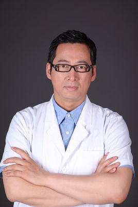 Shi Tan (谭石)合作论文数Department of Ultrasound, Peking University Third Hospital28
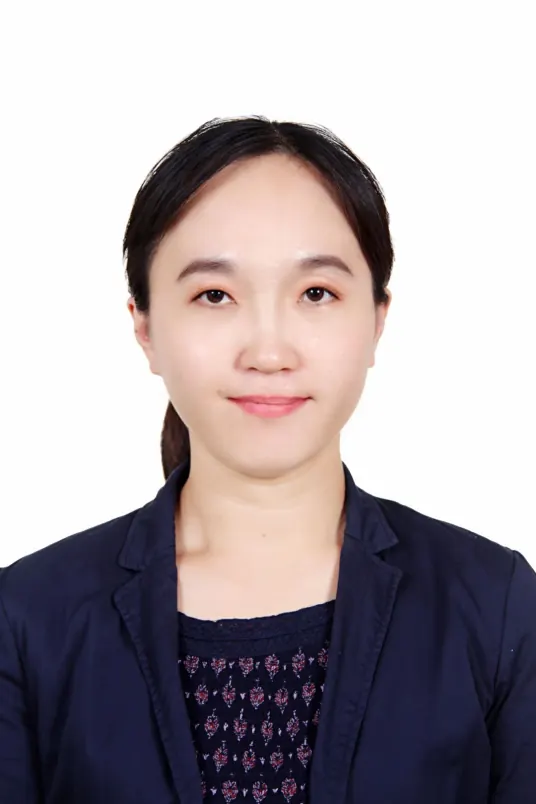 Jie Jiang (蒋洁)合作论文数Department of Ultrasound, Peking University Third Hospital27
 Zhiqiang Li (李志强)合作论文数Department of Ultrasound Medicine, Peking University Third Hospital23
 Bo Zhao (赵博)合作论文数Department of Ultrasound Medicine, Peking University Third Hospital21LiYing Miao (苗立英)合作论文数20Ying Fu合作论文数17
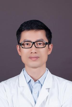 Xiaolong Liang (梁晓龙)合作论文数Peking University Third Hospital16
 Yan Sun (孙彦)合作论文数Department of Ultrasound, Peking University Third Hospital16
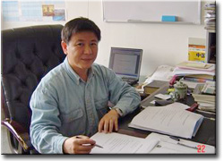 Wu Zhang (张武)合作论文数School of Computer Engineering and Science,Shanghai University,Shanghai,China15Weiwei Shen (沈伟伟)合作论文数14Yutao Lei (雷玉涛)合作论文数14
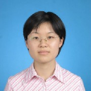 Fang Mei (梅放)合作论文数School of Basic Medical Sciences, Peking University Health Science Center14
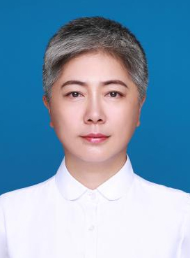 Huiyu Ge (葛辉玉)合作论文数Beijing Chao-Yang Hospital, Capital Medical University;Molecular Imaging and Minimally Invasive Therapy Laboratory, Peking University13Suhui Sun合作论文数9Yunxia Hao合作论文数9
 Guanying Gao合作论文数9HuaBin Zhang (张华斌)合作论文数9Fu Qiang合作论文数9
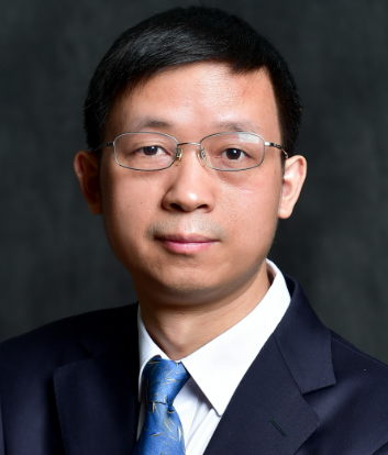 Jianwen Luo (罗建文)合作论文数School of Biomedical Engineering, Tsinghua University9
 Shuai Fu (付帅)合作论文数Department of Ultrasound, Peking University Third Hospital8LIN Zhuo-hua (林卓华)合作论文数7Qiang FU (傅强)合作论文数7Jinxia Zhang合作论文数7
 Yan Xu合作论文数Linklogis7Ming-Yu Bai合作论文数6Youjing Sun合作论文数6Xiaohui Sun (孙晓慧)合作论文数6Xifeng Ge (葛喜凤)合作论文数6Rui Tang合作论文数6Ruiqi Wu合作论文数6
 Yang Sun合作论文数Columbia University in the City of New York, Columbia University6WeiQiang Ran (冉维强)合作论文数6JingYing Yang (杨敬英)合作论文数6Jinhua Shao (邵金华)合作论文数6
 Jing Bai (白净)合作论文数School of Medicine, Tsinghua University;Department of Biomedical Engineering, Tsinghua University6Lijuan Guo (郭丽娟)合作论文数6Weijun Lin (林伟军)合作论文数6
 Linxue Qian (钱林学)合作论文数Department of Ultrasound, Beijing Friendship Hospital, Capital Medical University6Lihong Sun合作论文数6
 Wen Chen (陈文)合作论文数College of Biochemical Engineering, Beijing Union University6




