Aims Perivascular endoscopic ultrasound augmented radiofrequency ablation with sugar (EUS-sugar-RFA) facilitated distal pancreatectomy and increases pancreatic tissues necrosis in a swine model. Analyze the sugar-RFA modulation applied with a EUS needle in the pancreatic perivascular tissue in an ex vivo swine pancreas.
La sleeve gastroplastie endoscopique (SGE) est une des procédures endoscopiques mini-invasives visant une perte de poids. Son efficacité en termes de perte pondérale est avérée. Il persiste, toutefois, un manque de connaissances concernant les bénéfices apportés par le traitement et son impact sur les comorbidités liées à l’obésité. Cette étude vise à évaluer l’effet de la SGE sur la perte pondérale et l’impact de cette dernière sur la qualité de vie et les comorbidités liées à l’obésité. Il s’agit d’une série de cas consécutifs issus d’une étude observationnelle prospective réalisée dans un centre spécialisé disposant d’un parcours de soins standardisés pour la prise en charge multimodale de l’obésité. Les patients bénéficiant d’une SGE et d’un suivi endoscopique et biologique à 6 et 12 mois après cette intervention, ont été inclus dans l’étude. La perte pondérale, les comorbidités et les paramètres biologiques, enregistrés prospectivement à 6 et 12 mois après l’intervention ont été analysés rétrospectivement. La variation de l’indice de masse corporelle (IMC), la perte pondérale absolue (AWL), la proportion de perte de poids excédentaire (% EWL) et la proportion de perte pondérale totale (% TWL) à 6 et 12 mois ont été évaluées. La réduction des différentes comorbidités liées à l’obésité (hypertension artérielle (HTA), diabète de type 2 (DT2), reflux gastro-œsophagien (RGO), syndrome d’apnée obstructive du sommeil (SAOS) et dyslipidémie a été évaluée à 6 et 12 mois. La variation de la glycémie, des tests sanguins hépatiques et lipidiques a également été analysée à 6 et 12 mois. D’octobre 2016 à juillet 2021, sur 227 patients ayant subi une SGE, 99 (43,6 %) ont bénéficié d’un suivi endoscopique et biologique complet à 6 et 12 mois. L’IMC initial était de 42,7 ± 7,8 kg/m2 et l’âge de 45 ± 12,7 ans. Soixante-quatorze (74,8 %) étaient des femmes. Les pertes pondérales (% TWL) et pertes de poids excessif (% EWL) étaient respectivement de 16,6 % ± 7,4 et de 43,3 % ± 21,2 à 6 mois, de 16,6 % ± 9,6 et de 42,9 % ± 25,6 % à un an. Une réduction significative à 6 et 12 mois a été observée pour les taux de DT2 (30,8 % et 32,7 %), de l’HTA (18,4 % et 22,1 %), du RGO (28 % et 25,7 %), du SAOS (15,8 % et 25,5 %) et la dyslipidémie (69,2 % et 77,2 %) (p < 0,001). La réduction de la glycémie était significative entre la période préopératoire et 6 mois (p < 0,01) et entre la période préopératoire jusqu’à 12 mois (p < 0,01). La réduction des triglycérides et du cholestérol total entre la période préopératoire et 6 mois était significative (p < 0,01). Entre la période préopératoire et 12 mois, cette même réduction a également été significative (p < 0,01) (p = 0,017). Pour les tests hépatiques, la réduction de l’ASAT était significative à 6 et 12 mois après la SGE (p = 0,048) (p = 0,048). Cette même réduction a été observée pour l’ALAT à 6 et 12 mois après l’intervention (p < 0,01) et (p < 0,01) respectivement. La SGE est une procédure chirurgicale bien tolérée et sûre, efficace en termes de perte pondérale et de réduction des comorbidités liées à l’obésité à 6 mois et 1 an. Cette procédure pourrait ainsi être adoptée sur un plan clinique élargi afin de promouvoir plus amplement un traitement efficace contre l’obésité morbide. Endoscopic sleeve gastroplasty (ESG) is one of the a novel endoscopic bariatric treatments. Gastric volume reduction and delayed gastric emptying are the mechanisms which drive weight loss. However, potential benefits for comorbidities in relation to weight loss after an ESG are still being investigated. This study aims to evaluate the effect of ESG procedures on major obesity-associated comorbidities, and on some biological parameters. Patients who underwent follow-up endoscopy (at 6 and 12 months) after ESG and were compliant with the bariatric protocol were included. Weight loss, comorbidities, and biological parameters were prospectively collected and retrospectively analyzed at 6 months and 1 year after ESG. BMI variation, absolute weight loss (AWL), excess weight loss (%EWL), and total weight loss (%TWL) were assessed at 6 and 12 months. The reduction in different obesity-associated comorbidities was calculated (arterial hypertension, type 2 diabetes, gastroesophageal reflux disease, sleep obstructive apnea syndrome (SOAS) and dyslipidemia at 6 and 12 months. The variations in glycemia, hepatic and lipid blood tests were also analyzed at 6 and 12 months. From October 2016 to July 2021, of the 227 patients who underwent ESG, 99 (43.6%) had a follow-up EGD at 6 and 12 months. %TWL and %EWL were respectively 16.6% ±7.4 and 43.3% ± 21.2 at 6 months, 16.6% ± 9.6 and 42.9% ± 25.6 at one year. Significant reduction rates at 6 and 12 months were observed in T2DM (30.8% and 32.7%), HTN (18.4% and 22.1%), GERD (28% and 25.7%), SOAS (15.8% and 25.5%) and dyslipidemia (69.2% and 77.2%) (p < 0.001). Moreover, glycemic levels were significantly reduced between the preoperative period and 6 months (1.11 ± 0.22 mg/L vs. 1.01 ± 0.17 mg/L, P < 0.01), and between the preoperative period and 12 months (1.11 ± 0.22 mg/L vs. 1.06 ± 0.32 mg/L, P < 0.01). A significant reduction was also observed in triglycerides and total cholesterol levels at 6 months (1.52 ± 0.74 mmol/L vs. 1.14 ± 0.52 mmol/L, P < 0.01) (1.94 ± 0.4 mmol/L vs. 1.85 ± 0.36 mmol/L, P < 0.01) and at 12 months (1.52 ± 0.74 mmol/L vs. 1.18 ± 0.67 mmol/L, P < 0.01) (1.94 ± 0.4 mmol/L vs. 1.82 ± 0.39 mmol/L, P = 0.017) and in ASAT (27.2 ± 11.7 IU/L vs. 23.7 IU/L ; P = 0.048) (27.2 ± 11.7 IU/L vs. 24.7 ± 14.65 IU/L, P = 0.048) and ALAT levels (34 ± 21.32 IU/L vs. 22.3 ± 10.4 IU/L, P < 0.01 and 34 ± 21.32 IU/L vs. 27.07 ± 25 IU/L, P < 0.01) at 6 and 12 months after ESG, respectively. ESG is an effective procedure in terms of weight loss and reduction in obesity-related comorbidities at 6 months and 1 year of follow-up. This procedure has the potential for expanded clinical application, which could broaden the obesity care treatment armamentarium.
Training programs are essential to introduce new methods for bile duct clearance. Visual examination via cholangioscopy is ideal to diagnose and treat biliary tract diseases such as cancer and choledocholithiasis. However, surgeons rarely use cholangioscopes. Specific training is required to master laparoscopic and percutaneous cholangioscopy. This study aims to assess skill acquisition and retention during cholangioscopy training in the Image-Guided Therapies Masterclass. This prospective study enrolled 17 physicians undergoing training in interventional treatments of biliary diseases. A novel disposable cholangioscope and access kit were used with a biliary tract model including two simulated common bile duct (CBD) stones. The curriculum required visualization of all critical structures before removal of one stone with a Dormia basket. After informed consent, demographic data and time to exercise completion were recorded on each of two subsequent training days. Task-specific questions were measured at the completion of training using a Likert scale (strongly disagree to strongly agree, 1–5 points). All participants successfully completed the task (6F/11 M, age 36 ± 5 years; 13 surgeons, 4 interventional radiologists; median experience with percutaneous procedures 2 years, range 0–20). Significant improvement in mean task completion time was observed (day 1: 172 ± 59 s, day 2: 89 ± 45 s; P < 0.0001). All task-specific questions were answered with a median rating of 5/5: “The platform facilitates cholangioscopy” and “This training method accelerates gain in proficiency and is useful for residents/fellows” (IQR 5–5), “This platform is useful to measure the proficiency level” and “There is an application for simulation in percutaneous surgery training” (IQR 4.5–5), “The platform is user-friendly” and “The model quality recreates realistic scenarios” (IQR 4–5). Cholangioscopic bile duct exploration and stone retrieval were achieved by all participants using a dedicated training program and physical simulator. Significant skill progress was observed during 2 days of dedicated training.
Introduction: Endoscopic sleeve gastroplasty (ESG) is one of the new minimally invasive endo-scopic treatments aimed at inducing weight loss. Its effectiveness in terms of weight loss is proven. Gastric volume reduction and delayed gastric emptying are the mechanisms that drive weight loss. However, potential benefits for co-morbidities in relation to weight loss after ESG are still being investigated. This study aims to evaluate the effect of ESG procedures on major obesity-associated co-morbidities, and on some biological parameters. Patients and methods: This is a series of consecutive cases from a prospective observational study carried out in a specialized center that follows a standardized care pathway for the multimodal management of obesity. Patients who have undergone ESG with endoscopic and laboratory follow-up at six and twelve months after this intervention were included in the study. Prospectively recorded data on weight loss, co-morbidities and laboratory parameters at six and twelve months after surgery was analyzed retrospectively. Changes in body mass index (BMI), absolute weight loss (AWL), percent of excess weight loss (%EWL) and percent total weight loss (%TWL) were assessed at six and twelve months. Reduction in various obesity-related co-morbidities (arterial hypertension [AHT], type 2 diabetes mellitus [T2DM], gastroesophageal reflux disease [GERD], obstructive sleep apnea syndrome [OSAS] and dyslipidemia was also evaluated at six and twelve months. Changes in blood glucose, liver function tests and lipid blood tests were also analyzed at six and twelve months.Results: From October 2016 to July 2021, 99 of the 227 patients who underwent ESG in our unit (43.6%) subsequently underwent a complete endoscopic and laboratory follow-up at six and twelve months. The initial BMI was 42.7 +/- 7.8 kg/m2 and age was 45 +/- 12.7 years. Seventy-four patients (74.8%) were female. Total weight loss (%TWL) and excess weight loss (%EWL) were 16.6 +/- 7.4% and 43.3 +/- 21.2%, respectively, at six months, 16.6 +/- 9.6% and 42.9 +/- 25.6%, respectively, at one year. At six and twelve month follow-up, a statistically significant reduc-tion was observed for the rates of T2DM (30.8 and 32.7%), hypertension (18.4 and 22.1%), GERD (28 and 25.7%), OSAS (15.8 and 25.5%) and dyslipidemia (69.2 and 77.2%) (P < 0.001). A statistically significant difference was found in the reduction in blood glucose between the pre-operative period and six months post-operatively (P < 0.01) and between the pre-operative period and twelve months post-operatively (P < 0.01). The reduction in triglycerides and total cholesterol between the pre-operative values and at six months was statistically significant (P < 0.01) as was the reduction at twelve months (P < 0.01) (P = 0.017). For liver function tests, the reduction in AST was statistically significant at six and twelve months after ESG (P = 0.048) (P = 0.048) as was ALT (P < 0.01) (P < 0.01) respectively. From October 2016 to July 2021, of the 227 patients who underwent ESG, 99 (43.6%) had follow-up gastro-duodenoscopy at 6 and 12 months. %TWL and %EWL were respectively 16.6 +/- 7.4% and 43.3% +/- 21.2 at 6 months, 16.6 +/- 9.6% and 42.9 +/- 25.6% at one year. Statistically significant reduction rates at 6 and 12 months were observed in T2DM (30.8 and 32.7%), AHT (18.4 and 22.1%), GERD (28 and 25.7%), OSAS (15.8 and 25.5%) and dyslipidemia (69.2 and 77.2%) (P < 0.001). Moreover, glycemic levels were statistically significantly reduced between the pre-operative period and 6 months post-operative (1.11 +/- 0.22 mg/L vs. 1.01 +/- 0.17 mg/L, P < 0.01), and between the pre-operative period and 12 months post-operative (1.11 +/- 0.22 mg/L vs. 1.06 +/- 0.32 mg/L, P < 0.01). A statistically significant reduction was also observed in triglycerides and total cholesterol levels at 6 months (1.52 +/- 0.74 mmol/L vs. 1.14 +/- 0.52 mmol/L, P < 0.01) (1.94 +/- 0.4 mmol/L vs. 1.85 +/- 0.36 mmol/L, P < 0.01) and at 12 months (1.52 +/- 0.74 mmol/L vs. 1.18 +/- 0.67 mmol/L, P < 0.01) (1.94 +/- 0.4 mmol/L vs. 1.82 +/- 0.39 mmol/L, P = 0.017) and in AST (27.2 +/- 11.7 IU/L vs. 23.7 IU/L; P = 0.048) (27.2 +/- 11.7 IU/L vs. 24.7 +/- 14.65 IU/L, P = 0.048) and ALAT levels (34 +/- 21.32 IU/L vs. 22.3 +/- 10.4 IU/L, P < 0.01 and 34 +/- 21.32 IU/L vs. 27.07 +/- 25 IU/L, P < 0.01) at 6 and 12 months after ESG, respectively.Conclusion: ESG is a well-tolerated and safe surgical procedure that is effective in terms of weight loss and reduction of obesity-related co-morbidities at six months and one year. This procedure could thus be adopted on a broader clinical scale and be more widely promoted as an effective treatment for morbid obesity.(c) 2022 Published by Elsevier Masson SAS.
Il existe des controverses quant au bénéfice du protocole ERAS en chirurgie colorectale pour les patients âgés. L'objectif était d'identifier toute différence chez les patients âgés par rapport aux plus jeunes. Analyse rétrospective de la base de données ERAS (2014–janvier 2021). Les patients ont été répartis en 3 groupes: <65 ans(groupe1), 65–75 ans(groupe2), >75 ans(groupe3). Paramètres évalués: comorbidités, complications postopératoires, taux de fistules, mortalité à 30 jours, durée de séjour en soins intensifs, réinterventions, durée de séjour après chirurgie primaire, réadmission et durée totale d'hospitalisation(TTH). 837 (524 hommes) patients ont été évalués; 415groupe1; 241groupe2, 181groupe3. Comorbidités: diabète 9,6 %(groupe 1), 27 %(groupe 2), 26 %(groupe 3). Cardiomyopathie: 11,3 %(groupe1), 35,2 %(groupe2), 49 %(groupe3), maladie pulmonaire 6,5 %(groupe1)14,9 %(groupe2), 11,6 %(groupe3); tabagisme 24,8 %(groupe1), 14,5 %(groupe2), 3,3 %(groupe3). Taux de complications: 47,2 %(groupe1), 50 %(groupe2) et 49,1 %(groupe3) (p = 0,694). Fistule: 5 %(groupe1), 4,1 %(groupe2), 3,2 %(groupe3)(p = 0,616). Mortalité à 30 jours: 0. Séjour en UIC: 20 jours-4,8 % (groupe1), 19 jours–7,8 %(groupe2), 12 jours–6,8 %(groupe3)(p = 0,27). Durée de séjour en soins intensifs:0,25 ± 1,51(0–16)(groupe 1); 0,51 ± 2,48(0–25)(groupe 2); 0,36 ± 2,14 (0–25)(groupe 3)(p = 0,262). Durée de séjour après chirurgie primaire(jours): 7,51 ± 7,85(0–56)(groupe1); 7,73 ± 9,94(0-98)(groupe2); 7,97 ± 9,73(0–112)(groupe3)(p = 0,06). Réinterventions: 45 (10,8 %)(groupe1), 25 (10,4 %)(groupe2), 11 (6,1 %)(groupe3)(p = 0,177). Réhospitalisation: 42 (10,1 %)(groupe1), 21 (11,6 %)(groupe2), 16 (8,8 %)(groupe3). Durée moyenne du séjour après réhospitalisation: 5,2 jours(groupe1), 7,7 jours(groupe2), 11,7 jours (groupe3)(p = 0,02). Durée totale d'hospitalisation(jours): 8,05 ± 8,3(0–56)(groupe1); 8,41 ± 10,1(0–98)(groupe2); 9 ± 11,1(1–112)(groupe3)(p < 0,006). Il n'y avait pas de différence significative dans tous les paramètres évalués entre les patients âgés et jeunes, sauf pour la durée totale du séjour et en cas de réadmission. Les patients âgés n'ont pas connu plus de complications. Le programme ERAS ne nécessite aucune adaptation pour les personnes âgées.
Background Because of the complexity of the intra-abdominal anatomy in the posterior approach, a longer learning curve has been observed in laparoscopic transabdominal preperitoneal (TAPP) inguinal hernia repair. Consequently, automatic tools using artificial intelligence (AI) to monitor TAPP procedures and assess learning curves are required. The primary objective of this study was to establish a deep learning-based automated surgical phase recognition system for TAPP. A secondary objective was to investigate the relationship between surgical skills and phase duration. Methods This study enrolled 119 patients who underwent the TAPP procedure. The surgical videos were annotated (delineated in time) and split into seven surgical phases (preparation, peritoneal flap incision, peritoneal flap dissection, hernia dissection, mesh deployment, mesh fixation, peritoneal flap closure, and additional closure). An AI model was trained to automatically recognize surgical phases from videos. The relationship between phase duration and surgical skills were also evaluated. Results A fourfold cross-validation was used to assess the performance of the AI model. The accuracy was 88.81 and 85.82%, in unilateral and bilateral cases, respectively. In unilateral hernia cases, the duration of peritoneal incision ( p = 0.003) and hernia dissection ( p = 0.014) detected via AI were significantly shorter for experts than for trainees. Conclusion An automated surgical phase recognition system was established for TAPP using deep learning with a high accuracy. Our AI-based system can be useful for the automatic monitoring of surgery progress, improving OR efficiency, evaluating surgical skills and video-based surgical education. Specific phase durations detected via the AI model were significantly associated with the surgeons’ learning curve.
Anastomotic fistula is the most fearsome complication following colorectal surgery. Numerous studies have demonstrated the interest of postoperative CRP assay as an early diagnostic marker. Must the critical threshold for biological inflammatory markers remain the same, whether resection be colic or rectal?This is a study based on a cohort constituted between 2011 and 2014, including 497 patients with planned colorectal resection. C-reactive protein and pro-calcitonin were measured daily from day before surgery to D4. All postoperative intra-abdominal complications were considered as an anastomotic fistula. Detection thresholds were calculated from the area under the ROC curve.An intra-abdominal septic complication occurred in 16.9% of the patients having undergone rectal resection vs. 9.9% of those having had colectomy (P = 0.03). In the absence of complications there was no significant difference between the two groups in terms of postoperative inflammatory response as determined by either CRP or PCT assay. Following rectal resection, optimal area under the curve (AUC = 0.87) corresponds to CRP on D4 for a threshold of 100 mg/L: sensitivity 83.3%, NPV 95.3%. For colons with the same CRP at 100 mg/L (AUC = 0.71): sensitivity 63.6%, NPV 93.9%.Notwithstanding riskier surgery, the detection threshold for an anastomotic fistula following rectal surgery remains the same: CRP > 100 mg/L at D4.
Purpose: We present the case of a 45 years old patient with a diagnosis of a single resectable 12mm histologically proved G1 NET of the pancreatic isthmus. Method: In her past medical hystory we notice a laparoscopic gastric by-pass was performed for morbid obesity. Pre-operative work-up included pancreatic MRI, thoracic CT-scan and blood exams. Chromogranine A was within limits. After multidisciplinary discussion either resection or surveillance were conceived considering size and grading, the patient preferred surgical resection in order to avoid life-long surveillance and because of the fear of having a pancreatic lesion potentially evolving. A robotic minimally invasive approach was then considered, the surgical strategy focused on the distal pancreatic stump anastomosis. Results: Intraoperative ultrasound confirmed the absence of other pancreatic lesions. A pancreatico-gastric anastomosis was performed on the excluded stomach, tutored by an Escat drain exteriorized according to Voelker. Post-operative course was uneventful, the patient was discharged on postoperative day 9 (POD9),Pathological findings: grade 1 well-differentiated neuroendocrine tumor, Ki-67 index <1%, R0 (complete resection) Escat drain clamped on POD 14 and withdrawn 1 month after the operation. Conclusion: robotic central pancreatectomy allowed a very precise dissection with the possibility of an ergonomic pancreatico-gastric anastomosis.
Purpose: We present the case of a 82 years-old patient affected by HCV liver fibrosis with a diagnosis of a single resectable 2cm HCC of the superior border of segment II. The patient underwent a robotic left lateral sectionectomy. Method: Pre-operative work-up included liver MRI, thoracic CT-scan and blood exams. Child-Pugh score A5, MELD score 8, Platelet count normal, AFP within normal values. Gastroscopy was normal. After multidisciplinary discussion a liver resection was indicated using a robotic minimally invasive approach. Results: During intraoperative ultrasound, after excluding other missed lesions, careful evaluation of the tumor is done in order to precise the relationship with the terminal part of the left hepatic vein. Kelly clamp crushing technique is performed with assistant driven irrigation of the instrument. When dissection needs to be more precise an original use of cavitron ultrasonic surgical aspirator (CUSA), introduced in the assistant trocar and guided by the robotic arm. Final section of the hepatic vein is made with scissors after vascular clamping. A double 5/0 polypropilene running suture is performed to close the venous stump. The specimen is then extracted by a Pfannenstiel incision. There were no intraoperative complications, the post-operative course was uneventful with patient discharged at POD 4.Pathological findings: pT1 well-differentiated hepatocellular carcinoma (Edmonson-Steiner grade 1), with no vascular invasion, R0 (complete resection). Conclusion: Robotic liver resection allows a precise dissection, vascular suturing is more ergonomic because of the articulating instruments tip.
La Société française de chirurgie digestive et la Société d’imagerie abdominale et digestive ont souhaité proposer des recommandations pour la pratique clinique portant sur la prise en charge de l’appendicite de l’adulte. L’analyse de la littérature a été effectuée selon la méthodologie de la Haute Autorité de santé (HAS). Une sélection manuelle des publications scientifiques a été effectuée à partir des références obtenues, puis un tri manuel des références listées dans les articles sélectionnés a été fait à la recherche d’articles pertinents supplémentaires. La recherche était limitée aux articles en langue anglaise ou française. Les articles se focalisant sur la population pédiatrique ont été exclus. Sur la base de l’analyse de la littérature, le groupe de travail a proposé, chaque fois que possible, des recommandations. Ces recommandations ont été relues et validées par un comité d’experts. Des recommandations ont été proposées au sujet de l’appendicite aiguë de l’adulte concernant les modalités diagnostiques cliniques, biologiques et radiologiques, la stratégie thérapeutique devant une forme non compliquée ou compliquée, la technique chirurgicale et les spécificités en cas d’appendice macroscopiquement sain, d’iléite terminale et d’appendicite chez le sujet âgé et chez la femme enceinte. Ces recommandations pour la pratique clinique pourront aider le chirurgien dans l’optimisation de la prise en charge de l’appendicite aiguë de l’adulte. The French Society of Digestive Surgery (SFCD) and the Society of Abdominal and Digestive Imaging (SIAD) have collaborated to propose recommendations for clinical practice in the management of adult appendicitis. An analysis of the literature was carried out according to the methodology of the French National Authority for Health (HAS). A selection was performed from collected references and then a manual review of the references listed in the selected articles was made in search of additional relevant articles. The research was limited to articles whose language of publication was English or French. Articles focusing on the pediatric population were excluded. Based on the literature review, the working group proposed recommendations whenever possible. These recommendations were reviewed and approved by a committee of experts. Recommendations about appendicitis in adult patients were proposed with regard to clinical, laboratory and radiological diagnostic modalities, treatment strategy for uncomplicated and complicated appendicitis, surgical technique, and specificities in the case of macroscopically healthy appendix, terminal ileitis and appendicitis in the elderly and in pregnant women. These recommendations for clinical practice may be useful to the surgeon in optimizing the management of acute appendicitis in adults.
Purpose: We present the case of a 78 years-old patient affected by HCV cirrhosis with a diagnosis of a single resectable 2cm HCC of the upper and lateral border of the segment II. Methods: Pre-operative work-up included liver MRI, thoracic CT-scan and blood exams. Child-Pugh score A5, MELD score 8, Platelet count normal, AFP within normal values. Gastroscopy was zithin limits. After multidisciplinary discussion a liver resection was indicated using a robotic minimally invasive approach. Results: Intraoperative ultrasound was performed to exlude other missed lesions. Parenchymal transection is then realized by the kelly clamp crushing technique under intermittent vascular clamping (extracorporeal control). There were no intraoperative complications, the post-operative course was uneventful with patient discharged at POD 1. Pathological findings: pT1 moderately differentiated hepatocellular carcinoma (Edmonson-Steiner grade 2 to 3), R0 (complete resection). Conclusion: robotic liver resection can be performed safely, with a magnified vision and optimal control of the surgical field.
L’objectif de l’étude est de rechercher des facteurs prédictifs de mortalité post-opératoire en chirurgie colorectale urgente chez des patients octogénaires. Analyse rétrospective de facteurs épidémiologiques, cliniques et biologiques sur une cohorte de 111 patients ayant bénéficié d’une chirurgie colorectale en urgence entre janvier 2015 et décembre 2019. Le critère principal était la mortalité à 30 jours. Analyse univariée et multivariée des données réalisée selon un modèle de régression logistique. Un total de 111 patients : 43 hommes,68 femmes. L’âge moyen était de 85,5 ans (80–96 ans). Les principales indications opératoires étaient : sigmoïdite compliquée (34,2 %), cancer occlusif (31,5 %), colite ischémique (27,9 %). Un total de 67 % des patients étaient ASA3. Le score de Charlson moyen était de 5,9. Le score Possum était de 38 pour mortalité. La mortalité à 30 jours était de 25,2 % ;17 décès étaient survenus précocement (< 7 jours). L’analyse univariée retrouvait comme statistiquement significatifs : admission directe en réanimation (60,7 % vs 22,9 % ; p < 0,001), colite ischémique (46,7 %vs21 % ; p = 0,012), hyperlactatémie (71,4 % vs 33,7 % ; p = 0,001), sepsis (50 % vs 16,87 % ; p = 0,001). L’analyse multivariée retrouvait uniquement l’hyperlactatémie (> 2 mmol/L) comme significative (p = 0,049). Le suivi moyen était de 3,4 ans (1–6,2 ans). La survie à 1 et 3 ans était de 57 % et 48,6 %. La chirurgie colorectale en urgence présente une mortalité importante. La mortalité à 30 jours retrouvée (25,2 %) correspond aux données de la littérature et inférieure à celle attendue. L’âge ne doit pas être une contre-indication. L’hyperlactatémie (> 2 mmol/L) semble être un facteur indépendant prédictif de mortalité.
The implantation of biological prostheses in an at-risk environment has seen increasing use. Their markedly higher cost compared to synthetic prostheses makes it important to analyse their usefulness in terms of actual benefit and cost-effectiveness. This study aims to examine the relevance of bioprostheses during surgical repair of Grade II/III ventral hernias as classified by the Ventral hernia working group (VHWG).This study analysed the data of 119 patients requiring non-emergency repair of VHWG II/III grade hernias between 2010 and 2017. The results of patients who were treated with a bioprosthesis (n = 59) were compared to those receiving a synthetic prosthesis (n = 60). The primary outcome was surgical site infection (SSI) at 90 days. The secondary endpoints were hernia recurrence rate, cost of the prosthesis, duration of hospital stay and re-hospitalisation rate.The two groups were shown to be comparable by analysis of demographic, pre- and intraoperative data. The SSI rate was significantly higher in the bioprosthesis group (20% vs. 7%; P = 0.010), as was the recurrence rate (56% vs. 28%; P = 0.003) with a median follow-up of 40 months. The cost of the bioprosthesis was significantly higher than that of the synthetic prosthesis (€3363 vs. €249; P < 0.010).In this retrospective study, the use of a bioprosthesis for repair of VHWG II/III ventral hernias was associated with a higher rate of both SSI and hernia recurrence at a cost 13 times greater than the use of a synthetic prosthesis.
Purpose: Central hepatectomy (CH) is a technically demanding procedure normally performed in high-volume hepatobiliary surgery centres. The rationale for this technique is to avoid extensive right or left resection leaving insufficient future liver remnant volume (FLR),but ensuring, however, oncological resections with clear surgical margins. CH remains an under-used technique due to its complexity. In presence of large tumours venous vascular control, when needed, may be demanding.The aim of this surgical descriptive study is to present a safe surgical strategy for the treatment of large central neoplasms for which venous vascular control is difficult. Methods: We propose a surgical strategy to reduce intraoperative and postoperative complications. It consists in obtaining a progressive control of suprahepatic veins, in a counterclockwise fashion. Hepatic transection starting from the left (left border of the segment 4) to arrive on the terminal part of the middle hepatic vein. After his section, the transection line and the future specimen are pulled on the right in order to allow an easier controle of the temrinal part of the right hepatic vein. Finally, the right part of the transection line begins from the bottom upward, following the right hepatic vein. Results: We used this strategy in 10 patients with large central hepatic neoplasms with compression, but without infiltration, of the right suprahepatic vein, and associated to a difficult right vein controle before transection. This approach was not associated to any intra- or post-operative complications (bleeding, bile duct leakage,post operative liver failure, post operative haemorrhages). Conclusion: Central hepatectomy is a complexc surgical procedure that maybe associated to intraoperative venous hemorrage, especially in case of large tumours. The surgical strategy allows a safer control of the hepatic veins in difficult situations where total vascular liver exclusion is not possible for hemodynamic reasons.
La chirurgie ambulatoire (CA) en France était en constante augmentation, avec un taux observé de 50 % et un objectif fixé par le ministre de la Santé de 70 % en 2022, qui semble difficilement atteignable du fait d’un ralentissement persistant de sa croissance. Identifier les raisons motivant des hospitalisations pour les procédures ambulatoires courantes et freinant ainsi le taux de CA, afin d’y remédier par des mesures adaptées. Étude prospective unicentrique à l’unité de CA de l’institut hospitalo-universitaire de Strasbourg, en recensant toutes les procédures de chirurgie digestive réalisées en ambulatoire et les mêmes procédures réalisées en hospitalisation, entre janvier 2017 et octobre 2019. Pour identifier les raisons des hospitalisations, une analyse rétrospective des dossiers a été abandonnée, car trop incomplète, au profit d’une analyse prospective sur trois mois. Les motifs d’hospitalisations ont été classés en 5 catégories. En 2019, 30,2 % des actes chirurgicaux sont réalisés en ambulatoire. Soixante-sept pour cent des cholécystectomies en 2019 vs 54 % en 2017 sont réalisées en ambulatoire pour les hernies/éventrations : 56 % en 2019 vs 52 %. Pour les gestes proctologiques : 94 % en 2019 vs 61 %. Dans 68,7 % des cas, une raison d’hospitalisation était retrouvée : on notait 16,7 % d’isolement géographique ou social, 37,5 % pour comorbidités sévères, 16,7 % dus à un défaut organisationnel du service, du chirurgien ou à un refus du patient. On observe une progression dans la prise en charge ambulatoire des patients pour la majorité des actes « marqueurs ». Parmi les facteurs motivant l’hospitalisation, certains sont naturellement limitant mais il en existe sur lesquels on peut agir.
Background Laparoscopic right hemicolectomy (LRHC) is increasingly performed for the treatment of right colon disease. Nevertheless, standardization of the surgical technique regarding the performance of intracorporeal (IC) or extracorporeal (EC) anastomosis is lacking. The purpose of this study was to compare short-term postoperative outcomes in patients who had laparoscopic right colectomy either with IC or EC. Methods This was a retrospective, non-randomized and multicenter study conducted from January 2005 to December 2015 on the CLIMHET study group cohort from five tertiary centers in France. Data were collected for all patients with LRHC to compare patient characteristics, intraoperative data and postoperative outcomes in terms of medical and surgical complications, duration of hospitalization and mortality. A multivariate analysis was performed to compare the results in the two groups. Results Of the 597 patients undergoing LRHC, 150 had IC and 447 had EC. The incidence of medical complications (cardiac, vascular, and pulmonary complications) was lower in the IC group than in the EC group (13 vs 20%, p = 0.049). This difference remained significant in multivariate analysis after adjusting to field characteristics and patient histories ( p = 0.009). Additionally, a shorter hospital stay (7 vs 8 days, p = 0.003) was observed in the IC group as compared to the EC group. This difference remained significant in favor of the IC group in multivariate analysis ( p = 0.029). There was no difference between the groups as regards: surgical complications ( p = 0.76), time of mobilization ( p = 0.93), reintervention rate ( p = 1) and 90-day mortality ( p = 0.47). Conclusions Our results show that IC anastomosis in LRHC is associated with fewer medical complications and shorter hospital stays compared to EC anastomosis.
In this video, we show a robotic central pancreatectomy for a well-differentiated neuroendocrine tumor. This is the case of a 50-year-old patient admitted to the emergency department for acute pancreatitis. CT-scan and MRI demonstrate the presence of a hypervascularized lesion of approximately 15mm in diameter in the pancreatic isthmus. Octreoscan does not evidence any intense uptake. The colon and the omentum are detached and the stomach is suspended laparoscopically. The robot is docked using a lateral approach. A retropancreatic passage is achieved on the mesenteric-portal axis. An intraoperative ultrasonography is performed to visualize the tumor and delimitate the resection margins. After the dissection, the anastomosis is performed between the distal part of the pancreatic remnant and the posterior gastric wall. The postoperative period was characterised by a grade B pancreatic fistula. Pathologist report confirmed the presence of a well-differentiated neuroendocrine tumor. The patient was discharged on postoperative day 22.
Clinique. La dénutrition est un véritable problème de santé publique engendrant d’importantes complications parmi lesquelles les infections nosocomiales(IN). Néanmoins, la dénutrition ne fait classiquement pas partie des facteurs de risque reconnus d’IN. L’objectif de cette étude a donc été d’évaluer l’impact de la dénutrition sur la survenue d’IN, et notamment d’estimer dans quelles proportions la dénutrition pouvait être considérée comme un facteur de risque indépendant d’IN. L’étude était prospective, monocentrique, composée de patients adultes hospitalisés dans 2 pôles médico-chirurgicaux du CHU de Strasbourg. L’enquête s’est déroulée sur cinq semaines successives, de février à mars 2017. Les patients ont bénéficié d’une évaluation de l’état nutritionnel par une diététicienne selon les recommandations HAS 2003–2007, avec le recueil de l’IMC, la perte de poids, l’albuminémie, l’EVA des ingesta et calcul du NRI. La survenue d’une IN a été ensuite recherchée puis validée par un médecin expert de l’Equipe opérationnelle d’hygiène du CHU selon les références de l’Enquête nationale de prévalence des IN. Sur le plan statistique, les comparaisons de moyennes ont été réalisées à l’aide d’un test t ou d’une Anova selon les cas, les comparaisons des distributions des variables qualitatives par un test du Chi2 et une régression logistique utilisée pour déterminer les variables prédictives d’une IN. Parmi les 609 patients éligibles, 339 ont bénéficié d’une évaluation nutritionnelle complète. Au total, 49,9 % des patients étaient dénutris avec 35,4 % et 14,5 % respectivement modérément et sévèrement dénutris. Un total de 57 patients ont présenté une IN, soit une incidence de 16,8 %. Il existe une plus forte incidence des IN en fonction de la gravité de la dénutrition (5,9 % non dénutris vs 23,3 % modérément dénutris vs 38,8 % sévèrement dénutris ; p < 0,0001). Les odds ratio (OR) de développer une IN chez les patients dénutris modérés et sévères sont respectivement de 4,9 (p < 0,0001) et 10,1(p < 0,0001). Les OR sont également majorés en fonction de la durée d’hospitalisation de 7–14 jours ou ≥ 14 jours avec, respectivement, des valeurs de 7,1 et de 33,3 (p < 0,0001). L’analyse multivariée démontre que la dénutrition est un des facteurs de risque prépondérant au développement d’une IN et proportionnel à sa sévérité [OR 2,3 (p = 0,063) pour les dénutris modérés ; OR 5,3 (p < 0,001) pour les dénutris sévères]. Les autres facteurs retenus sont la présence d’une durée d’hospitalisation prolongée [OR 7,0 (p < 0,0001) pour 7–14 jours ; OR 17,4 (p < 0,0001) pour ≥ 14 jours], la présence d’un cathéter (KT) central [OR 4,7 (p < 0,0001)] ou d’une chirurgie récente [OR 2,6 (p = 0,012)]. Plus, la durée d’hospitalisation se prolonge moins bon est l’état nutritionnel (p < 0,001). Au vu de nos résultats, la dénutrition semble être un facteur de risque indépendant et important de développement d’une IN au même titre qu’une durée d’hospitalisation prolongée ou la présence d’un KT central. L’impact de la dénutrition sur la survenue d’une IN est amplifié par une durée d’hospitalisation qui se prolonge, ces deux éléments étant souvent intriqués en pratique clinique courante, avec l’aggravation de l’état nutritionnel des patients dont l’hospitalisation se prolonge.
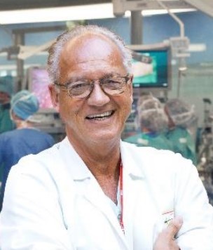 Jacques Marescaux合作论文数Institut de Recherche contre les Cancers de l'Appareil Digestif153
 Michel Vix合作论文数47
 Serge Evrard合作论文数34
 Joel Leroy合作论文数24
 Patrick Pessaux合作论文数20Antonio D'Urso合作论文数“Francesco Durante” Department of Surgery, University of Rome “La Sapienza”,16Silvana Perretta合作论文数Department of Gastrointestinal and Endocrine Surgery, University of Strasbourg15
 Luc Soler合作论文数Visible Patient S.A.S;Faculty of Medicine, University of Strasbourg;Digestive Cancer Research Institute14P Keller合作论文数13R. Memeo合作论文数11Bernard Dallemagne合作论文数IRCAD, Res Inst Digest Canc11Marc Aprahamian合作论文数11V Tassetti合作论文数10
 Marius Nedelcu合作论文数8L Mendoza-Burgos合作论文数7
 Stylianos Tzedakis合作论文数Faculté de Médecine, Université de Paris7C Damge合作论文数6C Napolitano合作论文数6
 Catherine F. Roy合作论文数6F Rubino合作论文数5M. Ignat合作论文数5Marichal J F合作论文数5
 Emanuele Felli合作论文数5
 Francesco Rubino合作论文数King's College London5
 Nicholas Ayache合作论文数Inria;AI Institute “3IA Côte d’Azur”5Y. Russier合作论文数5
 Tullio Piardi合作论文数5Nicolas Demartines合作论文数5P Sonzini合作论文数4V. De Blasi合作论文数4F. Alexandre合作论文数4
 Edoardo Maria Muttillo合作论文数Sant'Andrea University Hospital, Sapienza University of Rome4
 Alfonso Lapergola合作论文数Visceral and Digestive Surgery Department, New Civil Hospital (NHC),;Digestive System Cancer Research Institute (IRCAD), 1, place de l'Hôpital, 67091 Strasbourg, France. Electronic address: alfonso.lapergola@chru-strasbourg.fr.;Digestive System Cancer Research Institute (IRCAD),, New Civil Hospital (NHC),4Petitjean P合作论文数4
 Erik Dutson合作论文数Huggins Hospital4J M Clément合作论文数3I. Jourdan合作论文数3S Vartolomei合作论文数3F Harder合作论文数3
 Heithem Jeddou合作论文数3L. Marx合作论文数3G. Mathis合作论文数3N. Ferreira合作论文数3
 Stefano Chiodini合作论文数3Costantino F合作论文数3Francoise Heibel合作论文数3Hahn E G合作论文数3D'Agostino J合作论文数3Dhumane P合作论文数3




