Physiological factors, such as cerebral blood flow, blood pressure, respiratory rate, and body temperature are critical determinants of ischemic stroke pathophysiology, infarct development, and overall outcomes, and vice versa. However, despite long-standing guidelines recommending rigorous monitoring of key physiological factors, this aspect is not always incorporated into experimental designs and is generally underreported. Further, even when collected it is not always clear whether the data are integrated into the interpretation of results, potentially affecting translation. This review outlines the importance of monitoring, reporting and interpreting key physiological parameters in experimental stroke models, including the cardiovascular and respiratory systems, metabolism, temperature, circadian rhythm and intracranial pressure. Our aim is to provide researchers with an understanding of 1) different aspects of physiology that can affect experimental outcomes; 2) why and when it is important to monitor animal physiology in the context of ischemic stroke; 3) potential options for monitoring equipment and procedures; and 4) why accurate and transparent reporting and deliberate incorporation of physiological data into interpretation will enhance experimental rigor and strengthen future translational outcomes.
Snake envenomation activates the immune system through permeability-increasing factors that generate an acute vascular inflammatory response by opening large inflammation-activated pores (IAPs) in the microvasculature. These events facilitate the exudation of plasma from blood into body tissues. However we show that IAPs also allow macromolecules, including venom toxins, to flood directly into the bloodstream, even against an outflow of plasma solutes. Such inflammation-facilitated macromolecular absorption (IFMA) acts together with lymphatic absorption and has a physiological role in removing interstitial molecules, as evidenced by our dextran studies. IFMA will function in vascular absorption of interstitial molecules, potentially up to the radius of IAPs, which we determined to be 21 nm (95% confidence interval (CI) 18-24 nm). This absorption depends on factors, including the interstitial-vascular concentration gradient, the reflection coefficient of each molecule and microvascular pressure. Molecules absorbed will include snake venom toxins and various cellular breakdown products that arise during processes such as the inflammatory phase of wound healing. Most, if not all, venom toxins will be absorbed, as these typically have a hydrodynamic radius (r0) of 1-6 nm, which is well below that of IAPs. Notably once in the circulation venoms cause distributed inflammation, enhancing venom toxin movement from the bloodstream into the tissues. These mechanisms markedly increase the absorption of often lethal snake toxins and ensure their dissemination throughout body tissues, facilitating prey capture and adding to make snakebite extremely dangerous to humans. These findings provide mechanistic insight into current empirical snakebite first aid and present directions that may improve these procedures.Key points Snake venoms induce acute vascular inflammation, manifested by the opening of large pores termed inflammation-activated pores (IAPs), which we experimentally measure to have a radius of 21 nm. These pores mediate well-known exudation of plasma proteins but simultaneously provide a pathway for absorption of macromolecules, including venoms, a process that operates in parallel with the lymphatic system in clearing interstitial macromolecules. Although utilized in snakebite envenomation, such inflammation-facilitated macromolecular absorption (IFMA) is likely to have important physiological or pathophysiological roles, candidates including clearance of cellular breakdown products during the inflammatory phase of wound healing. Once in the vasculature venoms induce distributed inflammation of the IAPs, facilitating the exudation of venom toxins into tissues. The findings together with a model based on our experimental data provide new directions for improving snakebite first aid and present experimental evidence for a mechanism that operates to clear interstitial macromolecules.
Abstract Aim Despite advances in endovascular recanalization for ischemic stroke, many patients experience poor outcomes. Adjunct cerebroprotective therapies are needed to improve recovery. The mammalian target of rapamycin complex 1 (mTORC1) inhibitor rapamycin has shown neuroprotective effects in preclinical stroke models. However, most studies administered rapamycin prior to or during stroke onset, limiting translational relevance. The aim of this study was to determine whether rapamycin administered immediately after recanalization improves infarct size and functional outcome, and whether these effects are associated with changes in cerebral blood flow (CBF) or blood‐brain barrier (BBB) integrity. Methods Male Wistar Han rats were subjected to transient middle cerebral artery occlusion (tMCAO) for 90 min. Animals were randomized using the sealed envelope method to receive intravenous rapamycin (250 μg/kg, n = 9) or vehicle (n = 9) immediately after recanalization. Infarct volume, CBF, and BBB integrity were assessed using magnetic resonance imaging (MRI) at 72 h, alongside validated neurological tests. Group comparisons were performed using unpaired Student's t‐tests. Results Rapamycin significantly reduced infarct volume compared with vehicle (44.77 ± 30.93 mm³ vs. 113.44 ± 60.19 mm³, p = 0.0114) and improved Garcia neurological scores (12.78 ± 1.04 vs. 11.67 ± 0.87, p = 0.0295). In the adhesive removal test, rapamycin‐treated animals showed shorter time to notice the stimulus (45.04 ± 11.91 s vs. 72.33 ± 12.17 s, p = 0.0002). Rapamycin had no significant effect on CBF, BBB disruption, or edema at 72 h (all p > 0.05). The p‐mTOR/mTOR ratio did not differ significantly between groups at day 3 (0.55 ± 0.32 vs. 0.90 ± 0.41, p = 0.1880). Discussion Rapamycin administered after recanalization improves functional outcomes and reduces infarct size, without altering sustained perfusion or BBB permeability. These findings highlight a perfusion‐independent, time‐sensitive cerebroprotective mechanism and support rapamycin's development as an adjunctive therapy in ischemic stroke.
Magnetic resonance imaging (MRI) of glucose metabolism shows significant potential for identifying disease biomarkers and monitoring therapeutic responses in neurological conditions. Here, we present a protocol utilizing chemical exchange-sensitive spin-lock (CESL) MRI with the glucose analogue 2-deoxy-D-glucose (2DG) in the rat brain. We employed this method to characterize metabolic changes in ischemic tissue in a rat model of stroke. However, the utility of the technique is not limited to stroke and may be adapted to other disease models with minimal modifications. Previous research has demonstrated that CESL MRI is sensitive to various glucose analogs, including regular D-glucose, which is suitable for human application. Consequently, our protocol provides a foundation for a wide range of future applications in both basic and translational research, with potential utility in animal models and, eventually, human studies.
In patients with ischaemic stroke, retrograde perfusion of the penumbra by leptomeningeal collateral vessels (LMCs) strongly predicts clinical outcome, suggesting that enhancing LMC flow can offer a novel therapeutic approach. Using in vivo measurements and computational modelling it is shown that LMCs experience elevated fluid shear stress that is significantly higher than that in other blood vessels during ischaemic stroke in rats and humans. We exploit this to selectively enhance flow in LMCs using shear-activated nanoparticle aggregates carrying the vasodilator nitroglycerin (NG-NPAs) that specifically release drug in regions of vessels with high wall shear stress (≥100 dyne cm-2). NG-NPAs significantly increased LMC-mediated penumbral perfusion, decreased infarct volume, and reduced neurological deficit without altering systemic blood pressure in a rat ischaemic stroke model. NG-NPAs also avoided common side effects of systemic nitrate administration, such as systemic hypotension, cerebral vascular steal, cortical vein dilation, or intracranial pressure elevation. Systemic administration of free NG at the maximal tolerated dose, which is ten times higher than the dose of NG used in the NG-NPAs, do not enhance LMC perfusion and dropped blood pressure. Thus, packaging NG within shear-activated NPAs can potentially enable this widely available vasodilator to become a highly effective therapeutic for ischaemic stroke.
Good leptomeningeal collateral vessels (LMC) are associated with smaller lesion volume and better patient outcomes from ischaemic stroke, but their architecture varies greatly between individuals. Statins can stimulate angiogenesis and show promise for stimulating cerebral collaterogenesis. Statins could thus improve LMC grade and ensure all patients receive positive outcomes from treatment. This potentially explains why statin treatment is effective given prior to stroke in pre-clinical models, but not prescribed afterwards in clinical trials. LMC-rich C57Bl/6 and LMC-poor BALB/c 12-week-old male mice were randomised to receive daily oral simvastatin (10 mg/kg) or vehicle for 4 weeks. The cerebrovasculature was silicone-perfused, allowing LMC numbers and dimensions to be analysed. Values for tortuosity, vascularity and LMC resistance were calculated. In BALB/c, estimated resistance was lower with simvastatin treatment (vehicle: 0.104 ± 0.09, simvastatin: 0.023 ± 0.02; p = 0.04) and LMC numbers were greater (vehicle: 1.6 ± 1.6, simvastatin: 4.0 ± 3.2; p = 0.04). In C57Bl/6, there was no difference to resistance, but LMCs were smaller in diameter (vehicle: 16.3 ± 1.8 μm, simvastatin: 14.0 ± 1.4 μm, p = 0.008). Statin-treated C57Bl/6 also had more arterial branchpoints (left hemisphere, vehicle: 363.8 ± 55 per cm2, simvastatin: 417.3 ± 58 per cm2, p = 0.007; right, vehicle: 315.1 ± 40 per cm2, simvastatin: 397.5 ± 43 per cm2, p < 0.0001). We have observed signs of collaterogenesis and angiogenesis, providing evidence that statins stimulate growth of LMCs. Greatest benefit was seen in LMC-poor BALB/c, suggesting that patients with poor LMC circulation stand to gain most from LMC-enhancing therapies. Patients receiving statins prior to stroke have likely developed better LMCs, leading to better stroke outcomes. These findings should stimulate investigation of further safe, widely-available LMC-enhancing therapies.
Rapid breakdown of cerebral glucose metabolism is a hallmark in stroke pathology. Metabolic activity delineates the penumbra from the infarct core, representing tissue that is potentially salvageable by therapeutic interventions. Tools to image dynamics of glucose and its spatial distribution could provide biomarkers of disease severity and of the success of therapeutic interventions. Here, we developed a new protocol to measure glucose transport and metabolism in a rat model of stroke using chemical exchange-sensitive spin-lock (CESL) MRI of the glucose analogue 2-deoxy-D-glucose (2DG). We further implemented a protocol that combines 2DG-CESL-MRI with perfusion and diffusion MRI to relate this new signal to established definitions of hypoperfused tissue, cytotoxic edema and the penumbra. We found that 2DG-CESL-MRI provides a biomarker of disturbed glucose transport and metabolism after stroke with high effect size. This is the first study to investigate CESL MRI of 2DG in the context of transport and metabolism imaging in rodent stroke.
Magnetic resonance imaging (MRI) of glucose metabolism shows significant potential for identifying disease biomarkers and monitoring therapeutic responses in neurological conditions. Here, we present a protocol utilizing chemical exchange-sensitive spin-lock (CESL) MRI with the glucose analogue 2-deoxy-D-glucose (2DG) in the rat brain. We employed this method to characterize metabolic changes in ischemic tissue within a rat model of stroke. However, the technique is not limited to stroke and may be adapted to other disease models with minimal modifications. Previous research has demonstrated that CESL MRI is sensitive to various glucose analogs, including regular D-glucose, which is suitable for human application. Consequently, our protocol provides a foundation for a wide range of future applications in both basic and translational research, with potential utility in animal models and, eventually, human studies.
OBJECTIVES:There is increasing evidence that poor leptomeningeal collateral blood flow in hypertensive animals is due to increased vascular myogenic tone, indicating that therapies to enhance collateral blood flow during ischemic stroke may be particularly effective. To develop such therapies, we need a greater understanding of the factors that regulate collateral blood flow in the setting of hypertension. Therefore, we aimed to quantify blood flow velocity, diameter and absolute blood flow in individual collateral vessels in an ischemic stroke model in spontaneously hypertensive rats (SHRs) and determine which factors had the greatest influence on blood flow. MATERIALS AND METHODS:We quantified collateral flow velocity and vessel diameter and calculated absolute collateral blood flow in SHRs (n = 5) during 70 min of middle cerebral artery occlusion (MCAO), using a fluorescent microsphere method. RESULTS:Average collateral blood flow significantly increased post-occlusion relative to baseline (pre-MCAO: 16.8 ± 7.1nL/min vs. post-MCAO: 146.4 ± 37.7nL/min, p = 0.02). Within animal linear regression analysis showed a strong positive correlation between changes in collateral blood flow versus changes in collateral diameter during stroke (r = 0.7-0.99, p = 0.3-0.002). In contrast, collateral blood flow was only weakly correlated with collateral blood flow velocity during stroke (r = -0.03-0.97, p = 0.9-0.1). CONCLUSIONS:Collateral blood flow and velocity significantly increased post-occlusion. Collateral flow was strongly influenced by vessel diameter, likely because of marked baseline vasoconstriction of collaterals which is flow-limiting.
BACKGROUND:Nitroglycerin has been of considerable interest as a treatment for ischaemic stroke. Recent clinical trials with nitroglycerin transdermal patches during the acute phase of stroke failed to improve functional outcomes. Systematic review and meta-analysis of the effectiveness of nitroglycerin in preclinical models of ischaemic stroke has not previously been reported, despite several clinical trials. OBJECTIVE:To conduct a systematic review and meta-analysis of preclinical evidence regarding the effect of nitroglycerin on infarct volume in animal models of ischaemic stroke. SUMMARY OF REVIEW:The protocol was registered in PROSPERO (CRD42023432644). Our search identified 238 publications. Three publications met inclusion criteria (including 10 comparisons of infarct size). Study quality was modest (median 6 out of 9), with no evidence of publication bias. Nitroglycerin did not significantly reduce infarct volume (NMD point estimate 20.2 % reduction, 95 % CI -1.52-52.7 %, p = 0.068). Subgroup analysis suggested greater efficacy of nitroglycerin with direct intracarotid administration to the ischaemic territory at the time of reperfusion. CONCLUSIONS:A small number of studies (three) were included in this review. Overall, nitroglycerin did not reduce infarct volume in experimental stroke models. However, nitroglycerin may be of benefit when administered directly into the ischaemic territory. Given nitroglycerin's short half-life, we propose this route may minimise harmful reduction of cerebral perfusion pressure resulting from hypotension following systemic administration.
The contraction and subsequent death of brain pericytes may play a role in microvascular no-reflow following the reopening of an occluded artery during ischemic stroke. Mammalian target of rapamycin (mTOR) inhibition has been shown to reduce motility/contractility of various cancer cell lines and reduce neuronal cell death in stroke. However, the effects of mTOR inhibition on brain pericyte contraction and death during ischemia have not yet been investigated. Cultured pericytes exposed to simulated ischemia for 12 h in vitro contracted after less than 1 h, which was about 7 h prior to cell death. Rapamycin significantly reduced the rate of pericyte contraction during ischemia; however, it did not have a significant effect on pericyte viability at any time point. Rapamycin appeared to reduce pericyte contraction through a mechanism that is independent of changes in intracellular calcium. Using a mouse model of middle cerebral artery occlusion, we showed that rapamycin significantly increased the diameter of capillaries underneath pericytes and increased the number of open capillaries 30 min following recanalisation. Our findings suggest that rapamycin may be a useful adjuvant therapeutic to reduce pericyte contraction and improve cerebral reperfusion post-stroke.
EDITORIAL article Front. Stroke, 07 November 2023Sec. Mechanisms, Models, and Biomarkers of Stroke Volume 2 - 2023 | https://doi.org/10.3389/fstro.2023.1323696
Intracranial pressure (ICP) elevation post-stroke has long been thought of as a cause of secondary deterioration after large, malignant infarction, and dramatic ICP elevation is frequently a pre-terminal event. However, there is an increasing body of evidence to suggest that ICP also rises after small stroke, typically within 24 h of the infarct. The timing of this rise suggests that it may play an important role in the collateral failure associated with early infarct expansion. Despite its increasingly recognized importance to patient outcome, very little is currently known about the underlying mechanisms of ICP elevation post-stroke. The traditional understanding suggests ICP elevation occurs solely due to cerebral edema, however this does not seem to be the case in mild-moderate infarction. Instead, recent studies suggest a role for changes in cerebrospinal fluid (CSF) volume. In this article, we will discuss recent mechanistic observations, as well as the consequences of ICP elevation post-stroke.
We have previously demonstrated that a cortical stroke causes persistent impairment of hippocampal-dependent cognitive tasks concomitant with secondary neurodegenerative processes such as amyloid-β accumulation in the hippocampus, a region remote from the primary infarct. Interestingly, there is emerging evidence suggesting that deposition of amyloid-β around cerebral vessels may lead to cerebrovascular structural changes, neurovascular dysfunction, and disruption of blood-brain barrier integrity. However, there is limited knowledge about the temporal changes of hippocampal cerebrovasculature after cortical stroke. In the current study, we aimed to characterise the spatiotemporal cerebrovascular changes after cortical stroke. This was done using the photothrombotic stroke model targeting the motor and somatosensory cortices of mice. Cerebrovascular morphology as well as the colocalization of amyloid-β with vasculature and blood-brain-barrier integrity were assessed in the cortex and hippocampal regions at 7, 28 and 84 days post-stroke. Our findings showed transient cerebrovascular remodelling in the peri-infarct area up to 28 days post-stroke. Importantly, the cerebrovascular changes were extended beyond the peri-infarct region to the ipsilateral hippocampus and were sustained out to 84 days post-stroke. When investigating vessel diameter, we showed a decrease at 84 days in the peri-infarct and CA1 regions that was exacerbated in vessels with amyloid-β deposition. Lastly, we showed sustained vascular leakage in the peri-infarct and ipsilateral hippocampus, indicative of a compromised blood-brain-barrier. Our findings indicate that hippocampal vasculature may represent an important therapeutic target to mitigate the progression of post-stroke cognitive impairment.
AimsSelective neuronal vulnerability of hippocampal Cornu Ammonis (CA)-1 neurons is a pathological hallmark of Alzheimer's disease (AD) with an unknown underlying mechanism. We interrogated the expression of tuberous sclerosis complex-1 (TSC1; hamartin) and mTOR-related proteins in hippocampal CA1 and CA3 subfields. MethodsA human post-mortem cohort of mild (n = 7) and severe (n = 10) AD and non-neurological controls (n = 9) was used for quantitative and semi-quantitative analyses. We also developed an in vitro TSC1 knockdown model in rat hippocampal neurons, and transcriptomic analyses of TSC1 knockdown neuronal cultures were performed. ResultsWe found a selective increase of TSC1 cytoplasmic inclusions in human AD CA1 neurons with hyperactivation of one of TSC1's downstream targets, the mammalian target of rapamycin complex-1 (mTORC1), suggesting that TSC1 is no longer active in AD. TSC1 knockdown experiments showed accelerated cell death independent of amyloid-beta toxicity. Transcriptomic analyses of TSC1 knockdown neuronal cultures revealed signatures that were significantly enriched for AD-related pathways. ConclusionsOur combined data point to TSC1 dysregulation as a key driver of selective neuronal vulnerability in the AD hippocampus. Future work aimed at identifying targets amenable to therapeutic manipulation is urgently needed to halt selective neurodegeneration, and by extension, debilitating cognitive impairment characteristic of AD.
Ischaemic stroke treatment is limited to recanalizing the occluded vessel, while there is no approved adjunctive cerebroprotective therapy to protect either the neurons and parenchyma or the neurovascular unit. Pharmacological inhibition of mammalian target of rapamycin-1 (mTORC1) with rapamycin has shown promise in reducing infarct volume and improving functional outcomes. However, previous studies that investigated the effects of rapamycin on the vasculature and cerebral blood flow (CBF), administered rapamycin prior to or during stroke induction, thus limiting the potential for clinical translation. Therefore we investigated whether rapamycin maintains its cerebrovascular protective effect when administered immediately after recanalization following 90 minutes stroke in Wistar rats. We show, that rapamycin significantly improved post-recanalization cerebral blood flow (CBF), suggesting a beneficial neurovascular effect of rapamycin. Rats treated with rapamycin had smaller infarct volumes and improved functional outcomes compared to the control animals at three days post-stroke. The mechanisms of the overall positive effects seen in this study are likely due to rapamycin’s hyperacute effects on the neurovasculature, as shown with increased CBF during this phase. This paper shows that rapamycin treatment is a promising adjunct cerebroprotective therapy option for ischemic stroke.
Introduction: Leptomeningeal collateral flow is a strong predictor of stroke outcome. Enhancing collateral flow is an appealing therapeutic approach. However, previous attempts to enhance collateral flow with vasodilators have failed, due to systemic hypotension and cerebrovascular steal from the penumbra. We have shown that during experimental stroke collaterals have fluid shear stress that is 3-7 times higher (100 dyne/cm 2 ) than other blood vessels. This unique feature of collateral vessels provides a way to selectively enhance flow, using nanoparticle aggregates loaded with nitroglycerin (NG-NPAs), that only release drug in areas of high shear stress (≥ 100 dyne/cm 2 ). Hypothesis: Shear-activated NG-NPAs will selectively enhance collateral perfusion and improve stroke outcome. Methods: Proximal occlusion of the middle cerebral artery (MCAo) was induced for 70 min in male Spontaneously Hypertensive Rats. Changes in cerebral blood flow in the collateral-supplied ischemic MCA territory, and in the contralateral homotypic region were measured by laser speckle contrast imaging. Animals were randomized to receive I.V. infusion of blank-NPA (B-NPA, control, 4mg in 2ml of saline, n=7) or NG-NPA (50μg NG in 4 mg NPA = 4μg/kg/min of NG, n=7), commencing 25 minutes after MCAo, until reperfusion. Infarct volume was measured at 24 h. Results: NG-NPA significantly increased collateral perfusion by 44% vs. 11% for B-NPA at 40 minutes post-infusion (p=0.026), without altering perfusion in the contralateral region (NG-NPA: +19%, B-NPA: +13% of pre-infusion baseline, p=0.99) and without reducing blood pressure (NG-NPA: 161 mmHg, B-NPA: 163 mmHg, p=0.99). NG-NPA significantly reduced infarct volume at 24 h (NG-NPA: 70 mm 3 , B-NPA: 121 mm 3 , p=0.005). Better collateral perfusion was correlated with smaller infarct volume (R 2 = 0.44, p=0.012). Conclusion: Shear-activated NG-NPA selectively enhanced collateral perfusion to penumbral tissue without inducing systemic side effects, which resulted in smaller infarcts in this model. Given the known importance of collateral flow, shear-activated NG-NPA show great promise as a potential therapy for ischemic stroke patients, both pre-reperfusion therapy and in those unsuitable for such therapies.
 Neil Spratt合作论文数School of Biomedical Sciences and Pharmacy, University of Newcastle;Hunter Medical Research Institute26
 Damian D McLeod合作论文数School of Biomedical Sciences &Pharmacy and the Centre for Brain &Mental Health Research, University of Newcastle;School of Biomedical Sciences & Pharmacy and the Centre for Brain & Mental Health Research, University of Newcastle13
 Lucy Murtha合作论文数School of Biomedical Sciences and Pharmacy, University of Newcastle and Hunter Medical Research Institute10
 Alastair Buchan合作论文数Radcliffe Department of Medicine, Medical Sciences Division, University of Oxford10
 Pepperall Debbie合作论文数9
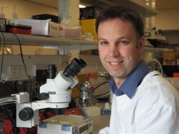 Brad A. Sutherland合作论文数University of Otago, University of Otago9
 Rebecca J Hood合作论文数School of Biomedicine, The University of Adelaide8
 Sara Azarpeykan合作论文数Massey University7
 Coupland Kirsten G.合作论文数6Yvonne Couch合作论文数Department of Pharmacology, Oxford University6
 Daniel Omileke合作论文数Sch Biomed Sci & Pharm, Univ Newcastle5
 Patabendige Adjanie合作论文数Institute of Infection and Global Health, University of Liverpool5
 Rohan Walker合作论文数School of Biomedical Sciences and Pharmacy, College of Health, Medicine and Wellbeing, University of Newcastle;Center for Advanced Training Systems, University of Newcastle;Hunter Medical Research Institute5A. Buchan合作论文数4Bryan A Adriaanse合作论文数4
 Steven William Bothwell合作论文数4
 Anna M. Schneider合作论文数4
 Gabriele DeLuca合作论文数Nuffield Department of Clinical Neurosciences, University of Oxford;Medical Sciences Division, University of Oxford4
 Gina Hadley合作论文数4Kristy Martin合作论文数3
 Sonia Sanchez Bezanilla合作论文数3
 Michael Nilsson合作论文数3
 Donald E. Ingber合作论文数Ingber Laboratory, Boston Children's Hospital;Harvard Medical School;Wyss Institute for Biologically Inspired Engineering, Harvard University;John A. Paulson School of Engineering and Applied Sciences, Harvard University3
 Deen Bhatta合作论文数School of Engineering and Applied Sciences, Harvard University3Neuhaus Ain A合作论文数3
 Lin Kooi Ong合作论文数School of Biomedical Sciences and Pharmacy, University of Newcastle3
 Oktay Uzun合作论文数Department of Materials Science and Engineering3C. Garcia Esperon合作论文数2Nikolce Mackovski合作论文数2
 Lachlan Shaun Brown合作论文数2Stefan P Koch合作论文数2
 Christopher Levi合作论文数Univ Newcastle, John Hunter Hosp, Dept Neurol, Newcastle, NSW, Australia2
 Ruslan Rust合作论文数Zilkha Neurogenetic Institute, Keck School of Medicine, University of Southern California2
 Boehm-Sturm Philipp合作论文数In-Vivo-NMR Laboratory, Max Planck Institute for Neurological Research2
 Schuenke Patrick合作论文数2Logan Caitlin L合作论文数2
 Collins-Praino Lyndsey合作论文数College of Physicians and Surgeons, Columbia University;College of Physicians and Surgeons, Columbia University;Columbia University, Columbia University2
 Bondarenko Evgeny合作论文数School of Biomedical Sciences, University of Newcastle;School of Psychology, University of Newcastle;University of Newcastle, University of Newcastle2
 Mark Woodward合作论文数Professorial Advisory Unit, The George Institute for Global Health;Faculty of Medicine, University of New South Wales, Sydney;Medical Sciences Division, University of Oxford2
 Mottahedin Amin合作论文数Department of Virology, Immunobiology and Parasitology, National Veterinary Institute2Eugene Nalivaiko合作论文数School of Medicine, Flinders University2
 Susanne Mueller合作论文数2
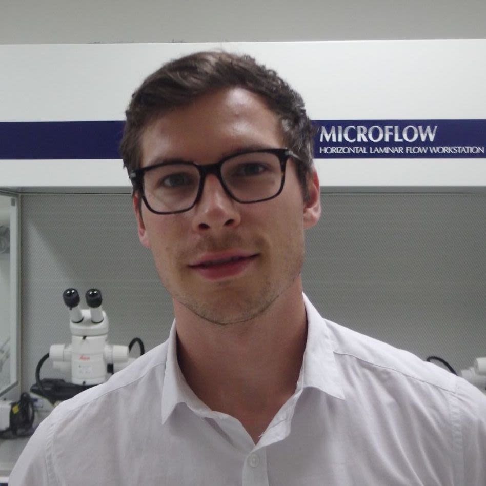 Paul Holloway合作论文数Radcliffe Department of Medicine, University of Oxford2
 Leif Schroder合作论文数Materials Sciences and Physical Biosciences Divisions, Lawrence Berkeley National Lab2
 Mirza Irfan Beig合作论文数School of Biomedical Sciences and Pharmacy, University of Newcastle and Hunter Medical Research Institute2Phillip Jobling合作论文数Centre for Neuroscience, Flinders University of South Australia2Marco Foddis合作论文数2
 Renee J Turner合作论文数Department of Neurology and M.I.N.D. Institute, University of California at Davis Medical Center2
 David Howells合作论文数School of Medicine, Faculty of Health, University of Tasmania2




