Vascular aging is characterized by the presence of chronic oxidative stress. Although cytosolic Sod 1 has a key role in the detoxification of superoxide (·O2−), little is known about its importance in vascular aging. We found that inhibition of Sod 1 had no effect on ·O2− generation. Furthermore, its expression decreased in an age-dependent manner. Interestingly, Sod 1 loses its membrane-association and is also lost from the caveolae with increasing age. Instead, a relocation of Sod 1 to the mitochondria takes place, presumably in an attempt to maintain mitochondrial integrity and to counter-balance age-associated oxidative stress. Unlike Sod 2, which is constitutively expressed in mitochondria to control ·O2− radical fluxes, Sod 1 is not inactivated by peroxynitrite and is not nitrated as a function of age. These novel insights into oxidative stress-associated vascular aging and the understanding about how redox-systems are regulated in old age may identify new targets to ameliorate aging as the greatest cardiovascular risk factor.
Background: The intracellular bacterium Chlamydia pneumoniae is suspected to play a role in formation and progression of atherosclerosis. Many studies investigated cell death initiation versus inhibition by Chlamydia pneumoniae in established cell lines but nothing is known in primary human aortic smooth muscle cells, a cell type among others known to be involved in the formation of the atherosclerotic plaque. Type of cell death was analyzed by various methods in primary aortic smooth muscle cells after infection with Chlamydia pneumoniae to investigate a possible pathogenic link in atherosclerosis.Results: Chlamydiae were found to be localized up to 72 h post infection in aortic smooth muscle cells either as single bacteria or inside of large inclusions. Quantification of host cell death by lactate dehydrogenase release assay revealed strictly dose and time dependent lysis for all tested isolates of Chlamydia pneumoniae. Phosphatidylserine exposure was detected by flow cytometry in Chlamydia pneumoniae infected cells. Ultrastructure of Chlamydia pneumoniae infected human aortic smooth muscle cells showed extensive membrane- and organelle damage, chromatin condensation but no nuclear fragmentation. DNA fragmentation as well as cell membrane permeability was analyzed by TUNEL and NHS-biotin staining and occurred exclusively in cells carrying Chlamydia pneumoniae spots but not in smooth muscle cells with inclusions. These morphological features of cell death were not accompanied by an activation of caspase-3 as revealed by analysis of enzyme activity but involved mitochondrial membrane depolarization as shown by TMRE uptake and release of cytochrome c from mitochondria.Conclusion: This study provides evidence that Chlamydia pneumoniae induce a spot like infection in human aortic smooth muscle cells, which results in a chimeric cell death with both apoptotic and necrotic characteristics. This aponecrotic cell death may assist chronic inflammation in atherosclerotic blood vessels.
Strains of Chlamydophila pneumoniae may be associated with respiratory disease or atherosclerosis. Two real-time quantitative PCR assays targeting the species-specific genes Cpn0278 and ArgR were developed to compare the in vitro growth of respiratory strains AR39 and K6 with that of atherosclerotic strain A03 and to quantify C. pneumoniae in clinical samples. A third real-time PCR assay was designed to assess contamination with Mycoplasma spp. The assays targeting C. pneumoniae detected DNA concentrations corresponding to 10(4) to 10(-4) inclusion-forming units (IFU)/reaction and were highly specific. AR39 exhibited the longest lag phase and period of exponential growth; K6 augmented growth rates at higher inocula; and A03 grew at highest rates. Contamination with Mycoplasma spp. of AR39 and A03 unlikely accounted for growth differences between them. Numbers of IFU in C. pneumoniae-positive respiratory secretions varied within 4 to 5 orders of magnitude. The assays described may prove valuable for pathogenicity studies.
After cloning and sequencing chicken connective tissue growth factor (cCTGF) we showed that in serum-deprived chicken embryo fibroblasts (CEF) the initial decline of cCTGF mRNA was followed by an increase. Human CTGF that was produced as a soluble protein in Escherichia coli by thioredoxin co-expression inhibited serum-deprivation induced apoptosis of CEF at I50 of ∼8 pM, demonstrating a function of CTGF at concentrations observed in supernatants of fibroblasts. CTGF was neither mitogenic for CEF nor for other cell types. In conclusion CTGF supports very efficiently survival of certain cells without stimulating their cell growth.
Vascular aging is mainly characterized by endothelial dysfunction. We found decreased free nitric oxide (NO) levels in aged rat aortas, in conjunction with a sevenfold higher expression and activity of endothelial NO synthase (eNOS). This is shown to be a consequence of age-associated enhanced superoxide (·O2−) production with concomitant quenching of NO by the formation of peroxynitrite leading to nitrotyrosilation of mitochondrial manganese superoxide dismutase (MnSOD), a molecular footprint of increased peroxynitrite levels, which also increased with age. Thus, vascular aging appears to be initiated by augmented ·O2− release, trapping of vasorelaxant NO, and subsequent peroxynitrite formation, followed by the nitration and inhibition of MnSOD. Increased eNOS expression and activity is a compensatory, but eventually futile, mechanism to counter regulate the loss of NO. The ultrastructural distribution of 3-nitrotyrosyl suggests that mitochondrial dysfunction plays a major role in the vascular aging process.
The kidney is an important target and source of the potent vasoconstrictor and mitogen endothelin-1 (ET-1). However, its exact role in acute renal failure (ARF) remains to be determined. ARF was induced in male Wistar-Kyoto rats (n = 7) in a 2-kidney, 2-clip model of 30-min clamping. Twenty-four hours after clamp release, contractions to angiotensin I (Angl) and II, ET-1, and big ET-1 were studied in isolated aortic and renal artery rings. Endothelium-dependent and -independent relaxations were assessed by acetylcholine and sodium nitroprusside. ET-1 clearance, tissue uptake, plasma levels, and vascular and kidney content were investigated. In addition, ET(A) and Et(B) receptor mRNA expression was determined. Sham-operated animals served as controls (n = 7). In ARF, ET-1 plasma levels and tissue content of the renal artery, the aorta, and the kidney markedly increased (P<0.01). Plasma half-life of radiolabeled 125I-ET-1 was markedly prolonged, whereas 125I-ET-1 tissue uptake decreased in the kidney in ARF. Contractions to AngI and AngII were blunted (P<0.05) and those to KCl were unchanged, whereas vascular responses to big ET-1 and ET-1 were enhanced in the renal artery and also in the aorta in ARF (P<0.05 to 0.001). Correspondingly, ET(A) and Et(B) receptor mRNA expression significantly increased in both vascular beds. In addition, endothelium-dependent relaxation to acetylcholine was diminished and inversely correlated with vascular ET-1 protein levels in the renal artery (r = -0.827, P<0.001) and the aorta (r = -0.812, P<0.001). In conclusion, the present study demonstrates that increase of circulating and tissue ET-1 protein levels and ET(A) and Et(B) receptor gene expression occurs, which induces endothelial dysfunction and enhanced vasoconstriction in different vascular beds in ARF.
 R Labugger合作论文数5B van der Loo合作论文数5
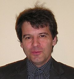 Markus Bachschmid合作论文数Department of Biology, University of Konstanz5Juliane Kilo合作论文数5
 Jeremy Neil Skepper合作论文数4
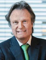 Thomas F. Lüscher合作论文数Center for Molecular Cardiology, University of Zurich;Heart Division, Royal Brompton and Harefield NHS Foundation Trust;Imperial College London4
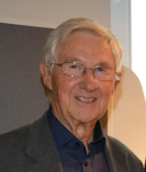 Volker Ullrich合作论文数Department of Biology, Universitat Konstanz4
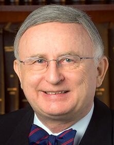 Tadeusz Malinski合作论文数Ohio University4
 Miriam Palacios-Callender合作论文数Wolfson Institute for Biomedical Research, University College London4Thomas F. Luescher合作论文数2
 Thomas Lüscher合作论文数Center for Molecular Cardiology, University of Zurich;Heart Division, Royal Brompton Hospital;Heart Division, Harefield Hospital2Stefan Schildknecht合作论文数1Raphael Hahn合作论文数1Lukas Altwegg合作论文数1
 Marten K J Schneider合作论文数1
 Georg Noll合作论文数HeartClinic Hirslanden1
 Martin Altwegg合作论文数Medisupport D-CH1
 Frank Ruschitzka合作论文数Klinik für Kardiologie, Universitätsspital Zürich;Medizinische Fakultät, Universität Zürich1
 Joachim E. Fischer合作论文数Institute for Behavioral Sciences, Federal Institute of Technology1
 Sidney G Shaw合作论文数1Thomas Quaschning合作论文数1P. Zumstein合作论文数1Athos Bonanomi合作论文数1
 Urs Ziegler合作论文数Center for Microscopy and Image Analysis, University of Zurich1Zaira Rickenbach合作论文数1Ralf Labugger合作论文数1Elisabeth Müssig合作论文数1
 Matthias Barton合作论文数Department of Cardiology,University of Zurich1
 Heinz Gehring合作论文数1Jorge D. Erusalimsky合作论文数1Claudia Dumrese合作论文数Institute of Anatomy, University of Zurich1Bernd Van Der Loo合作论文数1Claudia Dohm合作论文数1
 David Nadal合作论文数1
 Peter Groscurth合作论文数Institute of Anatomy, University of Zurich-Irchel;Fax: +41-1-2575702 CH;Institute of Anatomy, University of Zurich-Irchel1
 Christine Maurus合作论文数1Michael Walch合作论文数Institute of Anatomy, University of Zurich1Doris Grossenbacher合作论文数1




