Study Design Narrative review. Objectives Artificial intelligence (AI) is being increasingly applied to the domain of spine surgery. We present a review of AI in spine surgery, including its use across all stages of the perioperative process and applications for research. We also provide commentary regarding future ethical considerations of AI use and how it may affect surgeon-industry relations. Methods We conducted a comprehensive literature review of peer-reviewed articles that examined applications of AI during the pre-, intra-, or postoperative spine surgery process. We also discussed the relationship among AI, spine industry partners, and surgeons. Results Preoperatively, AI has been mainly applied to image analysis, patient diagnosis and stratification, decision-making. Intraoperatively, AI has been used to aid image guidance and navigation. Postoperatively, AI has been used for outcomes prediction and analysis. AI can enable curation and analysis of huge datasets that can enhance research efforts. Large amounts of data are being accrued by industry sources for use by their AI platforms, though the inner workings of these datasets or algorithms are not well known. Conclusions AI has found numerous uses in the pre-, intra-, or postoperative spine surgery process, and the applications of AI continue to grow. The clinical applications and benefits of AI will continue to be more fully realized, but so will certain ethical considerations. Making industry-sponsored databases open source, or at least somehow available to the public, will help alleviate potential biases and obscurities between surgeons and industry and will benefit patient care.
BACKGROUND AND OBJECTIVES:Neurosurgeons and hospitals devote tremendous resources to improving recovery from lumbar spine surgery. Current efforts to predict surgical recovery rely on one-time patient report and health record information. However, longitudinal mobile health (mHealth) assessments integrating symptom dynamics from ecological momentary assessment (EMA) and wearable biometric data may capture important influences on recovery. Our objective was to evaluate whether a preoperative mHealth assessment integrating EMA with Fitbit monitoring improved predictions of spine surgery recovery. METHODS:Patients age 21-85 years undergoing lumbar surgery for degenerative disease between 2021 and 2023 were recruited. For up to 3 weeks preoperatively, participants completed EMAs up to 5 times daily asking about momentary pain, disability, depression, and catastrophizing. At the same time, they were passively monitored using Fitbit trackers. Study outcomes were good/excellent recovery on the Quality of Recovery-15 (QOR-15) and a clinically important change in Patient-Reported Outcomes Measurement Information System Pain Interference 1 month postoperatively. After feature engineering, several machine learning prediction models were tested. Prediction performance was measured using the c-statistic. RESULTS:A total of 133 participants were included, with a median (IQR) age of 62 (53, 68) years, and 56% were female. The median (IQR) number of preoperative EMAs completed was 78 (61, 95), and the median (IQR) number of days with usable Fitbit data was 17 (12, 21). 63 patients (48%) achieved a clinically meaningful improvement in Patient-Reported Outcomes Measurement Information System pain interference. Compared with traditional evaluations alone, mHealth evaluations led to a 34% improvement in predictions for pain interference (c = 0.82 vs c = 0.61). 49 patients (40%) had a good or excellent recovery based on the QOR-15. Including preoperative mHealth data led to a 30% improvement in predictions of QOR-15 (c = 0.70 vs c = 0.54). CONCLUSION:Multimodal mHealth evaluations improve predictions of lumbar surgery outcomes. These methods may be useful for informing patient selection and perioperative recovery strategies.
INTRODUCTION: Facial nerve injury is disabling for patients with skull base brain tumors or head trauma. NAD+ is a key cofactor in metabolism and is an important component in axonal survival. Exogenous NAD+ prolongs axonal survival after axotomy in both in vitro and in vivo studies. Additionally, axonal survival is improved by blocking enzymes that decrease NAD+ levels (ex SARM1). Nicotinamide precursors like nicotinamide mononucleotide (NMN) have demonstrated neuroprotective benefits in aging and cellular death. METHODS: We developed a model of facial nerve injury by subjecting mice to 200 Hz of vibratory injury to the facial nerve distal to the stylomastoid foramen using SARM1KO and littermate controls, as well as mice implanted with intraventricular pumps containing 400 mM NMN or saline one week before facial nerve injury. EMG was performed using the NIM Nerve Monitoring System. Whisker movements were recorded and scored daily using Vibrissae observation scale. NAD+ metabolite levels were measured using liquid-chromatography/mass-spectrometry. NF200 immunofluorescence of facial nerves was performed. RESULTS: SARM1KO mice demonstrated robust axoprotection to vibratory injury for both 15 and 30 second durations and regained normal whisker movement in significantly less time compared to controls. Post-injury, SARM1 KO facial nerves required a lower stimulus current to elicit electrical responses in vibrissae muscles. We then used intracranial NMN delivery to test if raising NAD+ levels is axoprotective. NMN delivery increased brain NAD+ 1.5-fold. NMN significantly shortened the time to normal whisker movement, required a lower stimulus current for muscle response, and showed increased axonal integrity by NF200 staining. CONCLUSION: Continuous delivery of the NAD+ metabolite, NMN, allows for neuroprotective benefits in the vibratory neuropraxia nerve injury model, likely through sustained elevated NAD+ levels. These benefits are magnified through inhibition of the NADase enzyme, SARM1. This research demonstrates NMN as a neuroprotective pretreatment adjuvant in patients undergoing planned surgical invention where facial nerve injury is a possible outcome.
The pluripotency transcription factor SOX2 is essential for the maintenance of glioblastoma stem cells (GSC), which are thought to underlie tumor growth, treatment resistance, and recurrence. To understand how SOX2 is regulated in GSCs, we utilized a proteomic approach and identified the E3 ubiquitin ligase TRIM26 as a direct SOX2-interacting protein. Unexpectedly, we found TRIM26 depletion decreased SOX2 protein levels and increased SOX2 polyubiquitination in patient-derived GSCs, suggesting TRIM26 promotes SOX2 protein stability. Accordingly, TRIM26 knockdown disrupted the SOX2 gene network and inhibited both self-renewal capacity as well as in vivo tumorigenicity in multiple GSC lines. Mechanistically, we found TRIM26, via its C-terminal PRYSPRY domain, but independent of its RING domain, stabilizes SOX2 protein by directly inhibiting the interaction of SOX2 with WWP2, which we identify as a bona fide SOX2 E3 ligase in GSCs. Our work identifies E3 ligase competition as a critical mechanism of SOX2 regulation, with functional consequences for GSC identity and maintenance.
INTRODUCTION: Glioblastomas harbor inter and intratumoral genetic diversity, posing a challenge for targeted therapies. A major question is whether shared mechanisms might control the malignant phenotypes of genetically diverse glioblastoma cells. We reasoned that ubiquitin-dependent regulation of pluripotency-related transcription factor SOX2, which is indispensable for the maintenance of tumorigenic glioblastoma stem-like cells (GSC), may represent one such mechanism. TRIM26, an E3-ubiquitin ligase with immune-related functions, is highly expressed in glioblastoma tumors compared to normal brain. Immunoprecipitation followed by mass spectrometry suggested TRIM26 interacts with SOX2. We hypothesized that TRIM26 plays an essential role in GSCs by regulating SOX2 function. METHODS: Direct protein-protein interactions were assessed by in vitro binding assays. In Vitro ubiquitination assays were performed. Lentiviral TRIM26 overexpression and knockdown were used to test the role of TRIM26 in regulating SOX2 stability, activity, and ubiquitination. The functional relevance of TRIM26 in GSCs was assessed by in Vitro self-renewal and in Vivo tumorigenicity assays. RESULTS: We found that TRIM26 directly interacts with SOX2 via the C-terminal PRY-SPRY domain. Unexpectedly, TRIM26 overexpression resulted in decreased SOX2 polyubiquitination in cells. In line with this observation, TRIM26 knockdown in GSCs decreased SOX2 protein stability without changing SOX2 mRNA levels. Functionally, TRIM26 knockdown reduced SOX2 transcriptional activity, self-renewal, and in Vivo tumorigenicity in multiple genetically divergent GSC lines. Mechanistically, we discovered TRIM26 stabilizes SOX2 protein by competitively reducing the interaction of SOX2 with WWP2, a bonafide SOX2 E3 ligase in GSCs. Accordingly; WWP2 depletion in the setting of TRIM26 knockdown in GSCs rescued SOX2 protein levels, self-renewal, and tumorigenicity. CONCLUSION: Together, these results suggest that TRIM26 maintains GSCs by protecting SOX2 from WWP2-mediated ubiquitination and subsequent proteasomal degradation. These findings raise the intriguing possibility that modulating ubiquitin-dependent regulation of SOX2 in genetically heterogeneous GSCs may represent a unifying therapeutic strategy.
In spinal cord injury, nerve transfers represent a potential adjunct in the comprehensive clinical management of patients. Unlike tendon transfers, nerve transfers preserve the native muscle biomechanics and provide greater than a 1:1 functional exchange. Nerve transfers can provide improved upper extremity function by capitalizing on the preserved upper motor neurons below the zone of spinal cord injury. One goal in reconstruction is to restore movement. Major movements that have been targeted for restoration include elbow extension (to allow the patient to assist in transfers) and pinch, grasp, and release, which can aid in controlling a motorized wheelchair as well as in feeding oneself. Other major goals are restoration of hand sensation and diaphragm reinnervation to allow ventilator weaning and spontaneous respiration.
 Albert H. Kim合作论文数Department of Neurology and Neurological Surgery, Washington University School of Medicine3
 Wilson Z. Ray合作论文数Department of Neurosurgery, Washington University School of Medicine3
 John Ogunlade合作论文数2Tatenda Mahlokozera合作论文数Department of Neurological Surgery, Washington University School of Medicine, St. Louis, MO, USA.2
 Diane D Mao合作论文数Washington Univ, Sch Med, Dept Neurol Surg, St Louis, MO 63130 USA2Mounica Paturu合作论文数2
 Brian J Neuman合作论文数Dept Orthopaed Surg, Johns Hopkins Univ2
 Camilo A. Molina合作论文数Johns Hopkins Hospital2
 Jacob A. Greenberg合作论文数2Karan Joseph合作论文数1Nicholas Pallotta合作论文数1Salim Yakdan合作论文数1Tim Bui合作论文数1
 Kathleen Botterbush合作论文数1patrick a desouza合作论文数1Jingwen Zhang合作论文数1Nicholas A. Pallotta合作论文数1Ziqi Xu合作论文数1
 Madelyn R Frumkin合作论文数2Psychology and Brain Sciences.1
 Braeden C. Benedict合作论文数Joint Graduate Program in Bioengineering, UC Berkeley—UC San Francisco1
 Yo Sasaki合作论文数1Saad Javeed合作论文数Washington University in St. Louis School of Medicine1Xuan Qu合作论文数1
 Justin K Zhang合作论文数Dept Neurol Surg, Washington Univ1
 Alexander Yahanda合作论文数Department of Neurological Surgery, Washington University School of Medicine1Hao Chen合作论文数1Yang Wei合作论文数Division of Biostatistics, Washington University School of Medicine1Petti Allegra A合作论文数Genome Inst, Washington Univ1
 Adam Kimball Bevan合作论文数1
 Ghogawala Zoher合作论文数Dept Neurosurg, Alan & Jacqueline Stuart Spine Res Ctr, Lahey Clin Fdn1
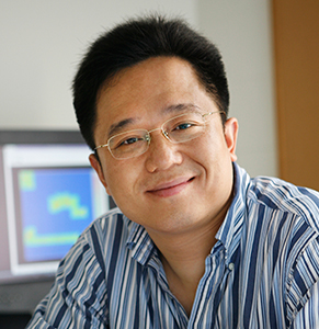 Chenyang Lu合作论文数Department of Computer Science & Engineering, James McKelvey School of Engineering, Washington University in St. Louis1
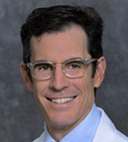 Michael Kelly合作论文数Division of Orthopedics & Scoliosis, Rady Children's Hospital-San Diego1
 Gujar Amit D合作论文数College of Pharmacy, The University of Louisiana at Monroe1Taiwo Rukayat合作论文数1
 Salehi Afshin合作论文数1Bhuvic Patel合作论文数1
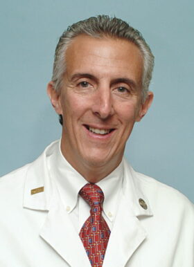 Jay F. Piccirillo合作论文数Department of Otolaryngology-Head and Neck Surgery, School of Medicine, Washington University in St Louis1
 Thomas L. Rodebaugh合作论文数Department of Psychological and Brain Sciences Washington University in St. Louis1Gavin P. Dunn合作论文数1Hiroko Yano合作论文数1Jacob M. Buchowski合作论文数1
 Nima Mosammaparast合作论文数Brigham and Women's Hospital, Harvard Medical School1
 Michael Steinmetz合作论文数Clarus Ventures1
 Munish C. Gupta合作论文数Department of Orthopedics, Washington University in St. Louis, School of Medicine1
 B. Pennicooke合作论文数1
 Burel R. Goodin合作论文数West Haven Veteran’s Affairs Medical Center1




