Objective Gene expression (transcriptomics) studies have revealed potential mechanisms of interstitial lung disease, yet sample sizes of studies are often limited and between-subtype comparisons are scarce. The aim this study was to identify and validate consensus transcriptomic signatures of interstitial lung disease subtypes. Methods We performed a systematic review and meta-analysis of fibrotic interstitial lung disease transcriptomics studies using an individual participant data approach. We included studies examining bulk transcriptomics of human adult interstitial lung disease samples and excluded those focusing on individual cell populations. Patient-level data and expression matrices were extracted from 43 studies and integrated using a multivariable integrative algorithm to develop interstitial lung disease classification models. Results Using 1459 samples from 24 studies, we identified transcriptomic signatures for idiopathic pulmonary fibrosis, hypersensitivity pneumonitis, idiopathic nonspecific interstitial pneumonia and systemic sclerosis-associated interstitial lung disease against control samples, which were validated on 308 samples from eight studies (idiopathic pulmonary fibrosis area under receiver operating curve (AUC) 0.99, 95% CI 0.99-1.00; hypersensitivity pneumonitis AUC 0.91, 95% CI 0.84-0.99; nonspecific interstitial pneumonia AUC 0.94, 95% CI 0.88-0.99; systemic sclerosis-associated interstitial lung disease AUC 0.98, 95% CI 0.93-1.00). Significantly, meta-analysis allowed us to identify, for the first time, robust lung transcriptomics signatures to discriminate idiopathic pulmonary fibrosis (AUC 0.71, 95% CI 0.63-0.79) and hypersensitivity pneumonitis (AUC 0.76, 95% CI 0.63-0.89) from other fibrotic interstitial lung disease, and unsupervised learning algorithms identified putative molecular endotypes of interstitial lung disease associated with decreased forced vital capacity and diffusing capacity of the lungs for carbon monoxide % predicted. Transcriptomics signatures were reflective of both cell-specific and disease-specific changes in gene expression. Conclusion We present the first systematic review and largest meta-analysis of fibrotic interstitial lung disease transcriptomics to date, identifying reproducible transcriptomic signatures with clinical relevance.
Fibrotic interstitial lung diseases (ILDs) result from excessive deposition of extracellular matrix (ECM) proteins in the lung, causing irreversible damage to the lung architecture. Clinical management of ILDs differs depending on the diagnosis, but differentiation between subtypes can be difficult and better clinical biomarkers are needed. In this study, we use a 166-gene NanoString assay to investigate whether there are ILD subtype-specific transcripts in whole blood. We identified one transcript, killer cell lectin like receptor 1 (KLRF1), as differentially expressed between idiopathic pulmonary fibrosis (IPF) and systemic sclerosis-associated ILD (SSc-ILD), and identified two transcripts (VCAN, LTK) associated with IPF expression against other ILD subtypes. These findings were validated by examining their expression in ILD lung, with KLRF1 expression significantly higher in SSc-ILD compared to IPF and hypersensitivity pneumonitis (HP) samples. Taken together, this pilot study provides support for the use of the peripheral transcriptome in identifying diagnostic biomarkers of ILD with biological relevance.
Abstract The therapeutic effects for cancer immunotherapy are largely hampered by intratumoral CD4+CD25+ regulatory T cells (Tregs) which leads to poor outcome in many cancer patients upon treatment of immune checkpoint inhibitors. Moreover, the balance between effector T (Teff) cells and Treg cells in the tumor microenvironment (TME) impacts on tumor progression and anti-tumor immunity. Depleting tumor-infiltrating Tregs by selectively targeting CD25 to reduce the ratios of regulatory to effector T cells (Treg/Teff) without disturbing IL-2 signalling is a promising strategy to advance anti-tumor immunity. Hence, we evaluated the in vivo anti-tumor effects of Non-IL-2 blocking Treg-depleting anti-hCD25 mAb versus IL-2-blocking anti-hCD25 mAb (Basiliximab) in an engineered hCD25 (IL-2RA) knockin mouse model.Firstly, we developed the genetically engineered CD25 (IL-2RA) humanized mouse model by ES cell targeting, in which mouse Cd25 exons 2-6 are replaced by human counterparts corresponding to the extracellular domain leaving the intracellular regions intact. Then, we performed flow cytometry analysis to confirm the surface expression of hCD25 on naive Treg cell from peripheral blood and spleen in homozygous hCD25 knockin mice. In addition, we found the hCD25 expression is upregulated on activated Treg cell derived from splenocytes for both heterozygous and homozygous hCD25 knockin mice. Furthermore, the female homozygous hCD25 knockin mice subcutaneously engrafted with MC38 syngeneic tumors received two different therapeutic anti-CD25 mAb treatment in parallel and we found the Non-IL-2 blocking Treg-depleting anti-human CD25 mAb (anti-hCD25NB) displayed more potent anti-tumor response compared to IL-2-blocking Treg-depleting anti-human CD25 mAb (anti-hCD25BL), supporting that keeping IL-2 signaling on effector T cells are required to enhance effector activation and anti-tumor immunity. Notably, the study groups treated with anti-hCD25NB plus anti-mPD-1 showed synergistic therapeutic effects as expected. As a matter of fact, both CD4+CD25+ T cells and CD4+CD25+FoxP3+ Treg cells reduced significantly upon treatment of anti-hCD25NB and anti-hCD25BL in spleen and tumors, but no big change in the number of CD4+FoxP3+ Treg cells.Likewise, anti-hCD25NB in combination with anti-mPD-1 leads to more significant CD4+CD25+ Treg depletion in spleen and tumors along with increased CD8+Granzyme B+ cytotoxic T cells.Collectively, our CD25 (IL-2RA) humanized mouse provides a powerful model to assess the in vivo preclinical therapeutic effects of CD25+ Treg-depleting antibodies for cancer immunotherapy. Citation Format: Daniel He. Non-IL-2 blocking Treg-depleting anti-human CD25 mAb primes potent anti-tumor immunity and synergizes anti-tumor effects of anti-PD-1 in a novel hIL-2RA knockin model. [abstract]. In: Proceedings of the American Association for Cancer Research Annual Meeting 2023; Part 1 (Regular and Invited Abstracts); 2023 Apr 14-19; Orlando, FL. Philadelphia (PA): AACR; Cancer Res 2023;83(7_Suppl):Abstract nr 5092.
Blood vessels is required for the growth of solid tumors and cutting off the blood supply to tumor is a potential novel strategy to retard tumor growth, although which may consequently result in pan-hypoxia and metastasis paradoxically. VEGFA is identified as the major angiogenic factor and VEGFR2 binds to all VEGFA isoforms and VEGFR2 (KDR) is a dominant mediator in regulating the angiogenic and permeability-enhancing effects of VEGFA. Importantly, in numerous solid tumors, VEGFA and VEGFR2 are overexpressed to promote intratumoral angiogenesis to support local tumor survival, tumor growth and distant metastasis. Given that VEGFA/VEGFR2 interaction plays a crucial role in promoting intratumoral angiogenesis and tumor growth, it is appealing to develop novel potent anti-angiogenic agents for cancer treatment by direct against VEGFA-VEGFR2 interaction. However, the preclinical assessment of anti-angiogenesis agents are limited due to lack of suitable preclinical models for human-specific therapeutic antibodies which usually don’t cross-bind to mouse targets. To this end, we developed the KDR (VEGFR2) humanized mouse model (hKDR model), in which the extracellular regions of mouse VEGFR2 corresponding to exons 4-15 are replaced by human counterparts via ES cell-based homologous recombination. As a result, hKDR humanized mouse expresses a chimeric VEGFR2 receptor containing the human extracellular domain plus mouse trans-membrane and intracellular domains. Firstly, we characterized the chimeric h/m KDR expression on the endothelial cells as well as in intratumoral blood vessels derived from hKDR knockin mice. Subsequently, we investigated the anti-tumor responses of anti-human VEGFR2 antibody (Cyramza, Ramucirumab) in hKDR mice bearing B16-F1 and MC38 syngenic solid tumors and we observed that Cyramza inhibited the tumor growth in vivo remarkably in a dose-dependent way, indicating the binding of anti-human VEGFR2 antibody to the chimeric h/m KDR receptor. Notably, we further generated hKDR knockin model in Rag2 knockout background (hKDR/Rag2KO) by crossbreeding which lack mature T cells and B cells. The female hKDR/Rag2KO mice were subcutaneously inoculated with human hepatocellular carcinoma LI0612 PDX model and human gastric cancer GA2419 PDX model to assess the anti-tumor effects of anti-angiogenic agents in xenograft solid tumors. The data suggested that Cyramza displayed moderate tumor growth inhibition (TGI) in LI0612 model at 10 mg/kg and weak TGI in GA2419 model at 10 mg/kg, respectively. Our findings support the notion that normalization of tumor vasculature is critical for tumor inhibition based on anti-angiogenic therapy and may synergize the anti-tumor effects with the immune checkpoint blockade or promote the cancer immunotherapy of immunomodulators in the future trials. Citation Format: Daniel He. Anti-angiogenic mAb, anti-human VEGFR2(Cyramza) retards tumor growth in KDR (VEGFR2) humanized mice engrafted with syngeneic vs. xenograft solid tumors. [abstract]. In: Proceedings of the American Association for Cancer Research Annual Meeting 2023; Part 1 (Regular and Invited Abstracts); 2023 Apr 14-19; Orlando, FL. Philadelphia (PA): AACR; Cancer Res 2023;83(7_Suppl):Abstract nr 4590.
A subgroup of patients with interstitial lung disease (ILD) have a degenerative phenotype known as progressive pulmonary fibrosis (PPF) irrespective of etiology, which is seen in roughly 30% of ILD cases. Anti-fibrotic medications slow progression of PPF, thus highlighting the need to reliably predict future progression when it is still preventable. The objective of this study was to examine whether whole blood gene expression profiles could predict a progressive phenotype in ILD patients. Whole blood RNA-seq samples of a retrospective cohort of treatment-naïve ILD patients were analyzed for differentially expressed genes (DEGs). After adjusting for sex and baseline predicted forced vital capacity (FVC%), we identified 17 DEGs (FDR<0.25) in association with FVC% slope over 6 months (n=42), including PGAM1P8, TSPAN33, and LRFN4 (FDRs = 0.01, 0.19, 0.20), which were also significantly associated with FVC% slope over 12 months (n=63). When examining FVC% slope over 24 months (n=68), we identified 70 DEGs including FMOD, PCSK6, TGFB1I1, and VEGFC (FDRs = 0.18, 0.18, 0.25, 0.25), all of which have previously been associated with fibrotic processes in ILDs. Using a linear mixed effects model to examine the effect of baseline gene expression on FVC% trajectory, we identified 1 gene (lnc-COL4A1-1) that was significantly associated with FVC% decline at 6, 12, and 24 months (FDRs = 0.02, 0.06, 0.14). Taken together, these data suggest that blood expression of genes associated with fibrosis are associated with long-term decline in FVC%, while genes associated with immune activation (TSPAN33 for B-cells and LRFN4 for monocytes) may precede short-term declines in FVC%.
BACKGROUND:Long-COVID (LC) encompasses diverse symptoms lasting months after the initial SARS-CoV-2 infection. Symptoms can be debilitating and affect the quality of life of individuals with LC and their families. Although the symptoms of LC are well described, the aetiology of LC remains unclear, and consequently, patients may be underdiagnosed. Identification of LC specific biomarkers is therefore paramount for the diagnosis and clinical management of the syndrome. This scoping review describes the molecular and cellular biomarkers that have been identified to date with potential use for diagnosis or prediction of LC. METHODS:This review was conducted using the Joanna Briggs Institute (JBI) Methodology for Scoping Reviews. A search was executed in the MEDLINE and EMBASE databases, as well as in the grey literature for original studies, published until October 5th, 2022, reporting biomarkers identified in participants with LC symptoms (from all ages, ethnicities, and sex), with a previous infection of SARS-CoV-2. Non-English studies, cross-sectional studies, studies without a control group, and pre-prints were excluded. Two reviewers independently evaluated the studies, extracted population data and associated biomarkers. FINDINGS:23 cohort studies were identified, involving 2163 LC patients [median age 51.8 years, predominantly female sex (61.10%), white (75%), and non-vaccinated (99%)]. A total of 239 candidate biomarkers were identified, consisting mainly of immune cells, immunoglobulins, cytokines, and other plasma proteins. 19 of the 239 candidate biomarkers identified were evaluated by the authors, by means of receiver operating characteristic (ROC) curves. INTERPRETATION:Diverse cellular and molecular biomarkers for LC have been proposed. Validation of candidate biomarkers in independent samples should be prioritized. Modest reported performance (particularly in larger studies) suggests LC may encompass many distinct aetiologies, which should be explored e.g., by stratifying by symptom clusters and/or sex. FUNDING:Dr. Tebbutt has received funding from the Canadian Institutes of Health Research (177747) to conduct this work. The funding source was not involved in this scoping review, or in the decision to submit this manuscript for publication.
Recurrent primary biliary cholangitis (rPBC) is frequent following liver transplantation and associated with increased morbidity and mortality. It has been argued that rPBC behaves like an infectious disease because more potent immunosuppression with tacrolimus is associated with earlier and more severe recurrence. Prophylactic ursodeoxycholic acid is an established therapeutic option to prevent rPBC, whereas the role of second line therapies, such as obeticholic acid and bezafibrate in rPBC, remains largely unexplored. To address the hypothesis that a human betaretrovirus plays a role in the development of PBC, we have tested antiretroviral therapy in vitro and conducted randomised controlled trials showing improvements in hepatic biochemistry. Herein, we describe the utility of combination antiretroviral therapy to manage rPBC in two patients treated with open label tenofovir/emtricitabine-based regimens in combination with either lopinavir or raltegravir. Both patients experienced sustained biochemical and histological improvement with treatment, but the antiretroviral therapy was associated with side effects.
Significance The human genome contains many thousands of genes that produce a large diversity of long noncoding RNAs (lncRNAs). Some lncRNA genes produce microRNAs (miRNAs), which are regulators of protein expression. It has been unclear whether lncRNA genes that produce miRNAs, termed lnc-pri-miRNAs, have important function independent of their miRNAs. We find multiple lnc-pri-miRNA genes that regulate cell proliferation even when miRNA production machinery is knocked down. lnc-pri-miRNA LOC646329 produces miR-29a/b1, and the function of LOC646329 in cell proliferation can be genetically and phenotypically separated from its cognate miRNAs. The miRNA-independent function of LOC646329 and other lnc-pri-miRNAs relates to their activation of genes in physical proximity. These results shed light on the function and intermingled complexity of the noncoding genome.
Abstract Background: CCR8 is a C-C chemokine receptor with 7-transmembrane regions which belongs to the G-protein coupled receptor family. Recently, tumor-infiltrating CCR8+ CD4+ Foxp3+ regulatory T (Treg) cells have been identified as a major immune-suppressive subset in the tumor microenvironment (TME), with CCR8 upregulated in tumor-infiltrating Tregs in patients with breast, lung and colorectal cancer. Among the tumor-infiltrating Treg cells, only 8%-10% of Tregs are CCR8 positive, which are regarded as the master driver populations for Treg-mediated immunosuppression. Thus, CCR8 is increasingly believed to be a potential therapeutic target with monoclonal antibodies specific to human CCR8 (hCCR8) inducing depletion of tumor-resident CCR8+ Treg cells, reverse immunosuppressive signals to enhance the anti-tumor role of effector T cells, particularly in combination with other immune checkpoint inhibitors. Given the lack of cross-binding to mouse targets of human-specific antibodies, we developed a hCCR8 knock-in mouse model (CCR8 HuGEMM) to evaluate the in vivo therapeutic efficacy of hCCR8 antibodies. Methods: Since there is only one coding exon (exon 2) in both hCCR8 and mouse CCR8 genes, CCR8 HuGEMM was developed by CRISPR/Cas9 engineering to substitute the mouse CCR8 exon 2 for hCCR8 exon 2 in C57BL/6 mice. The hCCR8 expression in CCR8 HuGEMM was confirmed by flow cytometry analysis. The non-tumor-bearing CCR8 HuGEMM mice were then treated with anti-hCCR8 antibodies to investigate the immune cell phenotyping in blood and spleen. Finally, the CCR8 HuGEMM mice were inoculated with either MC38-OVA or Hepa1-6 syngeneic tumors in parallel to evaluate the anti-tumor responses of anti-hCCR8 antibodies in vivo. Results: The surface expression of hCCR8 on CD4+ and CD8+ T cells, derived from thymus as well as on FoxP3+ Treg cells from CCR8 HuGEMM mice, was characterized by flow cytometry analysis. Monocytic myeloid-derived suppressor cells (MDSCs) (CD3-CD11b+Ly6Chi) populations decreased significantly in non-tumor-bearing CCR8 HuGEMM mice upon treatment of anti-hCCR8 antibody compared to the control mice, which indicates that anti-hCCR8 may play a critical role in abrogating the immunosuppressive role of MDSC and further retard the MDSC-mediated Treg induction. Furthermore, MC38-OVA and Hepa1-6 syngeneic tumors were inoculated into the homozygous CCR8 HuGEMM mice and the mice were treated with 10mg/kg anti-hCCR8,4 days post tumor engraftment. The anti-hCCR8 alone led to ~30% tumor growth inhibition (TGI) in MC38-OVA tumor model, and ~50% tumor growth inhibition (TGI) in Hepa1-6 tumor model. Similarly, we found the tumor-resident CCR8+ CD4+ FoxP3+ populations decreased significantly post-treatment which suggests that CCR8+ Tregs potentially play an important immunosuppressive role in the TME. Conclusions: Our CCR8 HuGEMM model provides an important predictive preclinical model to evaluate the efficacy of hCCR8 antibodies alone or in combination regimens with other immune modulators. Citation Format: Daniel He, Henry Li. In vitro and in vivo characterization of CCR8 humanized mouse model (HuGEMM™) [abstract]. In: Proceedings of the AACR-NCI-EORTC Virtual International Conference on Molecular Targets and Cancer Therapeutics; 2021 Oct 7-10. Philadelphia (PA): AACR; Mol Cancer Ther 2021;20(12 Suppl):Abstract nr P003.
Conventional vaccine design has been based on trial-and-error approaches, which have been generally successful. However, there have been some major failures in vaccine development and we still do not have highly effective licensed vaccines for tuberculosis, HIV, respiratory syncytial virus, and other major infections of global significance. Approaches at rational vaccine design have been limited by our understanding of the immune response to vaccination at the molecular level. Tools now exist to undertake in-depth analysis using systems biology approaches, but to be fully realized, studies are required in humans with intensive blood and tissue sampling. Methods that support this intensive sampling need to be developed and validated as feasible. To this end, we describe here a detailed approach that was applied in a study of 15 healthy adults, who were immunized with hepatitis B vaccine. Sampling included ~350 mL of blood, 12 microbiome samples, and lymph node fine needle aspirates obtained over a ~7-month period, enabling comprehensive analysis of the immune response at the molecular level, including single cell and tissue sample analysis. Samples were collected for analysis of immune phenotyping, whole blood and single cell gene expression, proteomics, lipidomics, epigenetics, whole blood response to key immune stimuli, cytokine responses, in vitro T cell responses, antibody repertoire analysis and the microbiome. Data integration was undertaken using different approaches—NetworkAnalyst and DIABLO. Our results demonstrate that such intensive sampling studies are feasible in healthy adults, and data integration tools exist to analyze the vast amount of data generated from a multi-omics systems biology approach. This will provide the basis for a better understanding of vaccine-induced immunity and accelerate future rational vaccine design.
Additional file 4: Table S3. DESeq2 output of differentially expressed genes following ASO knockdown of lncGRS-1 compared to negative control ASO in GBM U87, DIPG SF8628, and NHA cells.
Background CD137 (4-1BB) is a powerful T cell co-stimulatory molecule belonging to the TNF receptor superfamily, which promotes cytotoxic T cell survival and memory formation upon CD137L ligation. CD137 has become an attractive immuno-oncology therapeutic target with multiple agonistic antibodies in clinical trials, including urelumab and utomilumab, with promising response in combination with anti-PD1 immunotherapies such as nivolumab. Clinical applications of CD137 agonistic antibodies are hampered, however, by dose-limiting off-tumor liver toxicity (urelumab) or lower efficacy (utomilumab). The cause of liver toxicity is reported primarily to be due to Fcγ receptor mediated cross linking;1–3 CD137 agonistic antibodies may also trigger hepatotoxicity through activation of IL-27 secreting liver Kupffer cells and monocytes.4 The remaining challenge in decoupling efficacy from liver toxicity is the lack of preclinical mouse models which can be used to assess both efficacy and the immune-related adverse events (irAE) of human CD137 agonistic antibodies. Methods To mimic the clinical outcomes of urelumab, we utilized humanized CD137 knock-in mice in Balb/c background (Balb/c CD137 HuGEMM) to evaluate its efficacy with CT26.WT syngeneic tumors. Liver toxicity was analyzed by monitoring fasting serum ALT/AST levels at different time points. Results Urelumab showed moderate anti-tumor response at the dose level of 5 mg/kg, while serum ALT/AST levels showed no difference compared to isotype control suggesting that, due to the different binding capacity of the human IgG4 Fc domain to mouse FcγR, the human version of the agonistic antibody cannot fully recapitulate its effect on HuGEMM mice. Therefore, a chimeric antibody with mouse IgG1 Fc domain (urelumab-mIgG1) was created to dissect the potential role of FcγR mediated cross linking on both efficacy and liver toxicity; an urelumab-mIgG1-DANA variant with D265A/N297A mutation to abolish Fc effector function was also included as a dominant negative control. We found that urelumab-mIgG1 showed further enhanced efficacy compared to urelumab alone through FcγR mediated cross linking, while urelumab-mIgG-DANA showed compromised anti-tumor response. With regards to liver toxicity, urelumab-mIgG1 caused chronic liver inflammation and hepatocyte damage indicated by immune cell infiltration in the liver and significantly elevated serum ALT levels, which was abolished by the urelumab-mIgG1-DANA variant. The study also compared urelumab treatment in CD137 HuGEMM head-to-head with the mouse surrogate agonistic antibody (3H3) in wild-type BALB/c mice. 3H3 showed robust tumor growth inhibition as well as dramatic ALT elevation. Conclusions We faithfully recapitulated the clinically observed tumor growth inhibition and liver toxicity of urelumab by using a chimeric version of urelumab in CD137 HuGEMM, indicating the importance of both the mouse model and antibody version in evaluation of efficacy and irAE. Ethics Approval Animal experiments were conducted in accordance with animal welfare law, approved by local authorities, and in accordance with the ethical guidelines of CrownBio (Taicang). References Claus C, Ferrara C1, Xu W, et al. Tumor-targeted 4-1BB agonists for combination with T cell bispecific antibodies as off-the-shelf therapy. Sci Transl Med 2019; 11:eaav5989. Qi X, Li F, Wu Y, et al. Optimization of 4-1BB antibody for cancer immunotherapy by balancing agonistic strength with FcγR affinity. Nat Commun 2019;10:2141–2151. Compte M, Harwood SL, Muñoz IG, et al. A tumor-targeted trimeric 4-1BB-agonistic antibody induces potent anti-tumor immunity without systemic toxicity. Nat Commun. 2018;9:4809–4821. Bartkowiak T, Jaiswal AR, Ager CR, et al. Activation of 4-1BB on liver myeloid cells triggers hepatitis via an interleukin-27-dependent pathway. Clin Cancer Res 2018;24:1138–1151.
Background Long non-coding RNAs (lncRNAs) exhibit highly cell type-specific expression and function, making this class of transcript attractive for targeted cancer therapy. However, the vast majority of lncRNAs have not been tested as potential therapeutic targets, particularly in the context of currently used cancer treatments. Malignant glioma is rapidly fatal, and ionizing radiation is part of the current standard-of-care used to slow tumor growth in both adult and pediatric patients. Results We use CRISPR interference (CRISPRi) to screen 5689 lncRNA loci in human glioblastoma (GBM) cells, identifying 467 hits that modify cell growth in the presence of clinically relevant doses of fractionated radiation. Thirty-three of these lncRNA hits sensitize cells to radiation, and based on their expression in adult and pediatric gliomas, nine of these hits are prioritized as lncRNA Glioma Radiation Sensitizers (lncGRS). Knockdown of lncGRS-1, a primate-conserved, nuclear-enriched lncRNA, inhibits the growth and proliferation of primary adult and pediatric glioma cells, but not the viability of normal brain cells. Using human brain organoids comprised of mature neural cell types as a three-dimensional tissue substrate to model the invasive growth of glioma, we find that antisense oligonucleotides targeting lncGRS-1 selectively decrease tumor growth and sensitize glioma cells to radiation therapy. Conclusions These studies identify lncGRS-1 as a glioma-specific therapeutic target and establish a generalizable approach to rapidly identify novel therapeutic targets in the vast non-coding genome to enhance radiation therapy.
Death from sepsis in the neonatal period remains a serious threat for millions. Within 3 days of administration, bacille Calmette-Guérin (BCG) vaccination can reduce mortality from neonatal sepsis in human newborns, but the underlying mechanism for this rapid protection is unknown. We found that BCG was also protective in a mouse model of neonatal polymicrobial sepsis, where it induced granulocyte colony-stimulating factor (G-CSF) within hours of administration. This was necessary and sufficient to drive emergency granulopoiesis (EG), resulting in a marked increase in neutrophils. This increase in neutrophils was directly and quantitatively responsible for protection from sepsis. Rapid induction of EG after BCG administration also occurred in three independent cohorts of human neonates.
Background Blood has proven to be a useful resource for molecular analysis in numerous biomedical studies, with peripheral blood mononuclear cells (PBMCs) and whole blood being the major specimen types. However, comparative analyses between these two major compartments (PBMCs and whole blood) are few and far between. In this study, we compared gene expression profiles of PBMCs and whole blood samples obtained from research subjects with or without mild allergic asthma. Methods Whole blood (PAXgene) and PBMC samples were obtained from 5 mild allergic asthmatics and 5 healthy controls. RNA from both sample types was measured for expression of 730 immune-related genes using the NanoString nCounter platform. Results We identified 64 uniquely expressed transcripts in whole blood that reflected a variety of innate, humoral, and adaptive immune processes, and 13 uniquely expressed transcripts in PBMCs which were representative of T-cell and monocyte-mediated processes. Furthermore, analysis of mild allergic asthmatics versus non-asthmatics revealed 47 differentially expressed transcripts in whole blood compared to 1 differentially expressed transcript in PBMCs (FDR < 0.25). Finally, through simultaneous measurement of PBMC proteins on the nCounter assay, we identified CD28 and OX40 ( TNFRSF4 ), both of which are critical co-stimulatory molecules during T-cell activation, as significantly upregulated in asthmatics. Conclusions Whole blood RNA preserved in PAXgene tubes is excellent for producing gene expression data with minimal variability and good sensitivity, suggesting its utility in multi-centre studies requiring measurement of blood gene expression.
Cholinergic synapse pathway gene polymorphisms may play a role in regulating a type of asthmatic airway response triggered upon allergen challenge http://bit.ly/2lJx1VG.
Systems biology can unravel complex biology but has not been extensively applied to human newborns, a group highly vulnerable to a wide range of diseases. We optimized methods to extract transcriptomic, proteomic, metabolomic, cytokine/chemokine, and single cell immune phenotyping data from <1 ml of blood, a volume readily obtained from newborns. Indexing to baseline and applying innovative integrative computational methods reveals dramatic changes along a remarkably stable developmental trajectory over the first week of life. This is most evident in changes of interferon and complement pathways, as well as neutrophil-associated signaling. Validated across two independent cohorts of newborns from West Africa and Australasia, a robust and common trajectory emerges, suggesting a purposeful rather than random developmental path. Systems biology and innovative data integration can provide fresh insights into the molecular ontogeny of the first week of life, a dynamic developmental phase that is key for health and disease.
Objective To determine whether the maximum hemodynamic response to scalp interictal epileptic discharges (IEDs) corresponds to the region where IEDs originate and from where they propagate. Methods We studied 19 patients who underwent first an EEG-fMRI showing responses in the gray matter, and then intracranial EEG (iEEG). We coregistered the hemodynamic responses to the iEEG electrode contacts and analyzed IEDs in the iEEG channel adjacent to a maximum response (labeled the main channel), in relation to IEDs in other channels during a widespread intracranial IED event. IEDs in the main channel were aligned at their peak, and IEDs in each channel were averaged time-locked to these instants. The beginning and peak of IEDs in the averaged trace were identified, blinded to the identity of the main channel. The latency of IEDs was computed between the earliest and all other channels. Results The median latency of IEDs in the main channel was significantly smaller than in other channels for either the peak (15.5 vs 67.5 milliseconds, p = 0.00037) or the beginning (46.5 vs 118.4 milliseconds, p = 0.000048). The latency of IED was significantly correlated to the distance from the maximum hemodynamic response (p < 0.0001 for either the peak or the beginning). Conclusion IED adjacent to a maximum hemodynamic response, which often corresponds to the seizure onset zone, is more likely to precede IEDs in remote locations during a widespread intracranial discharge. Thus, EEG-fMRI is a unique noninvasive method to reveal the origin of IEDs, which we propose to label the spike onset zone.
 Benoit G. Bruneau合作论文数School of Medicine, University of California, San Francisco13
 Tebbutt Scott J合作论文数9
 Max Horlbeck合作论文数Division of Genetics, Boston Children's Hospital;EpiChroma Clinic8
 Casey P. Shannon合作论文数PROOF Centre of Excellence, Vancouver, BC, Canada;UBC James Hogg Centre for Heart Lung Innovations;PROOF Centre of Excellence, UBC James Hogg Centre for Heart Lung Innovations8
 Jonathan Weissman合作论文数Department of Biology, Massachusetts Institute of Technology;Whitehead Institute;Howard Hughes Medical Institute8Katherine S. Pollard合作论文数7
 Sean Thomas合作论文数Department of Genome Sciences, University of Washington7
 Martina Malatesta合作论文数6
 S John Liu合作论文数 University of California San Francisco, San Francisco, CA, USA6
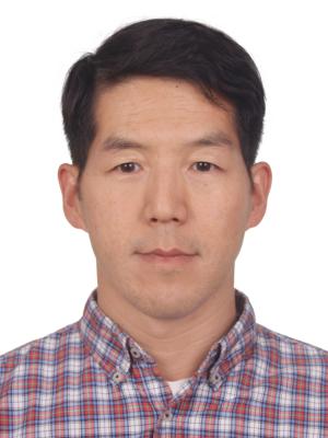 Daniel Lim合作论文数Duke Kunshan University;Center for Analytic Philosophy, Renmin University of China6
 Aaron Diaz合作论文数University of California, University of California5Birk Harjus合作论文数5Sung Hong合作论文数4
 Chinten James Lim合作论文数Faculty of Medicine, University of British Columbia4
 Parna Saha合作论文数Biomed Sci Grad Program, Univ Calif San Francisco4
 Jason Fish合作论文数Department of Laboratory Medicine and Pathobiology, Temerty Faculty of Medicine, University of Toronto4Dang Lan T H合作论文数Department of Cell and Systems Biology, University of Toronto4
 Joseph F. Costello合作论文数Helen Diller Family Comprehensive Cancer Center, University of California, San Francisco;Department of Neurological Surgery, University of California, San Francisco4
 Mark A Akeson合作论文数Baskin School of Engineering, University of California , Santa Cruz4
 Frank J. Attenello合作论文数Department of Neurosurgery, Johns Hopkins University School of Medicine4Mark P White合作论文数3Molong Li合作论文数3
 Tobias Kollmann合作论文数University of Duisburg-Essen;netSTART GmbH3
 Amrit Singh合作论文数3
 Mark Koontz合作论文数3Kyounghee Seo合作论文数Univ Calif San Francisco, San Francisco, CA 94143 USA3Rym Ben-Othman合作论文数Univ British Columbia, Dept Pediat, Vancouver, BC, Canada3
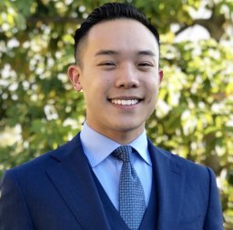 Brian V Lien合作论文数Department of Neurological Surgery, University of California3
 Kyle Walsh合作论文数Department of Population Health Sciences, Duke University School of Medicine3
 Sua Myong合作论文数Department of Biophysics, Krieger School of Arts & Sciences, Johns Hopkins University3
 Arnold R Kriegstein合作论文数Department of Neurology, School of Medicine, University of California, San Francisco3
 Tomasz Nowakowski合作论文数School of Medicine, University of California, San Francisco3
 Melike Pekmezci合作论文数3Wrensch Margaret合作论文数3
 Erik Ullian合作论文数School of Medicine, University of California, San Francisco3
 Miten Jain合作论文数Department of Bioengineering, College of Engineering, Northeastern University;Department of Physics, College of Engineering, Northeastern University3
 Alex Pollen合作论文数Weill Institute for Neurosciences, School of Medicine, University of California San Francisco3
 John K. Wiencke合作论文数Helen Diller Family Comprehensive Cancer Center, University of California, San Francisco;Department of Neurological Surgery, University of California, San Francisco;Division of Epidemiology, University of California, San Francisco3Cai Bing合作论文数3
 Andrew Mancini合作论文数3Alex Kreig合作论文数3
 H. Tomas Rube合作论文数Department of Physics, University of Illinois;Institute for Genomic Biology, University of Illinois3
 Nelly Amenyogbe合作论文数University of British Columbia, University of British Columbia3Liu Lei合作论文数3Theodoris Christina V合作论文数Gladstone Institute of Cardiovascular Disease, University of California3
 Hugh Olsen合作论文数UC Santa Cruz Genomics Institute, University of California3
 Jan H. Lui合作论文数University of California, University of California3
 Deepak Srivastava合作论文数Srivastava Lab, Gladstone Institute of Cardiovascular Disease, University of California San Francisco;Department of Pediatrics, University of California San Francisco;Department of Biochemistry and Biophysics, University of California San Francisco3




