Transcranial focused ultrasound (tFUS) has various applications in non-invasive treatments like neuromodulation, opening the blood-brain barrier, and tissue ablation. The objective of this work is to accurately model the propagation of waves in tFUS using the spectral-element method (SEM) and to better understand the types of physics that affect the wavefield within the laboratory measurements. Simulating transcranial ultrasound is non-trivial because of the skull's high acoustic impedance, the P-S conversions at the skull boundary, and significant attenuation in the diploe layer. Within this framework, we aim to qualitatively and quantitatively compare different skull model simulations. A coupled acoustic-viscoelastic wave equation is solved using the SEM. First, the reference simulation is carried out with a heterogeneous attenuating solid skull. Then, three simpler simulations are carried out by (1) eliminating heterogeneities in the skull, (2) neglecting the effects of shear waves, and (3) ignoring the effects of attenuation. We quantified the differences between each simulation and the reference model, with the acoustic skull simulation showing the highest percentage deviation of around 35% while the homogeneous and non-attenuating model shows deviation of around 20%. The higher deviation in the acoustic model is seen because of the prominent shear wave mode conversion at the elastic skull boundary. This comparison helps identify potential errors from omitting specific skull properties in simulations and guides researchers on which properties can be safely simplified based on their application.
Objective.To demonstrate the potential of full-waveform inversion (FWI) for high-resolution ultrasonic imaging usingin-vivodata.Approach.Acoustic FWI is applied toin-vivomeasurements, accounting for the nonlinear relationship between the ultrasonic wavefield and model parameters. Two key components are investigated: (1) estimation of an effective source wavelet by inverting the source-time function using calibration data in water, and (2) reduction of nonlinearity and sensitivity to the initial model using a graph-space optimal transport misfit functional. A perturbation-based resolution analysis is employed to quantify local spatial smearing.Main results.The proposed approach enables improved spatial resolution in reconstructed models and allows accurate identification of anatomical features inin-vivodata.Significance.These results provide further evidence for the applicability of FWI toin-vivoultrasonic imaging and highlight methodological components that influence reconstruction quality, offering insight into its potential for clinical applications.
High-frame-rate volumetric optoacoustic tomography with hexagonal illumination enabled noninvasive whole-heart murine imaging at 200 volumes/s. NIR-II CuS nanoagents improved full-heart coverage, penetration, quantification, and assessment of cardiac disorders and therapeutic effects.
To understand microvascular resilience, OMNIMap performs functional mapping of the microvascular network using extended-focus optical coherence microscopy. We reveal a conserved topological organization that mitigates individual capillary stalls, preserving network perfusion after ischemic stroke.
Optoacoustic (OA) imaging has emerged as a powerful investigation tool, with demonstrated applicability in oncology, neuroscience, and cardiovascular biology. However, its clinical translation is limited with the existing OA systems, which often rely on bulky and expensive acquisition hardware mainly optimized for pulse-echo ultrasound (US) imaging. Despite the fact that OA imaging has different requirements for receive bandwidths and timing synchronization with external laser sources, there is a strong need for unified OA-US imaging platforms, as pulse-echo US remains the standard tool for visualizing soft tissues. To address these challenges, we propose a new data acquisition architecture for ultrafast OA and US imaging that fully covers the requirements for large channel counts, wide bandwidth, and software-defined operation. LtL combines state-of-the-art wideband analog front-ends, a Zynq UltraScale+ MPSoC integrating FPGA fabric with an Application Processing Unit, and a 100 GbE Remote Direct Memory Access (RDMA) backend enabling raw-data streaming at up to 95.6 Gb/s. The architecture avoids local buffers followed by burst transfers, which commonly constrain sustainable frame rate and recording intervals, thus achieving true continuous and sustained streaming of raw data. We validate the core elements of the LtL architecture using a 16-channel demonstration system built from commercial evaluation boards. We further verify the signal chain for up to 256-channel scalability, confirming the wide bandwidth capabilities to support state-of-the-art data transmission speeds.
Endovascular interventions are essential for treating cerebrovascular diseases, yet their monitoring methods commonly rely on ionizing radiation and contrast agents, posing unnecessary risks to patients and clinicians. We present a multifunctional optoacoustically augmented magnetic guidewire (OptoMaG) that integrates optoacoustic imaging with magnetic navigation to enable radiation-free, image-guided interventions. The ~250-micrometer flexible guidewire incorporates a 460-nanometer luminescent core with an enhanced optoacoustic signature and a FePt magnetic tip for precise, steerable control. Proof-of-concept studies show that OptoMaG can be actively navigated with external magnetic fields to traverse a 3D human-scale cerebrovascular phantom and accurately reach target brain sites. Beyond navigation, the FePt tip enables localized thermal ablation under remote radiofrequency stimulation, highlighting its theranostic potential for tumor treatment. In addition, OptoMaG functions as a light source for photodynamic therapy, selectively activating photosensitizers to destroy tumor cells while preserving healthy tissue. Collectively, OptoMaG provides a safe, radiation-free platform merging real-time navigation with targeted therapeutic capabilities.
This Feature Issue in Biomedical Optics Express, "Photoacoustic Imaging and Sensing: Beyond Fundamentals to Translation" is a collection of seasonal research activities from fundamental science to clinical and industrial translation in the field of photoacoustic imaging.
Comprehensive understanding of brain functions necessitates high-speed imaging of neuronal and vascular dynamics across extensive volumes. Functional neuroimaging investigations with two-photon microscopy are commonly hindered by its limited depth of field which restricts imaging rates across multiple planes. We introduce needle-shaped beam two-photon microscopy (NB-2PM), a versatile platform for high-throughput neurovascular imaging at sub-cellular resolution across multiple depths. It employs customized diffractive optical elements to generate single- or multi-plane needle beams with up to 10 times elongated depth of field relative to Rayleigh lengths and engineered axial energy distribution to effectively offset light attenuation with depth. The proposed method was applied to snapshot volumetric vascular imaging and multi-plane neurovascular dynamic recordings of resting state and stimulus-evoked activity in mice. NB-2PM can seamlessly be integrated into existing microscopy systems, thus providing a scalable platform for gaining comprehensive insights into the functional architecture of murine brain.
Optoacoustic (OA) imaging has emerged as a powerful hybrid modality for investigating brain structure and function by combining optical absorption contrast with ultrasonic detection. This approach enables high-resolution imaging beyond the optical diffusion limit while preserving the strong molecular sensitivity of optical methods, thereby providing a unique platform for mapping cerebral vasculature, hemodynamics, oxygen metabolism, and disease-specific labels across spatial scales ranging from microvascular networks to whole-brain volumes. Here we present a systematic review on OA brain imaging, including both technological advances and biomedical applications while also providing a quantitative overview of the evolving research landscape. Specifically, we map the development of the field by analyzing worldwide research activity, including the geographical distribution of contributing institutions, yearly publication trends. This analysis provides a comprehensive perspective on the state of the art and emerging research directions. We then summarize recent advances in OA microscopy and tomography dedicated to brain imaging and discuss emerging multimodal platforms integrating fluorescence imaging, electrophysiology, ultrasound, and magnetic resonance imaging to enable multiparametric interrogation of brain dynamics. Applications in animal models of major neurological disorders are reviewed, highlighting functional and molecular biomarkers of disease progression and therapy. Finally, we discuss key challenges facing the field, including spectral quantification, transcranial imaging, system standardization, and clinical translation. Overall, this review serves as a comprehensive reference for capturing the multi-scale trajectory of OA neuroimaging.
Optoacoustic (OA) imaging combines optical absorption contrast with ultrasonic detection, enabling high-resolution mapping of cerebral vasculature, hemodynamics, oxygen metabolism, and molecular labels across scales from capillaries to whole brains. This systematic review covers technological advances and biomedical applications while providing a quantitative overview of global research trends, including publication activity and institutional distribution. We summarize recent progress in OA microscopy and tomography for brain imaging, as well as multimodal platforms integrating fluorescence, electrophysiology, ultrasound, and MRI. Applications in animal models of neurological disorders—brain tumors, ischemic stroke, traumatic brain injury, epilepsy, and neurodegenerative diseases—are reviewed, highlighting OA imaging’s ability to reveal functional and molecular biomarkers of disease progression and treatment response. Finally, we discuss key challenges: spectral quantification, transcranial imaging, system standardization, and clinical translation. Continued advances in instrumentation, reconstruction algorithms, and probes are expected to accelerate both preclinical and clinical adoption.
Accurate blood flow mapping over mesoscale fields of view is essential for understanding physiological and pathological processes, yet conventional optical methods often rely on bulky high-speed cameras that generate massive datasets with excessive computation burden. Here, we introduce Event2Flow, a compact and data-efficient framework leveraging event-based vision sensors, which asynchronously capture brightness changes with sub-millisecond latency and minimal data redundancy. Event2Flow supports multiple contrast mechanisms for flow measurement, including speckle fluctuation and particle tracking. By correlating the event count with flow velocity through simulations and experiments, we first demonstrate its application in laser speckle imaging for noninvasive mapping of mouse ear vasculature and ethanol-induced hemodynamic changes. When integrated with widefield fluorescence localization microscopy and point spread function engineering, Event2Flow further enables kilohertz-rate particle tracking for rapid 3D velocity quantification in transcranial brain imaging and snapshot flow direction estimations using event polarity. Overall, Event2Flow offers a scalable alternative to conventional high-speed imaging systems for vascular and neuroimaging applications.
Efficacy of antiangiogenic treatments is often linked to the complex interplay between tumor vascularization and oxygenation. Yet their relationship remains difficult to assess in vivo due to limitations of conventional clinical imaging techniques. We used a combination of noninvasive optoacoustic (OA) angiography and diffuse optical spectroscopy (DOS) to investigate the effects of the antiangiogenic therapy on vascular structure and oxygenation in subcutaneous xenograft model of Colo320 colon adenocarcinoma. Axitinib, a tyrosine kinase inhibitor targeting VEGF receptors, was administered into animals at 50 mg/kg, five days per week for four weeks. Raster-scan OA imaging was performed using 532 nm pulsed laser source and a wideband polyvinylidene difluoride (PVDF) detector. DOS measurements were conducted using a fiber-optic-based reflectance system. Immunohistochemical (IHC) analysis for CD31 and the hypoxia marker pimonidazole was used for validation. Axitinib treatment resulted in a thirtyfold reduction in the median tumor volume. OA imaging revealed reductions in volumetric vessel fraction and projected vessel area, while DOS showed a transient increase in blood oxygen saturation. IHC confirmed a decrease in microvessel density post-treatment and indicated larger hypoxic areas in treated tumors compared to controls at the experimental endpoint. The newly introduced approach thus facilitates experimental studies aiming at optimization of antiangiogenic treatment regimens and their subsequent combination with other treatment modalities, such as radiation therapy, where effectiveness may strongly depend on the vascular network condition and tumor oxygenation levels.
Transcranial ultrasound applications require accurate simulations to predict intracranial acoustic pressure fields. The current gold standard typically consists of calculating a longitudinal ultrasound wave propagation using a fluid skull model, which is based on full head CT images for retrieving the skull's geometry and elastic constants. Although this approach has extensively been validated for deep brain targets and routinely used in transcranial ultrasound ablation procedures, its accuracy in shallow cortical regions remains unexplored. In this study, we explore the shear wave effects associated with transcranial focused ultrasound propagation, both numerically and experimentally. The intracranial acoustic pressure was measured at different incidence angles at the parietal and frontal regions in an ex vivo human skull. The fluid-like skull model was then compared to the solid model comprising both longitudinal and shear waves. The results consistently show a larger error and variability for both models when considering an oblique incidence, reaching a maximum of 125% mean deviation of the focal area when employing the fluid skull model. Statistical assessments further revealed that ignoring shear waves results in an average ~40% overestimation of the intracranial acoustic pressure and inability to obtain an accurate intracranial acoustic pressure distribution. Moreover, the solid model has a more stable performance, even when small variations in the skull-transducer relative position are introduced. Our results could contribute to the refinement of the transcranial ultrasound propagation modeling methods thus help improving the safety and outcome of transcranial ultrasound therapy in the cortical brain areas.
Abstract Quantitative, volumetric imaging of cerebrovascular networks and microcirculation is essential for understanding brain function. However, rapid mesoscopic 3D imaging remains challenging because of fundamental trade-offs between spatiotemporal resolution, field of view, and sensitivity to functional parameters. Here we present a mesoscopic fluorescence imaging platform featuring a double-helix phase mask for real-time, depth-resolved measurements through the intact mouse skull. The compact phase-mask design is compatible with both laser-scanning and widefield microscopy. Using multifocal laser scanning, we demonstrate real-time volumetric in vivo imaging while discriminating calvarial from cerebral vasculature across 6.6×6.6×0.8 mm3 volume. Beyond high-resolution structural imaging, perfusion time-to-peak values are extracted from the laser-scanning configuration while accurate flow velocity/direction information is provided via widefield tracking of fluorescently labeled cells. We demonstrate the platform’s capabilities by analyzing brain-layer-specific perfusion dynamics and vascular topology in glioma-bearing mouse brains, offering unprecedented views for probing cerebrovascular alterations in both physiological and pathological contexts.
Delivering optoacoustic (OA) imaging to point‑of‑care and resource‑limited settings requires systems that are both compact and affordable without compromising image quality. Conventional OA design treats hardware configuration and image reconstruction as separate, iterative tasks, often resulting in redundant components and suboptimal performance. Here we introduce a deep learning‑based codesign framework that unifies these processes through a differentiable forward model, enabling joint optimization of transducer layout and reconstruction algorithms. This approach yields ultrasparse circular and hemispherical arrays that use only 0.8–12.5% of conventional detector counts while maintaining state‑of‑the‑art spatial and temporal fidelity. Guided by the learned designs, we built a 32‑element handheld OA system powered by low‑cost laser diodes, achieving ultrafast volumetric imaging at 2 kHz. By integrating artificial intelligence‑driven optimization directly into system architecture, this work establishes a pathway towards compact, economical OA instruments with strong translational potential. Deep learning-based codesign enables ultrasparse, low-cost optoacoustic systems that deliver high-fidelity, ultrafast volumetric imaging in a compact, translational form.
Brain stimulation is increasingly recognized as an effective and important therapeutic intervention for many brain diseases. Distance between the scalp and other brain regions is a pivotal variable for neurostimulation planning and the development of new techniques, but alterations in the distance between the scalp and other regions in brain diseases are largely unknown. In this study, we developed an automatic pipeline to calculate scalp-to-region distance (SRD) values from T1 MR images and applied it to a total of 1382 participants, including patients with autism spectrum disorder (ASD), Parkinson's disease (PD), Alzheimer's disease (AD), and cognitively normal controls (CNs). Cloud points were uniformly sampled on the automatically extracted scalp surface and cortex surface, on which the point-wise distance maps were generated. The brain was then coregistered with the BCI-DNI atlas, and SRD value for each brain region was extracted. Analysis of covariance (ANCOVA) was performed for SRD in each brain region, with age and sex as covariates. Compared with CNs, ASD patients showed widespread SRD decreases across the brain with prominent involvement of the frontal lobe, especially the orbitofrontal cortex and adjacent regions. In contrast, in AD patients, significantly increased SRD values were observed in various regions of the frontal gyrus. No significant SRD alteration was found in PD patients after correction. The automatic SRD calculation pipeline and the different patterns of SRD alterations in these diseases might be helpful for future neurostimulation planning in clinical practice.
Tumor angiogenesis promotes tumor growth, metastasis and disease progression. Different cancer types vary in their angiogenic potential, which may influence prognosis and response to therapy. In the present work, we established xenograft models of three of the most aggressive types of human cancers: glioblastoma U87MG, gastric cancer MKN-45, and pancreatic cancer MIA PaCa-2, in immunodeficient mice. The study of vascular network by optoacoustic microangiography revealed the highest degree of vascularization in U87MG xenografts, and the lowest in MIA PaCa-2 xenografts. As shown by PAS-CD31 dual staining, U87MG-derived tumors also showed the highest expression of the endothelial marker CD31 as well as the highest vasculogenic mimicry capacity. In line with this, metabolic imaging by fluorescence Lifetime Imaging Microscopy (FLIM) of nicotinamide adenine dinucleotide (NADH) revealed that MIA PaCa-2 xenografts were the most glycolytic, whereas U87MG had higher levels of oxidative phosphorylation, and MKN-45 showed intermediate values. Therefore, when creating animal models with xenografted tumors, it is important to understand the angiogenic potential of cancer cells, especially for studying drug candidates with an antiangiogenic effect. Also, the combination of optoacoustics and immunohistochemical analysis with FLIM imaging allows for a comprehensive assessment of both vascularization and the metabolic state of the tumor, which can help predict the therapeutic response.
Gold nanoparticles (AuNPs) absorbing light in the near-infrared (NIR) range offer unparalleled benefits for both optoacoustic (OA) imaging and photothermal therapy (PTT), stemming from their ability to transform optical energy into heat. These unique theranostic capabilities are further complemented by the high sensitivity of OA signals to temperature variations. However, AuNPs typically experience rapid photodegradation when exposed to high laser intensities, which hinders their efficient monitoring with OA. To address this critical limitation, we synthesized silica-coated gold nanorods (AuNRs) featuring enhanced photostability and an absorption peak in the second NIR window (NIR-II, 1064 nm) for optimal tissue penetration. Their comprehensive evaluation under exposure to nanosecond-pulsed and continuous-wave (CW) radiation revealed that the synthesized AuNRs are photostable under laser energy densities required for efficient therapy under OA imaging guidance, which was confirmed with electron microscopy images. Real-time volumetric OA mapping of PTT-induced temperature variations was verified using simultaneous thermal camera readings, whilst post-mortem experiments in mice corroborated the viability of this theranostic approach in deep biological tissues.
A unique hybrid system delivering real-time 3D optoacoustic and ultrasound images was developed to expedite clinical translation. We implemented 64-element plane-wave excitation, demonstrating improvement in ultrasound contrast without sacrificing field-of-view.
Ultrasound imaging is an essential part of the modern clinical routine. However, its dependence on costly multichannel electronics limits its use in chronic monitoring of disease. Single-detector compressed-sensing approaches have been proposed to simplify the signal acquisition pipeline, but they suffer from reduced acoustic sensitivity due to reliance on multiple scattering topologies. We propose foldable origami structures with built-in ultrasound sensing capabilities for single-pixel imaging that increase the acoustic sensitivity by leveraging a foldable transducer geometry. By detecting ultrasound fields at various origami folding states, target images in two- and three-dimensions are recovered using model-based reconstruction techniques. We simulated the Foldable Origami-based Compressive Ultrasound Sensing (FOCUS) concept and inverse designed the origami geometry for maximum imaging performance. We quantified the performance of the FOCUS concept with the reconstruction accuracy of synthetic target images including point-scatterers and vessel-like structures, reaching an average structure similarity index measure of 0.63 and $$\:{L}_{2}$$ error of 11.89. We showed that the optimized FOCUS pattern remains effective even when exposed to geometric distortions and electrical noise. Our approach can tailor the FOCUS design to various targets, scales, and applications, potentially transforming ultrasound imaging devices through miniaturized single-channel electronics.
 X. Luís Dean-Ben合作论文数Institute for Biomedical Engineering, University of Zurich263
 Vasilis Ntziachristos合作论文数School of Medicine and Health, Technical University of Munich;Institute of Biological and Medical Imaging, Helmholtz Zentrum Münche148
 Héctor Estrada合作论文数Unidad Asociada ICMM CSIC UPV, Univ Politecn Valencia57
 Zhenyue Chen (陈振跃)合作论文数School of Physics Science and Engineering, Tongji University45
 Ni Ruiqing合作论文数41Sven Gottschalk合作论文数41
 Johannes Rebling合作论文数40
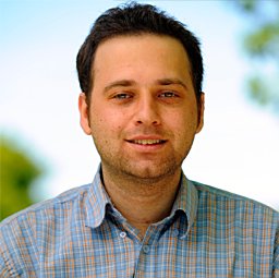 Amir Rosenthal合作论文数Institute for Biological and Medical Imaging, Helmholtz Zentrum München und Technische Universität München35
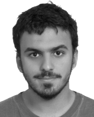 Ali Ozbek合作论文数Univ Zurich, Inst Biomed Engn, CH-8057 Zurich, Switzerland30
 Shy Shoham合作论文数Department of Ophthalmology, Grossman School of Medicine, New York University/Department of Neuroscience and Physiology, Grossman School of Medicine, New York University28Quanyu Zhou合作论文数Inst Pharmacol & Toxicol, Univ Zurich24
 Michael Reiss合作论文数University of Zurich23
 Andreas Buehler合作论文数Department of Radiation Oncology, University of Heidelberg23
 Stefan Morscher合作论文数22
 Neal Burton合作论文数21
 Cagla Ozsoy合作论文数Univ Zurich, Fac Med, Zurich, Switzerland20
 Jan Klohs合作论文数Swiss Federal Institute of Technology, University of Zurich20
 Pavel Subochev合作论文数Laboratory of Ultrasonic and Optoacosutic Diagnostics, Institute of Applied Physics20
 Sandeep Kumar Kalva合作论文数Nanyang Technol Univ, Sch Chem & Biomed Engn, Singapore, Singapore19
 Roger M. Nitsch合作论文数Institute for Regenerative Medicine, University of Zurich;Neurimmune AG19p d einziger合作论文数18
 Subhamoy Mandal合作论文数16Adrian Taruttis合作论文数Institute for Biological and Medical Imaging, Technische Universität München and Helmholtz Zentrum München16
 Claudio Vinegoni合作论文数Center for Systems Biology, Massachusetts General Hospital, Harvard University;Harvard Medical School16
 Elena Merčep合作论文数Tech Univ Munich, Fac Med, Munich, Germany15
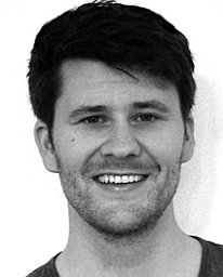 Fehm Thomas F合作论文数Institute of Biological and Medical Imaging (IBMI),, Helmholtz Zentrum München15Dan Adam合作论文数Editorial Board ? the IEEE Transactions on Biomedical Engineering15
 Moritz Kneipp合作论文数14Weiye Li合作论文数13
 Berkan Lafci合作论文数Swiss Fed Inst Technol, Inst Biomed Engn13
 Daniil Nozdriukhin合作论文数Ctr Photon Sci & Engn, Skolkovo Inst Sci & Technol13Oleksiy Degtyaruk合作论文数Helmholtz Zentrum Munchen13
 Justine Robin合作论文数ESPCI Paris, PSL Res Univ13
 Anna Orlova合作论文数Rudbeck Laboratory, Uppsala University13
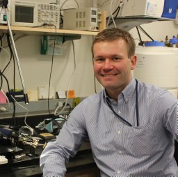 Stephan Kellnberger合作论文数13
 Benedict Edward Mc Larney合作论文数Molecular Imaging Therapy Service, Memorial Sloan Kettering Cancer Center12Hsiao-Chun Amy Lin合作论文数12Lu Ding合作论文数Tech Univ Munich, IngolstadterLandstr 1, D-85764 Neuherberg, Germany12
 Yu-Hang Liu合作论文数Singapore Inst Neurotechnol SINAPSE, Natl Univ Singapore12
 Gil Westmeyer合作论文数Department of Chemistry, Technische Universität München12
 Urs A. T. Hofmann合作论文数Inst Pharmacol & Toxicol, Univ Zurich11
 Nikolaos Deliolanis合作论文数Goethe-Universität Frankfurt A. Main11Turner Jake合作论文数11Erwin Bay合作论文数10Rui Ma合作论文数10
 Ivana Ivankovic合作论文数9Chaim Glück合作论文数9
 Francisco Oyaga合作论文数9
 Avihai Ron合作论文数Helmholtz Zentrum München9




