We developed acellular tissue engineered vessels (ATEV) using small intestine submucosa (SIS) incorporating heparin and a novel protein named H2R5. ATEVs were implanted into the arterial circulation of an ovine animal model, demonstrating high primary patency rates over a period of three months. Implanted grafts were infiltrated by host cells, the majority of which were monocytes/macrophages (MC/MΦ), as demonstrated by scRNA sequencing and immunostaining. They also developed functional endothelial and medial layers that deposited new extracellular matrix leading to matrix remodeling and acquisition of mechanical properties that were similar to those of native arteries. Notably, during this short implantation time, ATEVs turned into functional neo-arteries, as evidenced by the development of the vascular contractile function. Our findings underscore the potential of H2R5-functionalized ATEVs as promising candidates for tissue replacement grafts in a large pre-clinical animal model and highlight the contribution of macrophages in vascular regeneration.
Recently our group demonstrated that acellular tissue engineered vessels (A-TEVs) comprised of small intestinal submucosa (SIS) immobilized with heparin and vascular endothelial growth factor (VEGF) could be implanted into the arterial system of a pre-clinical ovine animal model, where they endothelialized within one month and remained patent. Here we report that immobilized VEGF captures blood circulating monocytes (MC) with high specificity under a range of shear stresses. Adherent MC differentiate into a mixed endothelial (EC) and macrophage (Mφ) phenotype and further develop into mature EC that align in the direction of flow and produce nitric oxide under high shear stress. In-vivo, newly recruited cells on the vascular lumen express MC markers and at later times they co-express MC and EC-specific proteins and maintain graft patency. This novel finding indicates that the highly prevalent circulating MC contribute directly to the endothelialization of acellular vascular grafts under the right chemical and biomechanical cues.
Cell-free small diameter vascular grafts, based on small intestinal submucosa functionalized with heparin and vascular endothelial growth factor (VEGF) are manufactured and implanted successfully into the arterial system of neonatal lambs, where they remain patent and grow in size with the host to a similar extent and with similar rate as native arteries. Acellular tissue engineered vessels (A-TEVs) integrate seamlessly into the native vasculature and develop confluent, functional endothelium that affords patency. The medial layer is infiltrated by smooth muscle cells, shows no signs of calcification, and develops contractile function. The vascular wall undergoes remarkable extracellular matrix remodeling exhibiting elastin fibers and even inner elastic lamina within six months. Taken together, the results suggest that VEGF-based A-TEVs may be suitable for treatment of congenital heart disorders to alleviate the need for repeated surgeries, which are currently standard practice.
The last two decades have seen many advances in regenerative medicine, including the development of tissue engineered vessels (TEVs) for replacement of damaged or diseased arteries or veins. Biomaterials from natural sources as well as synthetic polymeric materials have been employed in engineering vascular grafts. Recently, cell-free grafts have become available opening new possibilities for the next generation, off-the-shelf products. These TEVs are first tested in small or large animal models, which are usually young and healthy. However, the majority of patients in need of vascular grafts are elderly and suffer from comorbidities that may complicate their response to the implants. Therefore, it is important to evaluate TEVs in animal models of vascular disease in order to increase their predictive value and learn how the disease microenvironment may affect the patency and remodeling of vascular grafts. Small animals with various disease phenotypes are readily available due to the availability of transgenic or gene knockout technologies and can be used to address mechanistic questions related to vascular grafting. On the other hand, large animal models with similar anatomy, hematology and thrombotic responses to humans have been utilized in a preclinical setting. We propose that large animal models with certain pathologies or age range may provide more clinically relevant platforms for testing TEVs and facilitate the clinical translation of tissue engineering technologies by increasing the likelihood of success in clinical trials.
Recent advances in vascular tissue engineering have led to the development of cell-free grafts that are available off-the-shelf for on demand surgery. Challenges associated with cell-based technologies including cell sourcing, cell expansion and long-term bioreactor culture motivated the development of completely cell-free vascular grafts. These are based on decellularized arteries, decellularized cultured cell-based tissue engineered grafts or biomaterials functionalized with biological signals that promote in situ tissue regeneration. Clinical trials undertaken to demonstrate the applicability of these grafts are also discussed. This comprehensive review summarizes recent developments in vascular graft technologies, with potential applications in coronary artery bypass procedures, lower extremity bypass, vascular injury and trauma, congenital heart diseases and dialysis access shunts, to name a few.
Background: The Neonatal Resuscitation Program (NRP) recommends close monitoring of oxygenation during the resuscitation of newborns using a pulse oximeter. However, there are no guidelines for monitoring carbon dioxide (CO2) to assess ventilation. Considering that cerebral blood flow (CBF) correlates directly with PaCO2, continuous capnography monitoring of end-tidal CO2 (ETCO2) may limit fluctuations in PaCO2 and, therefore, CBF during resuscitation of asphyxiated infants. Objective: To evaluate whether continuous monitoring of ETCO2 with capnography during resuscitation of asphyxiated term lambs with meconium aspiration will prevent fluctuations in PaCO2 and carotid arterial blood flow (CABF). Methods: Fifty-four asphyxiated term lambs with meconium aspiration syndrome were mechanically ventilated from birth to 60 min of age. Ventilatory parameters were adjusted based on clinical observation (chest excursion) and frequent arterial blood gas analysis in 24 lambs (control group) and 30 lambs (capnography group) received additional continuous ETCO2 monitoring. Left CABF was monitored. We aimed to maintain PaCO2 between 35 and 50 mm Hg and ETCO2 between 30 and 45 mm Hg. Results: There was a significant correlation between ETCO2 and PaCO2 (R = 0.7, p < 0.001), between PaCO2 and carotid flow (R = 0.52, p < 0.001) and between ETCO2 and carotid flow (R = 0.5, p < 0.001). PaCO2 and CABF during the first 60 min of age showed significantly higher fluctuation in the control group compared to the capnography group. Conclusion: Continuous monitoring of ETCO2 using capnography with mechanical ventilation during and after resuscitation in asphyxiated term lambs with meconium aspiration limits fluctuations in PaCO2 and CABF and may potentially limit brain injury.
Objective: Hypertension is a leading cardiovascular risk factor worldwide, playing a significant role in ischemic heart disease, stroke and atherosclerosis. Here, we describe the development of a hypertensive ovine model that was employed to test the performance of autologous arteries or veins when transplanted under disease conditions. Methods: After inducing hypertension using the one-kidney-one-clip (1K1C) model, autologous carotid artery grafts (CAG) or facial vein grafts (FVG) were implanted into carotid arteries of hypertensive (HT) or normotensive sheep (NT). Autologous carotid artery (CA) and facial vein (FV) were used as control. Valuable real-time hemodynamic data was collected through instrumentation that was implanted during grafting. Results and Discussion: Our study revealed that autologous arteries demonstrated a significant decrease of their function when grafted under NT conditions, but HT showed no additional decline in function. On the contrary, grafted veins showed remodeling under NT, while under HT they performed poorly. Surprisingly, both CAG and FVG showed significantly reduced mechanical properties as well as collagen and elastin content following grafting, albeit FVG were affected to a higher extent by HT. Smooth muscle cell remodeling, as well as vascular function was also more significantly affected by HT in FVG as compared to CAG. Interestingly, transplanted FVG were infiltrated by pro-inflammatory macrophages in HT animals, but macrophages of an anti-inflammatory phenotype prevailed in NT animals.
The large number of coronary artery bypass procedures necessitates development of off-the-shelf vascular grafts that do not require cell or tissue harvest from patients. However, immediate thrombus formation after implantation due to the absence of a healthy endothelium is very likely. Here we present the successful development of an acellular tissue engineered vessel (A-TEV) based on small intestinal submucosa that was functionalized sequentially with heparin and VEGF. A-TEVs were implanted into the carotid artery of an ovine model demonstrating high patency rates and significant host cell infiltration as early as one week post-implantation. At one month, a confluent and functional endothelium was present and the vascular wall showed significant infiltration of host smooth muscle cells exhibiting vascular contractility in response to vaso-agonists. After three months, the endothelium aligned in the direction of flow and the medial layer comprised of circumferentially aligned smooth muscle cells. A-TEVs demonstrated high elastin and collagen content as well as impressive mechanical properties and vascular contractility comparable to native arteries. This is the first demonstration of successful endothelialization, remodeling, and development of vascular function of a cell-free vascular graft that was implanted in the arterial circulation of a pre-clinical animal model.
Objective: To engineer and implant vascular grafts in the arterial circulation of a pre-clinical animal model and assess the role of donor medial cells in graft remodeling and function.Approach and results: Vascular grafts were engineered using Small Intestinal Submucosa (SIS)-fibrin hybrid scaffold and implanted interpositionally into the arterial circulation of an ovine model. We sought to demonstrate implantability of SIS-Fibrin based grafts; examine the remodeling; and determine whether the presence of vascular cells in the medial wall was necessary for cellular infiltration from the host and successful remodeling of the implants. We observed no occlusions or anastomotic complications in 18 animals that received these grafts. Notably, the grafts exhibited unprecedented levels of host cell infiltration that was not limited to the anastomotic sites but occurred through the lumen as well as the extramural side, leading to uniform cell distribution. Incoming cells remodeled the extracellular matrix and matured into functional smooth muscle cells as evidenced by expression of myogenic markers and development of vascular reactivity. Interestingly, tracking the donor cells revealed that their presence was beneficial but not necessary for successful grafting. Indeed, the proliferation rate and number of donor cells decreased over time as the vascular wall was dominated by host cells leading to significant remodeling and development of contractile function.Conclusions: These results demonstrate that SIS-Fibrin grafts can be successfully implanted into the arterial circulation of a clinically relevant animal model, improve our understanding of vascular graft remodeling and raise the possibility of engineering mural cell-free arterial grafts. (C) 2015 Elsevier Ltd. All rights reserved.
We demonstrate the ability of immobilized vascular endothelial growth factor (VEGF) to capture endothelial cells (EC) with high specificity under fluid flow. To this end, we engineered a surface consisting of heparin bound to poly-l-lysine to permit immobilization of VEGF through the C-terminal heparin-binding domain. The immobilized growth factor retained its biological activity as shown by proliferation of EC and prolonged activation of KDR signaling. Using a microfluidic device we assessed the ability to capture EC under a range of shear stresses from low (0.5 dyne/cm(2)) to physiological (15 dyne/cm(2)). Capture was significant for all shear stresses tested. Immobilized VEGF was highly selective for EC as evidenced by significant capture of human umbilical vein and ovine pulmonary artery EC but no capture of human dermal fibroblasts, human hair follicle derived mesenchymal stem cells, or mouse fibroblasts. Further, VEGF could capture EC from mixtures with non-EC under low and high shear conditions as well as from complex fluids like whole human blood under high shear. Our findings may have far reaching implications, as they suggest that VEGF could be used to promote endothelialization of vascular grafts or neovascularization of implanted tissues by rare but continuously circulating EC.
We examined the effects of senescence on the proliferation and leiomyogenic differentiation potential of mesenchymal stem cells (MSCs) isolated from bone marrow (BM-MSCs) or hair follicles (HF-MSCs). To this end, we compared ovine HF-MSCs and BM-MSCs in terms of their proliferation and differentiation potential to the smooth muscle cell lineage. We discovered that HF-MSCs are less susceptible to culture senescence compared with BM-MSCs. We hypothesized that application of mechanical forces may enhance the contractility and mechanical properties of vascular constructs prepared from senescent MSCs. Interestingly, HF-MSCs and BM-MSCs responded differently to changes in the mechanical microenvironment, suggesting that despite phenotypic similarities, MSCs from different anatomic locations may activate different pathways in response to the same microenvironmental factors. In turn, this may also suggest that cell-based tissue regeneration approaches may need to be tailored to the stem cell origin, donor age, and culture time for optimal results.
The Neonatal Resuscitation Program (NRP) recommends upper and lower limits of preductal saturations (SpO2) extrapolated from studies in infants resuscitated in room air. These limits have not been validated in asphyxia and lung disease.Seven control term lambs delivered by cesarean section were ventilated with 21% O2. Thirty lambs with asphyxia with meconium aspiration were randomly assigned to resuscitation with 21% O2 (n = 6), 100% O2 (n = 6), or initiation with 21% O2 followed by variable FIO2 to maintain NRP target SpO2 ranges (n = 18). Hemodynamic and ventilation parameters were recorded for 15 min.Control lambs maintained preductal SpO2 near the lower limit of NRP target range. Asphyxiated lambs had low SpO2 (38 ± 2%), low arterial pH (6.99 ± 0.01), and high PaCO2 (96 ± 7 mm Hg) at birth. Resuscitation with 21% O2 resulted in SpO2 values below the target range with low pulmonary blood flow (Qp) compared to variable FIO2 group. The increase in PaO2 and Qp with variable FIO2 resuscitation was similar to control lambs.Maintaining SpO2 as recommended by NRP by actively adjusting inspired O2 leads to effective oxygenation and higher Qp in asphyxiated lambs with lung disease. Our findings support the current NRP SpO2 guidelines for O2 supplementation during resuscitation of an asphyxiated neonate.
The development of Tissue Engineered Vessels (TEVs) is advanced by the ability to routinely and effectively implant TEVs (4-5 mm in diameter) into a large animal model. A step by-step protocol for inter-positional placement of the TEV and real-time digital assessment of the TEV and native carotid arteries is described here. In vivo monitoring is made possible by the implantation of flow probes, catheters and ultrasonic crystals (capable of recording dynamic diameter changes of implanted TEVs and native carotid arteries) at the time of surgery. Once implanted, researchers can calculate arterial blood flow patterns, invasive blood pressure and artery diameter yielding parameters such as pulse wave velocity, augmentation index, pulse pressures and compliance. Data acquisition is accomplished using a single computer program for analysis throughout the duration of the experiment. Such invaluable data provides insight into TEV matrix remodeling, its resemblance to native/sham controls and overall TEV performance in vivo.
BACKGROUND:The oxygenation index (OI = mean airway pressure, MAP × FiO2 × 100 : PaO2) is used to assess the severity of hypoxic respiratory failure (HRF) and persistent pulmonary hypertension of the newborn (PPHN). An indwelling arterial line or arterial punctures are necessary to obtain PaO2 for the calculation of OI. Oxygenation can be continuously and noninvasively assessed using pulse oximetry. The use of the oxygen saturation index (OSI = MAP × FiO2 × 100 : SpO2) can be an alternate method of assessing the severity of HRF.OBJECTIVE:To evaluate the correlation between OSI and OI in the following: (1) neonates with HRF and (2) a lamb model of meconium aspiration syndrome.METHODS:Human neonates: a retrospective chart review of 74 ventilated late preterm/term neonates with indwelling arterial access and SpO2 values in the first 24 h of life was conducted. OSI and OI were calculated and correlated. Lamb model: arterial blood gases were drawn and preductal SpO2 was documented in 40 term newborn lambs with asphyxia and meconium aspiration. OI and OSI were calculated and correlated with pulmonary vascular resistance (PVR).RESULTS:Mean values of OSI and OI showed a correlation coefficient of 0.952 in neonates (mean value of 308 observations in 74 neonates) and 0.948 in lambs (mean value of 743 observations in 40 lambs). In lambs, with increasing PVR, there was a decrease in OI and OSI.CONCLUSION:OSI correlates significantly with OI in infants with HRF. This noninvasive measure may be used to assess the severity of HRF and PPHN in neonates without arterial access.
Background: Current neonatal resuscitation guidelines recommend tracheal suctioning of nonvigorous neonates born through meconium-stained amniotic fluid. Methods: We evaluated the effect of tracheal suctioning at birth in 29 lambs with asphyxia induced by cord occlusion and meconium aspiration during gasping. Results: Tracheal suctioning at birth ( n = 15) decreased amount of meconium in distal airways (53 ± 29 particles/mm 2 lung area) compared to no suction (499 ± 109 particles/mm 2 ; n = 14; P < 0.001). Three lambs in the suction group had cardiac arrest during suctioning, requiring chest compressions and epinephrine. Onset of ventilation was delayed in the suction group (146 ± 11 vs. 47 ± 3 s in no-suction group; P = 0.005). There was no difference in pulmonary blood flow, carotid blood flow, and pulmonary or systemic blood pressure between the two groups. Left atrial pressure was significantly higher in the suction group. Tracheal suctioning resulted in higher Pa o 2 /FiO 2 levels (122 ± 21 vs. 78 ± 10 mm Hg) and ventilator efficiency index (0.3 ± 0.05 vs.0.16 ± 0.03). Two lambs in the no-suction group required inhaled nitric oxide. Lung 3-nitrotyrosine levels were higher in the suction group (0.65 ± 0.03 ng/µg protein) compared with the no-suction group (0.47 ± 0.06). Conclusion: Tracheal suctioning improves oxygenation and ventilation. Suctioning does not improve pulmonary/systemic hemodynamics or oxidative stress in an ovine model of acute meconium aspiration with asphyxia.
Because of rise in cardiovascular disease throughout the world, there is increasing demand for small diameter blood vessels as replacement grafts. The present review focuses on the animal models that have been used to test small-diameter TEVs with emphasis on the attributes of each model. Small animal models are used to test short-term patency and address mechanistic hypotheses; and large, preclinical animal models are employed to test long-term patency, remodeling and function in an environment mimicking human physiology. We also discuss recent clinical trials that employed laboratory fabricated TEVs and showed very promising results. Ultimately, animal models provide a testing platform for optimizing vascular grafts before clinical use in patients without suitable autologous vessels.
 Stelios Andreadis合作论文数Department of Chemical and Biological Engineering, School of Engineering and Applied Sciences, State University of New York at Buffalo;Cell, Gene and Tissue Engineering Center, University at Buffalo;Angiograft, LLC.30
 Satyan Lakshminrusimha合作论文数UC Davis Children's Hospital;Department of Pediatrics, University of California21
 James A. Russell合作论文数Division of Critical Care Medicine, Department of Medicine, Faculty of Medicine, The University of British Columbia;Centre for Heart Lung Innovation, The University of British Columbia19
 Sylvia F Gugino合作论文数15
 Rita Ryan合作论文数School of Medicine and Biomedical Sciences, State University of New York14
 Bobby Mathew合作论文数12Row Sindhu合作论文数11Koenigsknecht Carmon合作论文数10
 Hui Meng合作论文数Department of Mechanical and Aerospace Engineering, School of Engineering and Applied Sciences, University at Buffalo, The State University of New York10
 Karen Wynn合作论文数Department of Psychology , Yale University9
 Eleni Metaxa合作论文数8
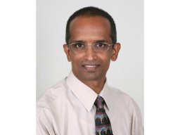 Kumar Vasantha H S合作论文数Department of Pediatrics, University of Connecticut School of Medicine8
 Robin Steinhorn合作论文数7Ling Gao合作论文数7Hao Fan Peng合作论文数Bioengineering Laboratory;Department of Chemical and Biological Engineering;University at Buffalo;State University of New York7John Kolega合作论文数6
 Jayasree Nair合作论文数6Koobatian Maxwell T合作论文数Department of Physiology &Buffalo School of Medicine, State University of New York;Buffalo School of Medicine, State University of New York6
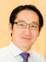 Yiemeng Hoi合作论文数Department of Mechanical and Industrial Engineering, University of Toronto6Evan M Schlaich合作论文数5Dj Geer合作论文数5
 Rawat Munmun合作论文数5Nielsen Lori C合作论文数5Frederick C. Morin合作论文数4Mocco J合作论文数4
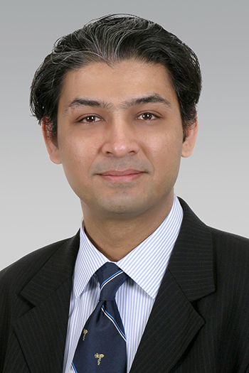 Adnan Siddiqui合作论文数Department of Neurosurgery, Jacobs School of Medicine and Biomedical Sciences, University at Buffalo;Canon Stroke and Vascular Research Center, University at Buffalo4
 Praveen Chandrasekharan合作论文数4Bruce A. Holm合作论文数4M Szymanski合作论文数4LC Nielsen合作论文数3RH Steinhorn合作论文数3
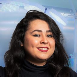 Bita Nasiri合作论文数SUNY Buffalo, Dept Chem & Biol Engn, Amherst, NY USA3
 Zhijie Wang合作论文数School of Biomedical Engineering, Colorado State University3
 Elad I. Levy合作论文数Department of Neurosurgery, Jacobs School of Medicine & Biomedical Sciences, University at Buffalo3Vali Payam合作论文数3
 Jinyu Liu (刘晋宇)合作论文数School of Public Health, Jilin University3Wang Huamei合作论文数3
 Changxing Ma合作论文数3Peter A. Nickerson合作论文数3David M. Wiley合作论文数2Nicolas L'Heureux合作论文数2Druck Reinhardt Druck Basel合作论文数2Christopher A D'Angelis合作论文数2Lauren C Goldie合作论文数2Peter J Amos合作论文数2P A Nickerson合作论文数2Hera Chaudhury合作论文数2Peter Christalla合作论文数2Fabio Savorgnan合作论文数2




