Epidural spinal cord stimulation has been shown to be a promising neurotechnology to improve upper limb function in people affected by stroke. It is well established that SCS targeting the dorsal root entry zone increases excitatory drive to α-motoneurons via the monosynaptic reflex pathway. However, the effects of SCS on inhibitory neural pathways remain unexplored. We hypothesized that SCS improves the neuromotor control of arm movement by strengthening both excitatory and inhibitory circuit function. We show in three individuals with post-stroke motor symptoms that SCS enhances postsynaptic Ia reciprocal inhibition. These changes correlate with enhanced muscle coordination and arm kinematics resulting in smoother and faster trajectories. Our study provides further insights into the neural targets of spinal cord stimulation and the use of this technology to treat post-stroke motor deficits.
Background Recent evidence suggests extra-cortical adaptations within the cerebellum may contribute to motor recovery in patients with cortical ischemic strokes. The molecular/cellular adaptations enabling this effect to have not been identified. Chloride transport proteins (NKCC1 and KCC2) are important regulators of neuronal transmission and may underlie adaptive changes following ischemic stroke. Objective Examine changes in cerebellar NKCC1 and KCC2 protein expression following cortical ischemic stroke. Methods Adult C57BL/6J male mice underwent sham or the left middle cerebral artery occlusion (tMCAo)-induced ischemic stroke. Changes of NKCC1 and KCC2 proteins within the deep cerebellar nuclei (DCN) were assessed by immunofluorescence staining. Results tMCAo induced selective infarct lesion in the left striatum and cortex of the stroke mice but not in other brain regions including cerebellum. The inwardly directed chloride transporter NKCC1 was equivocally expressed within bi-hemispheric DCN of both sham control and stroke mice. In contrast, the outwardly directed chloride transporter KCC2 protein expression was significantly higher in the bi-hemispheric DCN of stroke brains, compared to sham controls. Double immunostaining analysis revealed a statistically significant increase in KCC2 intensity within VGLUT-1+ neurons of the ipsilateral DCN of the stroke mice, but not in the VGAT+ neurons. Conclusions Ischemic cortical stroke stimulates KCC2 protein expression in the DCN VGLUT-1+ neurons, without a change in NKCC1 protein expression.
Here, we report the final outcomes of a pilot clinical trial testing preliminary efficacy and safety of cervical epidural spinal cord stimulation (SCS) for chronic post-stroke upper-limb hemiparesis (NCT04512690). We implanted seven participants with profound motor deficits (Fugl-Meyer Assessment [FMA] scores 15-35) using two leads implanted unilaterally in the cervical spinal cord for 4 weeks. Under SCS ON, motor function immediately improved regardless of impairment severity (average +32% strength and +5.6 FMA-points). Notably, 3/7 participants with residual corticospinal connectivity to finger muscles regained hand/finger function with SCS. Despite performing only 8.6hrs of motor activity (5.5hrs with SCS ON), participants improved by average +6.6 FMA-points at the end of the study compared to baseline and spasticity decreased in all participants. While all benefited, our preliminary analysis indicates that spared sensory function may be a determinant of responsiveness to SCS. No serious adverse events occurred.
INTRODUCTION: Preoperative nutritional status is essential for predicting postoperative outcomes. Studies have shown that prealbumin levels can be a prognostic indicator for surgical outcomes in spine surgery. METHODS: A retrospective cohort study of 697 patients was conducted between April 1st, 2018, to October 31st, 2022. Collected data included patient demographics, PPL, chronic steroid use, index surgery, length of surgery, postoperative hospital length of stay, 30-day return, and pseudoarthrosis (verified by a board certified neuroradiologist via radiographic imaging). PPL were first analyzed as absolute values, and then categorized as binary values. The first cut point used for binary categorization was =30 and >30. We then repeated statistical analyses similarly using =25, =20, and =15 as additional cut points (all as binary values). RESULTS: Fifty-six patients had pseudoarthrosis (8%), while 641 (92%) did not. There was no statistical difference between patients who had pseudoarthrosis and patients who did not, in terms of age (62 vs. 62.9, p=0.298), BMI (29.9 vs. 29.8, p=0.441), and preoperative prealbumin level (26.6 vs 26.4, p=0.387). There was a significant difference regarding the length of surgery (337.9 mins vs. 253.8, p=0.008) and hospital length of stay (6.2 days vs. 4.2 days, p<0.001) in patients with pseudoarthrosis. Finally, there was no significant difference between PPL and developing pseudoarthrosis when the data was categorized as binary values with cut points at =30 (p=0.595), =25 (p=0.987), =20 (p=0.799), and =15 (p=0.333). CONCLUSIONS: Preoperative nutritional assessment is essential in spine surgery as it can predict postoperative outcomes and recovery. In our cohort of patients, low serum prealbumin levels did not correlate with a higher rate of pseudoarthrosis following lumbar spine surgery.
Cerebral white matter lesions prevent cortico-spinal descending inputs from effectively activating spinal motoneurons, leading to loss of motor control. However, in most cases, the damage to cortico-spinal axons is incomplete offering a potential target for therapies aimed at improving volitional muscle activation. Here we hypothesize that, by engaging direct excitatory connections to cortico-spinal motoneurons, stimulation of the motor thalamus could facilitate activation of surviving cortico-spinal fibers thereby immediately potentiating motor output. To test this hypothesis, we identify optimal thalamic targets and stimulation parameters that enhance upper-limb motor-evoked potentials and grip forces in anesthetized monkeys. This potentiation persists after white matter lesions. We replicate these results in humans during intra-operative testing. We then design a stimulation protocol that immediately improves strength and force control in a patient with a chronic white matter lesion. Our results show that electrical stimulation targeting surviving neural pathways can improve motor control after white matter lesions. Cerebral lesions result in loss of upper-limb motor functions. Here, the authors show that electrical stimulation of the motor thalamus can immediately and significantly improve strength and volitional force control improving arm and hand functions.
Spinal Muscular Atrophy (SMA) is an inherited neurodegenerative disease causing motoneuron dysfunction, muscle weakness and early mortality1,2. Three therapies can slow disease progression enabling people to survive albeit with lingering motoneuron dysfunction and severe motor impairments3,4. Here we introduce a neurotechnological approach that improved spinal motoneuron function, muscle strength and walking in three adults with SMA. Starting from preclinical evidence showing that motoneuron dysfunction in SMA originates from the loss of excitatory inputs from primary afferents5,6, we hypothesized that augmentation of sensory neural activity with targeted electrical stimulation could compensate for this loss thereby improving motoneuron function. To test this hypothesis we implanted three adults with SMA with epidural electrodes over the lumbosacral spinal cord to stimulate the sensory axons of the legs7,8. We stimulated participants for 4 weeks 2 hours per day while they executed walking and strength tasks. Remarkably, our neurostimulation regime led to robust improvements in strength, walking and fatigue paralleled by reduced neuronal hyperexcitability, increased sensory inputs and higher motoneuron firing rates. Our data indicates that targeted neurostimulation can reverse degenerative processes of circuit dysfunction thus promoting disease modifying effects in a human neurodegenerative disease.
Intracortical microstimulation (ICMS) is a method for restoring sensation to people with paralysis as part of a bidirectional brain-computer interface to restore upper limb function. Evoking tactile sensations of the hand through ICMS requires precise targeting of implanted electrodes. Here we describe the presurgical imaging procedures used to generate functional maps of the hand area of the somatosensory cortex and subsequent planning that guided the implantation of intracortical microelectrode arrays. In five participants with cervical spinal cord injury, across two study locations, this procedure successfully enabled ICMS-evoked sensations localized to at least the first four digits of the hand. The imaging and planning procedures developed through this clinical trial provide a roadmap for other brain-computer interface studies to ensure successful placement of stimulation electrodes.
Intracortical microstimulation (ICMS) is a method for restoring sensation to people with paralysis as part of a bidirectional brain-computer interface (BCI) to restore upper limb function. Evoking tactile sensations of the hand through ICMS requires precise targeting of implanted electrodes. Here we describe the presurgical imaging procedures used to generate functional maps of the hand area of the somatosensory cortex and subsequent planning that guided the implantation of intracortical microelectrode arrays. In five participants with cervical spinal cord injury, across two study locations, this procedure successfully enabled ICMS-evoked sensations localized to at least the first four digits of the hand. The imaging and planning procedures developed through this clinical trial provide a roadmap for other BCI studies to ensure the successful placement of stimulation electrodes.
(1) Background: Spinal cord injury (SCI) represents a major health challenge, often leading to significant and permanent sensorimotor and autonomic dysfunctions. This study reviews the evolving role of epidural spinal cord stimulation (eSCS) in treating chronic SCI, focusing on its efficacy and safety. The objective was to analyze how eSCS contributes to the recovery of neurological functions in SCI patients. (2) Methods: We utilized the PRISMA guidelines and performed a comprehensive search across MEDLINE/PubMed, Embase, Web of Science, and IEEE Xplore databases up until September 2023. We identified studies relevant to eSCS in SCI and extracted assessments of locomotor, cardiovascular, pulmonary, and genitourinary functions. (3) Results: A total of 64 studies encompassing 306 patients were identified. Studies investigated various stimulation devices, parameters, and rehabilitation methods. Results indicated significant improvements in motor function: 44% of patients achieved assisted or independent stepping or standing; 87% showed enhanced muscle activity; 65% experienced faster walking speeds; and 80% improved in overground walking. Additionally, eSCS led to better autonomic function, evidenced by improvements in bladder and sexual functions, airway pressures, and bowel movements. Notable adverse effects included device migration, infections, and post-implant autonomic dysreflexia, although these were infrequent. (4) Conclusion: Epidural spinal cord stimulation is emerging as an effective and generally safe treatment for chronic SCI, particularly when combined with intensive physical rehabilitation. Future research on standardized stimulation parameters and well-defined therapy regimens will optimize benefits for specific patient populations.
Abstract Cerebral white matter tract lesions prevent cortico-spinal descending inputs from effectively activating spinal motoneurons, leading to untreatable muscle paralysis. However, in most cases the damage to cortico-spinal axons is incomplete and the spared connections could be potentiated by neurotechnologies to restore motor function. Here we hypothesized that, by engaging direct excitatory connections to cortico-spinal motoneurons, deep brain stimulation (DBS) of the motor thalamus could facilitate activation of spared cortico-spinal fibers improving muscle activation of the paretic limb. We first identified, in monkeys, optimal stimulation targets and parameters that enhanced motor evoked potentials to arm, hand, and face muscles, as well as grip forces. This potentiation persisted after cerebral white matter lesions. We then translated these results to human subjects by identifying the corresponding optimal thalamic targets (VIM/VOP nuclei) and replicated the results obtained in monkeys. Finally, we designed a DBS protocol that immediately improved voluntary grip force control in a patient with a chronic traumatic brain injury. Our results suggest that targeted DBS of the motor thalamus may become an effective therapy for motor paralysis.
Within the sample of 181 patients with cervical CT, CT identified unstable injury with a sensitivity of 100% and specificity of 95%. CT identified operable injury at the CCJ with 86% sensitivity and 91% specificity. CT was considered the gold standard for identification of fractures. Together, the presence of CT imaging suggestive of unstable injury or persistent neurologic complaint had a 100% sensitivity and 81% specificity. Finally, across all patients MRI had 100% sensitivity and 89% specificity for detection of unstable injury requiring surgery.
Spinal cord stimulation (SCS) restores motor control after spinal cord injury (SCI) and stroke. This evidence led to the hypothesis that SCS facilitates residual supraspinal inputs to spinal motoneurons. Instead, here we show that SCS does not facilitate residual supraspinal inputs but directly triggers motoneurons action potentials. However, supraspinal inputs can shape SCS-mediated activity, mimicking volitional control of motoneuron firing. Specifically, by combining simulations, intraspinal electrophysiology in monkeys and single motor unit recordings in humans with motor paralysis, we found that residual supraspinal inputs transform subthreshold SCS-induced excitatory postsynaptic potentials into suprathreshold events. We then demonstrated that only a restricted set of stimulation parameters enables volitional control of motoneuron firing and that lesion severity further restricts the set of effective parameters. Our results explain the facilitation of voluntary motor control during SCS while predicting the limitations of this neurotechnology in cases of severe loss of supraspinal axons.
ABSTRACT Study Design A cross-sectional study. Objective The primary objective of this study is to compare the efficacy of continuous versus threshold drainage strategies for maintaining spinal cord perfusion pressure (SCPP) in patients with new traumatic spinal cord injuries (SCI). Setting Level 1 trauma center. Methods A retrospective study of 19 patients with traumatic SCIs. SCPP was optimized at the discretion of the managing clinician using either vasopressors to increase mean arterial pressure or cerebral spinal fluid (CSF) drainage to decrease intrathecal pressure. Six patients were managed with continuous drainage (CSF drained at regular intervals regardless of SCPP) and 13 had CSF drained only when SCPP fell below 65mmHg (i.e. threshold drainage). Intrathecal pressure, SCPP, mean arterial pressure, and vasopressor utilization were compared using univariate T-test statistical analysis. Results The cohort included over 1500 time points from 19 patients. While there was no difference in rates of sub-optimal SCPP (< 65mmHg; p = 0.257), patients managed with threshold drainage were more likely to exhibit critically-low SCPP (< 50 mmHg; p = 0.003) despite also having lower average intrathecal pressures (p < 0.001). There were no differences in average SCPP, MAP, or vasopressor utilization between the two groups (p > 0.05). Conclusions Acute SCI patients managed with continuous CSF drainage were less likely to exhibit critically-low SCPPs, previously shown to be associated with worse clinical recovery. A larger, prospective cohort is needed to validate the impact of CSF drainage strategies on long-term SCI outcomes.
OBJECTIVE:Prealbumin levels correlate with overall nutritional status, and low values are associated with poor wound healing. We investigated whether low preoperative prealbumin levels predict risk of endoscopic endonasal skull base surgery (EESBS) reconstruction failure, as demonstrated by postoperative cerebrospinal fluid (CSF) leak and/or infection.METHODS:Between October 2018 and February 2020, 98 patients with documented preoperative prealbumin levels were prospectively followed. The incidence of CSF leak and infection in patients with low prealbumin levels (≤20 mg/dL) was compared with those with normal prealbumin levels (>20 mg/dL). Numerous factors previously shown to influence CSF leak rates were assessed. Both univariate and multivariable analyses were performed to identify independent predictive factors.RESULTS:Within this prospectively gathered patient cohort composed of >95% "high-risk" expanded EESBS, 14 of 98 patients (14.3%) experienced a postoperative CSF leak. Factors univariately associated with postoperative complications at the 0.2 level of significance were used in a multivariable model. Low prealbumin levels (≤20 mg/dL) proved to be a strong independent predictive factor associated with a 5-fold increased risk of postoperative CSF leak (odds ratio 5.01, P = 0.01), and postoperative surgical-site infection (P = 0.0009). These associations remained after controlling for multiple other factors, including body mass index, surgical pathology, previous EESBS, risk assessment index, and high- versus low-flow intraoperative CSF leaks.CONCLUSIONS:Preoperative prealbumin levels are an independent predictor of EESBS associated CSF leak and infection. Future studies are needed to investigate the utility of screening and correcting prealbumin levels to limit postoperative complications.
OBJECTIVE:Patient feedback surveys provide important insight into patient outcomes, satisfaction, and perioperative needs. Recent critiques have questioned provider-initiated surveys and their capacity to accurately gauge patient perspectives due to intrinsic biases created by question framing. In this study, the authors sought to evaluate provider-independent, patient-controlled social media Instagram posts in order to better understand the patient experience following scoliosis correction surgery.METHODS:Twitter and Instagram were queried for posts with two tagged indicators, #scoliosissurgery or @scoliosissurgery, resulting in no relevant Twitter posts and 25,000 Instagram posts. Of the initial search, 24,500 Instagram posts that did not directly involve the patient's own experience were eliminated. Posts were analyzed and coded for the following criteria: the gender of the patient, preoperative or postoperative timing discussed in the post, and classified themes related to the patient's experiences with scoliosis correction surgery.RESULTS:Females made 87.6% of the Instagram posts about their experience following scoliosis correction surgery. The initial postoperative stage of surgery was mentioned in 7.6% of Instagram posts. The most common theme on Instagram involved offering or seeking online support from other patients, which constituted 85.2% of all posts. Other common themes included concern about the surgical scar (31.8%), discussing the results of treatment (28.8%), and relief regarding results (21.2%).CONCLUSIONS:Social media provided a platform to analyze unprompted feedback from patients. Patients were most concerned with their scoliosis correction surgery in the period of time 2 weeks or more after surgery. Themes that were most commonly found on Instagram posts were offering or seeking online support from other patients and concern about the surgical scar. Patient-controlled social media platforms, like Instagram, may provide a useful mechanism for healthcare providers to understand the patient experience following scoliosis correction surgery. Such platforms may help in evaluating postoperative satisfaction and improving postoperative quality of care.
ABSTRACT BACKGROUND Prophylactic anticoagulation helps prevent postoperative venous thromboembolism (VTE) and time to initiation postcraniotomy has relied on clinical judgment and practice patterns. OBJECTIVE To compare risks of postoperative VTE and hemorrhage among patients undergoing tumor resection with initiation of prophylactic anticoagulation on postoperative day 1 (POD1) vs POD2. METHODS Adult patients undergoing craniotomy for tumor between 2008 and 2018 were retrospectively reviewed. Outcomes were recorded from the Electronic medical record (EMR) including deep vein thrombosis (DVT), pulmonary embolism (PE), and hemorrhage. RESULTS Of a total of 1168 patients undergoing craniotomy, 225 initiated anticoagulation on POD1 and 389 initiated on POD2. Of the 171 glioblastoma (GBM) cases, 64 initiated on POD1 and 107 on POD2. There were 9 DVTs (1.5%), 1 PE (0.20%), overall VTE rate of 1.6%, and 7 hemorrhagic complications (1.10%), 4 being clinically significant. The GBM cohort contained 4 DVTs (2.3%) and 3 hemorrhagic complications (1.80%). There was no increased risk of VTE or hemorrhage with anticoagulation initiated on POD2 compared to POD1 in either cohort. Multivariate analysis in both cohorts did not reveal a significant association between DVT, PE, or hemorrhagic complications with age, body mass index, GBM pathology, or extent of resection. Interestingly, glioma patients older than 70 with subtotal resection had a higher likelihood of suffering intracranial hemorrhage when anticoagulation was started on POD1 (odds ratio 12.98). CONCLUSION Risk of VTE or hemorrhagic complication did not significantly differ with prophylactic anticoagulation started on POD1 vs POD2. Early anticoagulation may certainly be considered in high risk cases; however, 1 group where risk may outweigh benefit is the elderly glioma population receiving a subtotal resection.
Alan, Nima MD; Agarwal, Nitin MD; Fields, Daryl; Ozpinar, Alp MD; Kanter, Adam S MD; Okonkwo, David O MD, PhD; Hamilton, David Author Information
 Pirondini Elvira合作论文数7
 Peter C. Gerszten合作论文数School of Medicine Neurological Surgery, University of Pittsburgh7
 Marco Capogrosso合作论文数6
 Agarwal Nitin合作论文数6
 Nikhil Verma合作论文数NeuroMechatronics Lab, Carnegie Mellon University5
 David O. Okonkwo合作论文数Neurotrauma Clinical Trials Center, Department of Neurological Surgery, School of Medicine, University of Pittsburgh5
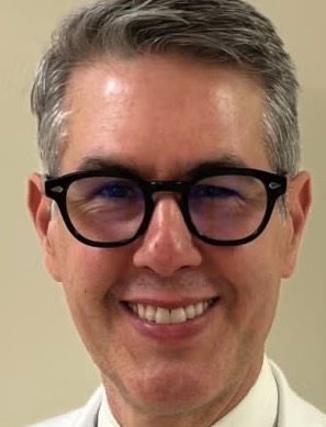 Jorge A. González-Martínez合作论文数Department of Neurological Surgery, School of Medicine, University of Pittsburgh5
 Jonathan Ho合作论文数4
 Erynn Sorensen合作论文数4Boos Amy合作论文数New Jersey Institute of Technology4
 Alan Nima合作论文数4Erinn M Grigsby合作论文数3Luigi Borda合作论文数3Arianna Damiani合作论文数Department of Bioengineering, University of Pittsburgh3Prakarsh Yadav合作论文数3
 Josep-Maria Balaguer合作论文数Rehab and Neural Engineering Labs, University of Pittsburgh3Lucy Liang合作论文数3
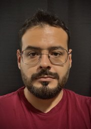 Erick Carranza合作论文数Dept Engn, Pontifical Catholic Univ Peru3
 Lee E Fisher合作论文数Case Western Reserve Univ.;c;Case Western Reserve Univ.3
 Douglas J. Weber合作论文数NeuroMechatronics Lab, Department of Mechanical Engineering, College of Engineering, Carnegie Mellon University;Department of Biomedical Engineering, College of Engineering, Carnegie Mellon University;Neuroscience Institute, Carnegie Mellon University;Reach Neuro, Inc3Buell Thomas J合作论文数Department of Neurosurgery, University of Virginia3D. Kojo Hamilton合作论文数3Ceci Verbaarschot合作论文数2Taylor G Hobbs合作论文数2Serena Donadio合作论文数2Sharath Anand合作论文数2Vahagn Karapetyan合作论文数2Gregory M Adams合作论文数2Theodora Constantine合作论文数2Sridula Kallakuri合作论文数2
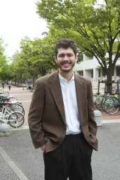 Roberto Martins de Freitas合作论文数Dept Mech Sci & Bioengn, Osaka Univ2
 Jessica Barrios-Martinez合作论文数Department of Neurological Surgery, University of Pittsburgh2
 Hunter R Schone合作论文数National Institutes of Mental Health, National Institutes of Health2Genís Prat-Ortega合作论文数Inst Invest Biomed August Pi I Sunyer IDIBAPS, Barcelona, Spain2Scott Ensel合作论文数Dept Bioengn, Univ Pittsburgh2
 Charles M. Greenspon合作论文数Department of Organismal Biology and Anatomy, University of Chicago2Hanna Algattas合作论文数2
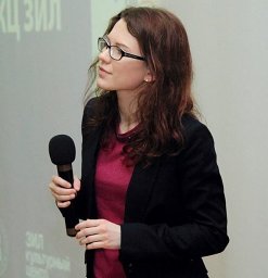 Elizaveta V. Okorokova合作论文数Committee on Computational Neuroscience, University of Chicago2
 George Wittenberg合作论文数University of Pittsburgh2
 John E. Downey合作论文数F. M. Kirby Research Center for Functional Brain Imaging, Kennedy Krieger Institute;F. M. Kirby Research Center for Functional Brain Imaging, Kennedy Krieger Institute2
 Gaunt Robert A合作论文数2
 Ozpinar Alp合作论文数School of Medicine, University of Pittsburgh2
 McDowell Michael M合作论文数Department of Neurological Surgery, University of Pittsburgh Medical Center2
 Youssofzadeh Vahab合作论文数School of Computing and Intelligent Systems, University of Ulster2
 Satzer David合作论文数Department of Neurosurgery, University of Minnesota2
 Michael Boninger合作论文数Department of Physical Medicine & Rehabilitation, School of Medicine, University of Pittsburgh2
 Brian Allen Coffman合作论文数Mind Research Network2
 Chan Hong Moon合作论文数2
 Jennifer L. Collinger合作论文数Human Engineering Research Laboratories, University of Pittsburgh2




