The repair of diabetic bone defects remains challenging due to disrupted bone immune homeostasis and increased susceptibility to infection. Under hyperglycemic conditions, the immune microenvironment fails to switch between the pro-inflammatory environment required for pathogen clearance and the anti-inflammatory environment for osteogenesis. Here, a glucose-responsive hydrogel (PEG-ZnO-sita) is developed by integrating zinc oxide (ZnO) and sitagliptin into a network formed by dopamine-modified tetra-armed poly(ethylene glycol) succinimidyl glutarate (DA-PEG-SG) and phenylboronic acid-modified gelatin (PBA-Gel). PEG-ZnO-sita, enabled by amidation, phenylboronic ester bonds, and metal-catechol coordination, confers rapid gelation, injectability, mechanical stability, self-healing, and robust tissue adhesion, adapting to the local complexity at the bone defect. In a diabetic microenvironment, the rapid breakage of boronate ester bonds triggers the early burst release of Zn2+ to inhibit infection by promoting M1 polarization, along with sitagliptin-intervening glucose regulation. Whereafter, the sustained Zn2+ and sitagliptin release induce M2 polarization and facilitate osteogenic differentiation, demonstrating a self-adaptive strategy in response to inflammatory environments. Both in vitro and in vivo studies confirm that PEG-ZnO-sita effectively promotes bone regeneration by local hyperglycemic microenvironment modulation and sequential regulation of macrophage polarization. So, this temporal immunoregulation of hyperglycemia-immune-osteogenic cascade presents a promising strategy for diabetic infected bone defects.
Objectives The purpose of this study was to investigate whether the eye-lip interaction influences facial attractiveness and the necessity to correct lip protrusion through orthodontic treatment. Materials and Methods A three-dimensional standard morphable facial model was constructed in Maya (Autodesk Inc., San Francisco, CA, USA) based on normative data of the Chinese population. Lip protrusion (Li to E-line) and eye size (palpebral fissure height) were quantitatively morphed into 9 and 3 graded levels, respectively, generating 27 variations. A total of 35 orthodontists, 37 dental residents, and 36 laypersons rated facial attractiveness using a visual analog scale and indicated whether orthodontic treatment was necessary. Multivariate linear regression, ANOVA, and multivariate logistic regression were performed for statistical analysis. Results Interaction effects between the eyes and lips on facial attractiveness were significant (Lip: Eye β = 0.62, P < 0.01). Among all groups, the eye–lip interaction coefficient was most pronounced in laypersons (β = 0.68, P < 0.01), followed by orthodontists (β = 0.59, P < 0.01) and dental residents (β = 0.59, P < 0.01). As eye size increased, the peak of the parabolic curve of aesthetic scores shifted rightward, indicating a preference for more protrusive lips. For orthodontic treatment recommendations, a 2 mm increase in palpebral fissure height was associated with 8% reduction in the odds of selecting treatment. (OR = 0.92, P < 0.01). Conclusions Larger eyes increased observers’ tolerance for lip protrusion and reduced willingness to seek orthodontic treatment. Laypersons were more sensitive to the eye-lip interaction than orthodontists and dental residents. Clinical Relavance Eye-lip interaction plays a notable role in the perception of lip protrusion and should be considered during patient-clinician communication and treatment planning discussions.
Background: Recent years have witnessed a surge in the integration of digital intelligence within dental education, with various technologies increasingly supporting clinical skill training and educational applications. This study aims to map the knowledge landscape of digital intelligence applications in dental education through a bibliometric analysis. Methods: A comprehensive literature search was conducted in the Web of Science Core Collection to identify relevant publications on digital intelligence in dental education. Bibliometric parameters were analysed qualitatively and quantitatively using Microsoft Excel 2019, Incites and CiteSpace software. Results: Of 606 initially retrieved publications, 424 were included in the analysis. Annual publication output remained relatively low from 2000 to 2020, followed by a pronounced increase in 2024 and 2025. Among the ten most-cited articles, the top seven were review articles. The most productive countries were the USA (n = 47, 12%), China (n = 40, 10%), and England (n = 28, 7%). The most active journals were the Journal of Dental Education, European Journal of Dental Education, and BMC Medical Education. Trend analysis via CiteSpace revealed an evolution in research focus from computer simulation (2010) to learning environments (2019) and, more recently, artificial intelligence (2024). Keyword clustering identified five thematic groups: artificial intelligence, computer-assisted instruction, skill assessment, haptic simulation, and simulation. Conclusions: This study offers insights and guidance for researchers interested in digital intelligence applications in dental education. With the rapid advancement of digital intelligence, dental education is poised to benefit substantially. Educators should strategically integrate digital tools and pedagogical concepts to enhance teaching methods and adapt to future educational demands.
Quantifying the biodistribution of lipid nanoparticles (LNPs) is critical for optimizing mRNA delivery systems, yet current approaches have inherent limitations. This study introduces a cost-effective method utilizing double-stranded DNA (dsDNA) barcodes and quantitative polymerase chain reaction (qPCR) for rapid analysis of a small library of mRNA-LNPs biodistribution and functional delivery in vivo. Three unique 100-bp dsDNA barcodes were designed to represent for three FDA-approved LNP formulations. Concurrently, these three formulations carrying luciferase mRNA were mixed with DNA-barcoding LNPs as a pool. Following intravenous administration of the pooled LNPs in mice, qPCR analysis revealed the highest abundance of DNA barcodes and accumulation of luciferase mRNA in spleen, with positive correlation between barcodes presence and mRNA localization across organs, validating DNA barcodes as reliable indicators of mRNA-LNPs biodistribution in vivo. Bioluminescence imaging further confirmed successful delivery and protein translation of luciferase mRNA facilitated by the LNPs in vivo. Integrating DNA barcodes for biodistribution analysis and luciferase mRNA for assessing functional delivery enabled comprehensive evaluation of LNP performance. This robust methodology provides valuable insights into the localization patterns and mRNA delivery capabilities of different LNP formulations, paving the way for the development of more effective and targeted mRNA-based therapeutics.
Introduction This study aimed to evaluate the short-term and long-term skeletal and dental asymmetry with miniscrew-assisted rapid palatal expansion (MARPE), rapid palatal expansion (RPE), and control group using cone-beam computed tomography (CBCT) analysis. Methods A total of 180 CBCT scans were analyzed for 60 patients divided into 3 groups: MARPE (mean age 13.7 ± 1.74 years), RPE (mean age 13.9 ± 1.14 years), and control (mean age 13.3 ± 1.49 years). CBCT scans were recorded at 3 points: pretreatment (T1), postexpansion (T2), and posttreatment (T3). The anterior nasal spine, posterior nasal spine, zygomaticomaxillary point, anterior nasal cavity width, posterior nasal cavity width (PNCW), external maxillary width, palatal width, and molar inclination were measured on the left and right sides in all CBCT scans. In addition, maxillary molar width (MMW) and maxillary canine width were also evaluated on the left and right sides. Statistical analysis was performed, and the parameters were tested within the groups from T1 to T2 and T1 to T3 using a paired t test. The mean changes were then compared among the 3 groups by a mixed analysis of variance test. Results The RPE group showed significantly greater MMW on the left and right sides at T2 (P <0.05). MARPE and RPE led to a significant increase in the anterior nasal spine (right and left), zygomaticomaxillary point (right and left), anterior nasal cavity width (right and left), PNCW (right and left), external maxillary width (right and left), palatal width (right and left), MMW (right and left), maxillary canine width (right and left), and posterior nasal spine (left) at T2 compared with control (P <0.05). RPE led to a significant increase in the molar inclination (right and left) compared with MARPE and controls at T2. In the long term, the PNCW (right and left) was greater in MARPE compared with RPE and controls (P <0.05). Conclusions Within-group comparisons showed that there was no significant asymmetry in the MARPE and control group in the short and long term. The asymmetry for the RPE group was isolated to the MMW in the short term but not observed in the long term, and the magnitude of the asymmetry was clinically not significant.
Diabetes mellitus is a global health threat, with chronic wounds, including oral mucosal wounds, being a severe complication. These wounds are characterized by delayed healing and increased inflammation due to hyperglycemia, affecting patients' quality of life. Current treatments for oral mucosal wounds cannot offer sustained management of these injuries in diabetic patients. Here, a glucose/ROS dual-responsive hydrogel incorporating sitagliptin was developed for the treatment of diabetic oral mucosal wounds. After chemical modification of tetra-armed poly(ethylene glycol) succinimidyl glutarate (tetra-PEG-SG) by dopamine (DA) and tetra-armed poly(ethylene glycol) amine (tetra-PEG-NH2) by phenylboronic acid (PBA), the resulting hydrogel was capable of rapid gelation, robust tissue adhesion, self-healing, antioxidant capacity, and dual response to glucose and reactive oxygen species (ROS), enabling the feasible injection and stable adherence in the moist oral environment while ensuring sustained therapeutic sitagliptin release. In vivo experiments on oral mucosal defects in diabetic mice revealed that the sitagliptin-loaded hydrogel could effectively reduce inflammation and promote wound healing. Collectively, this finding identifies a potential wound dressing as a therapeutic strategy for diabetic oral mucosal wounds.
Aim: The purpose of this study was to evaluate the accuracy and efficacy of a new wireframe template methodology in analyzing three-dimensional facial soft tissue asymmetry. Materials and methods: Three-dimensional facial soft tissue data were obtained for 24 patients. The wireframe template was established by identifying 34 facial landmarks and then forming a template on the face with the MeshLab 2020 software. The angle asymmetry index was automatically scored using the template. The mirroring and overlapping technique is accepted as the golden standard method to diagnose facial asymmetry by acquiring deviation values of one’s face. Consistency rates between the two methodologies were determined through a statistical comparison of the angle asymmetry index and deviation values. Results: Overall consistency rates in the labial, mandibular angle, cheek, chin, and articular regions were 87.5%, 95.8%, 87.5%, 91.7%, and 100%, respectively. Regions with consistency rates in three dimensions of more than 85% are the x-axis and the z-axis of all regions and the y-axis of the mandibular angle, chin, and articular region. Conclusions: Soft tissue facial asymmetry can be diagnosed accurately and effectively by using a three-dimensional soft tissue spatial angle wireframe template. Precise localization of asymmetry can be offered, and indiscernible tiny asymmetry can be identified.
Objectives: Facial asymmetry is a common problem seen in orthodontic clinics that may affect patient esthetics. In some instances, severe asymmetry that affects patient esthetics may cause psychological issues. An objective method is therefore required to help orthodontists identify asymmetry issues. Materials and methods: We used three-dimensional (3D) facial images and landmark-based anthropometric analysis to construct a 3D facial mask to evaluate asymmetry. The landmark coordinates were transformed using a symmetric 3D face model to evaluate the efficacy of this method. Patients with facial asymmetry were recruited to conduct mirror and overlap analysis to form color maps, which were used to verify the utility of the novel soft tissue landmark-based method. Results: The preliminary results demonstrated that the asymmetry evaluation method had a similar response rate compared to diagnosis using mirror and overlap 3D images, and could therefore identify 3D asymmetry problems. Conclusions: By using 3D facial scans and 3D anthropometric analysis, we developed a preliminary evaluation method that provides objective parameters to clinically evaluate patient facial asymmetry and aid in the diagnosis of asymmetric areas. Clinical relevance: This study presents a novel facial asymmetry diagnostic method that has the potential to aid clinical decisions during problem identification, treatment planning, and efficacy evaluation.
OBJECTIVE:This study aimed to investigate the biomechanical effects of clear aligner (CA) with different shape designs at extraction space (CAES) area during space closing. MATERIALS AND METHODS:A finite-element method (FEM) model of mandibular dentition, periodontal ligaments, attachments, and corresponding CA was established. The connecting rod design of CAES was modelled for the control group. Eight test groups with different heights of CAES from -4 mm to +4 mm were designed. Tooth displacement tendencies were calculated. The maximum principal stress in PDLs, teeth, and CAs was analysed. Both global coordinate system and local coordinate system were also used to evaluate individual tooth movements. RESULTS:Across all groups, stresses concentrated on the lingual outer surface of CAESs. For the lowered CAES groups, both the stress value and the stress distribution area at CAESs were increased. The lowered CAES groups showed reduced movement in anterior teeth and less tipping tendency of the canines. CONCLUSION:The shape of CAES has a biomechanical impact on anterior teeth movement and should be considered in aligner design. The results suggest that increasing the height of CAES can enhance anterior teeth retraction, while lowered CAES may facilitate controlled root movement. Changes in the shape of CAES represent a potential direction for biomechanical improvement of clear aligner in extraction cases and are worth exploring.
Abstract Background Molar distalization with clear aligners (CAs) is a common treatment. However, when the molars reach their target position and the distal movement of premolars begins, the mesial movement of molars might reduce the overall efficiency of molar distalization. This study aimed to investigate tooth movement patterns under different CA designs in the premolar distalization stage using a four-dimensional mechanical simulation method. Methods A finite element method (FEM) model encompassing the maxillary dentition, periodontal ligaments, attachments, and associated CAs was constructed. The simulation aimed to replicate a premolar distalization of 2 mm within 10 sequential steps. Buccal interradicular mini-implants were used. Three groups of CAs were designed: the conventional CA design group (Con group), the second molar half-wrap group (SMHW group) and the all-molar half-wrap group (MHW group). An iterative computational approach was employed to simulate prolonged tooth movement resulting from orthodontic forces. Additionally, morphological alterations in the CA throughout the staging process were simulated utilizing the thermal expansion method. Results Compared with the Con and SMHW groups, the MHW group presented significantly reduced mesial movement of the first and second molars. However, the MHW group presented the greatest displacement of canines and incisors. The distalization efficiency of premolars in the MHW group reached 95.5–96.5%, which was substantially greater than that in the Con group (84.5–85%) and the SMHW group (75–75.5%). Conclusions The four-dimensional mechanical simulation results indicate that during the process of premolar distalization with CA, removing the distal portion of the aligner covering the first and second molars (MHW group) can effectively reduce the mesial movement of molars. Consequently, this approach can increase the overall efficiency of molar distalization.
Inflammation-responsive hydrogels loaded with therapeutic factors are effective biomaterials for bone tissue engineering and regenerative medicine. In this study, a matrix metalloproteinase (MMP)-responsive injectable hydrogel is constructed by integrating an MMP-cleavable peptide (pp) into a covalent tetra-armed poly-(ethylene glycol) (PEG) network for precise drug release upon inflammation stimulation. To establish a pro-regenerative environment, phosphatidylserine (PS) is encapsulated into a scaffold to form the PEG-pp-PS network, which could be triggered by MMP to release a large amount of PS during the early stage of inflammation and retain drug release persistently until the later stage of bone repair. The hydrogel is found to be mechanically and biologically adaptable to the complex bone defect area. In vivo and in vitro studies further demonstrated the ability of PEG-pp-PS to transform macrophages into the anti-inflammatory M2 phenotype and promote osteogenic differentiation, thus, resulting in new bone regeneration. Therefore, this study provides a facile, safe, and promising cell-free strategy on simultaneous immunoregulation and osteoinduction in bone engineering.
Three-dimensional (3D) facial models have been increasingly applied in orthodontics, orthognathic surgery, and various medical fields. This study proposed an approach to reconstructing 3D facial models from standard orthodontic frontal and lateral images, providing an efficient way to expand 3D databases. A total of 23 participants (average age 20.70 ± 5.36 years) were enrolled. Based on the Hifi3D face model, 3D reconstructions were generated and compared with corresponding face scans to evaluate their accuracy. Root mean square error (RMSE) values were calculated for the entire face, nine specific facial regions, and eight anatomical landmarks. Clinical feasibility was further assessed by comparing six angular and thirteen linear measurements between the reconstructed and scanned models. The RMSE of the reconstruction model was 2.00 ± 0.38 mm (95% CI: 1.84–2.17 mm). High accuracy was achieved for the forehead, nose, upper lip, paranasal region, and right cheek (mean RMSE < 2 mm). The forehead area showed the smallest deviation, at 1.52 ± 0.88 mm (95% CI: 1.14–1.90 mm). In contrast, the lower lip, chin, and left cheek exhibited average RMSEs exceeding 2 mm. The mean deviation across landmarks was below 2 mm, with the Prn displaying the smallest error at 1.18 ± 1.10 mm (95% CI: 0.71–1.65 mm). The largest discrepancies were observed along the Z-axis (Z > Y > X). Significant differences (p < 0.05) emerged between groups in the nasolabial, nasal, and nasofrontal angles, while the other 13 linear and 3 angular measurements showed no statistical differences (p > 0.05). This study explored the feasibility of reconstructing accurate 3D models from 2D photos. Compared to facial scan models, the Hifi3D face model demonstrated a 2 mm deviation, with potential for enriching 3D databases for subjective evaluations, patient education, and communication. However, caution is advised when applying this model to clinical measurements, especially angle assessments.
Objectives We used three-dimensional (3D) virtual images to undertake a subjective evaluation of how different factors affect the perception of facial asymmetry among orthodontists and laypersons with the aim of providing a quantitative reference for clinics. Materials and methods A 3D virtual symmetrical facial image was acquired using FaceGen Modeller software. The left chin, mandible, lip and cheek of the virtual face were simulated in the horizontal (interior/exterior), vertical (up/down), or sagittal (forward or backward) direction in 3, 5, and 7 mm respectively with Maya software to increase asymmetry for the further subjective evaluation. A pilot study was performed among ten volunteers and 30 subjects of each group were expected to be included based on 80% sensitivity in this study. The sample size was increased by 60% to exclude incomplete and unqualified questionnaires. Eventually, a total of 48 orthodontists and 40 laypersons evaluated these images with a 10-point visual analog scale (VAS). The images were presented in random order. Each image would stop for 30 s for observers with a two-second interval between images. Asymmetry ratings and recognition accuracy for asymmetric virtual faces were analyzed to explore how different factors affect the subjective evaluation of facial asymmetry. Multivariate linear regression and multivariate logistic regression models were used for statistical data analysis. Results Orthodontists were found to be more critical of asymmetry than laypersons. Our results showed that observers progressively decreased ratings by 1.219 on the VAS scale and increased recognition rates by 2.301-fold as the degree of asymmetry increased by 2 mm; asymmetry in the sagittal direction was the least noticeable compared with the horizontal and vertical directions; and chin asymmetry turned out to be the most sensitive part among the four parts we simulated. Mandible asymmetry was easily confused with cheek asymmetry in the horizontal direction. Conclusions The degree, types and parts of asymmetry can affect ratings for facial deformity as well as the accuracy rate of identifying the asymmetrical part. Although orthodontists have higher accuracy in diagnosing asymmetrical faces than laypersons, they fail to correctly distinguish some specific asymmetrical areas.
Craniomaxillofacial bone defects are characterized by an irregular shape, bacterial and inflammatory environment, aesthetic requirements, and the need for the functional recovery of oral–maxillofacial areas. Conventional clinical treatments are currently unable to achieve high-quality craniomaxillofacial bone regeneration. Hydrogels are a class of multifunctional platforms made of polymers cross-linked with high water content, good biocompatibility, and adjustable physicochemical properties for the intelligent delivery of goods. These characteristics make hydrogel systems a bright prospect for clinical applications in craniomaxillofacial bone. In this review, we briefly demonstrate the properties of hydrogel systems that can come into effect in the field of bone regeneration. In addition, we summarize the hydrogel systems that have been developed for craniomaxillofacial bone regeneration in recent years. Finally, we also discuss the prospects in the field of craniomaxillofacial bone tissue engineering; these discussions can serve as an inspiration for future hydrogel design.
This literature review aims to determine the applications of optimization design in the field of stomatology, to investigate its current usages, methods and benefits. This review was performed following the Preferred Reporting Items for Systematic reviews and Meta-Analyses extension for Scoping Reviews (PRISMA-ScR) guidelines. The electronic literature search was performed through MEDLINE, Scopus, Web of Science databases with a 10-year time restriction: January 2012 till April 2022. Abundant studies focused on optimization design of dental implants, maxillofacial surgery fixation plates or implants, prosthodontics were published. Shape optimization has been commonly used in implant prostheses, and various studies have proved it to be an effective method to improve initial stability and reduce maximum stress. Shape optimization and topology optimization have been widely used in maxillofacial surgery to reduce strain, volume, and weight of internal fixation plates or bone block implants. The lack of further in vivo and in invitro tests is one of the main limitations of current published studies.
目的 通过影像学、模型分析探讨儿童及青少年髁突肋(软)骨移植后,使用正畸功能矫治器促进牙列发育,诱导建的治疗疗效.方法 本研究为回顾性研究,选择髁突肋(软)骨移植后的儿童、青少年患者进行正畸功能矫治共9例.利用头颅侧位片观察下颌生长;通过石膏模型观察咬合关系变化.配对样本差值符合正态分布时,使用配对t检验统计学分析;不符合时使用Wilcoxon符号秩检验.结果 治疗后患者颏部及口内咬合偏斜显著改善.头影测量显示患者下颌矢状向SNB角增加 4.13°±1.49°,P<0.001,ANB角减小 3.51°±1.95°,P<0.001.垂直向,MP-FH 角增加2.23°±1.29°,P<0.001;Y轴角增加量M(Q1,Q2)为 1.30°(0.80°,1.60°),P=0.0149.模型分析显示前牙覆盖减小(2.49±1.43)mm,P<0.001;上颌第一前磨牙/乳磨牙间宽度增加(4.97±2.13)mm,P<0.001;上颌第一恒磨牙间宽度增加(2.30±1.90)mm,P<0.001;下颌第一前磨牙/乳磨牙间宽度增加(2.60±1.25)mm,P<0.001.结论 接受髁突肋(软)骨移植术的儿童、青少年,术后正畸功能矫治可以促进患儿牙列发育,可诱导建,改善咬合功能.提示正畸-髁突手术联合治疗具有临床应用前景.
Fig. S1. Infectivity of KSHV in PDLSCs, GMSCs and DPSCs. Fig. S2. Persistent KSHV infection in PDLSCs. Fig. S3. Long-term cultured KSHV-infected PDLSCs preserve the potential of tubulogenesis and invasiveness. Fig. S4. Expression of oral MSC-specific and endothelial markers in mock- and KSHV-infected MSCs in mouse kidney capsule implant. Fig. S5. Gene expression profiling analysis of KSHV-infected oral MSCs. Fig. S6. Top 20 significantly enriched Gene Ontology (GO) categories of molecular function and biological process in KSHV-infected PDLSC. Fig. S7. Recalculation of the average linkage distance between KS and each cell group from published data. Fig. S8. Relationship of KSHV-infected MSC and an MEndT model (induced hAMSC) in gene expression profile. Table S1. Common DEGs in KSHV-PDLSC vs. PDLSC and KS signature Table S2. Consistently regulated genes between KSHV-PDLSC vs. PDLSC and MEndT hAMSC vs. hAMSC. Table S3. Common DEGs in KSHV-HDMEC vs. HDMEC and KS signature. Table S4. Common DEGs in KSHV-HMVEC vs. HMVEC and KS signature. Table S5. Common DEGs in KSHV-LEC vs. LEC and KS signature. Table S6. Common DEGs in KSHV-BEC vs. BEC and KS signature.
Mesenchymal stem cells (MSCs) influence T cells in health, disease and therapy through messengers of intercellular communication including extracellular vesicles (EVs). Apoptosis is a mode of cell death that tends to promote immune tolerance, and a large number of apoptotic vesicles (apoVs) are generated from MSCs during apoptosis. In an effort to characterize these apoVs and explore their immunomodulatory potential, here we show that after replenishing them systemically, the apoV deficiency in Fas mutant mice and pathological lymphoproliferation were rescued, leading to the amelioration of inflammation and lupus activity. ApoVs directly interacted with CD4+ T cells and inhibited CD25 expression and IL-2 production in a dose-dependent manner. A broad range of Th1/2/17 subsets and cytokines including IFNγ, IL17A and IL-10 were suppressed while Foxp3+ cells were maintained. Mechanistically, exposed phosphatidylserine (PtdSer/PS) on apoVs mediated the interaction with T cells to disrupt proximal T cell receptor signaling transduction. Remarkably, administration of apoVs prevented Th17 differentiation and memory formation, and ameliorated inflammation and joint erosion in murine arthritis. Collectively, our findings unveil a previously unrecognized crosstalk between MSC apoVs and CD4+ T cells and suggest a promising therapeutic use of apoVs for autoimmune diseases.
专科进修是继续医学教育的重要形式,有力地促进了医师职业发展.口腔正畸专科教育在我国起步相对较晚,一般作为口腔医学本科教育后的专科课程,医师对于正畸继续医学教育需求较高.北大口腔医院正畸科自上世纪70年代开始一直承担医学进修培训职责,来科进修医师累计超过500人.针对正畸学科发展新进展、医师临床新需求,北大口腔医院正畸科在进修医师培训教育中引入"导师制"模式,倡导理论学习、学术研究和临床特色有机融合,改革教学和考核方式,全方位提升医师的学习能力、科研能力和临床能力.
 Yanheng Zhou (周彦恒)合作论文数Beijing Tsinghua Changgung Hospital, Tsinghua University31
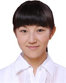 Ting Zhang (张婷)合作论文数School of Public Health, Southeast University12
 Hui-Chun Yan合作论文数8
 Jing Li (李菁)合作论文数8Feifei Lei合作论文数8
 Xiaoxing Kou (寇晓星)合作论文数Hospital of Stomatology, Sun Yat-sen University8
 Songtao Shi (施松涛)合作论文数Center for Stem Cell Biology and Tissue Engineering, Sun Yat-sen University8
 Ruili Yang (杨瑞莉)合作论文数Peking University School and Hospital of Stomatology8
 Chider Chen合作论文数Center for Craniofacial Molecular Biology, University of Southern California School of Dentistry;School of Dental Medicine, University of Pennsylvania7
 Tingting Yu (俞婷婷)合作论文数School of Basic Medical Sciences, Nanjing Medical University6Mingjin Zhang (张铭津)合作论文数5Yi Yu合作论文数5
 Danqing He (何丹青)合作论文数Peking University School of Stomatology5Li Qian合作论文数5
 Xing Wang (王兴)合作论文数College of Life Science and Technology, Beijing University of Chemical Technology5
 Yan Liu (刘艳)合作论文数Department of Pharmaceutics, School of Pharmaceutical Sciences, Peking University5
 Qiguo Rong (荣起国)合作论文数College of Engineering, Peking University5Liang Lyu合作论文数4
 Bochun Mao合作论文数4
 Boxi Yan合作论文数4
 Yiqiang Qiao (乔义强)合作论文数郑州大学第一临床学院4Wang Xue-Dong合作论文数4
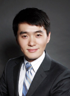 Xuedong Wang (王雪东)合作论文数Peking University Hospital of Stomatology4
 Wenjing Yu合作论文数3Yajing Tian合作论文数3Chuanlin Wang合作论文数3Liang Guo (郭亮)合作论文数3
 Yong Wang (王勇)合作论文数Hospital of Stomatology, Peking University3He Dan-Qing合作论文数3Yijiao Zhao合作论文数3
 Dan Luo (罗聃)合作论文数Beijing Institute of Nanoenergy and Nanosystems, Chinese Academy of Sciences3Y H Liu合作论文数2Yimei Zhang合作论文数2Karen Yotsumoto合作论文数2Aonan Wen合作论文数2Xiao Zhang合作论文数2
 Min Yu (余敏)合作论文数University of Chinese Academy of Sciences;Shenzhen Institutes of Advanced Technology, Chinese Academy of Science2
 Xueli Mao合作论文数2Yuki Nakao合作论文数2Yukari Watanabe合作论文数2Chikako Hayashi合作论文数2Hiroaki Yamato合作论文数2Shengjie Cui合作论文数2Takanori Shinjo合作论文数2Ci Zhang合作论文数2Runzhi Guo合作论文数2




