Objective:To summarize the indications, early functional outcome, complications and precautions of modular reverse shoulder tumor prosthesis in the treatment of proximal humerus tumor.Methods:From September 2018 to October 2019, there were 7 patients with proximal humeral tumor underwent tumor resection and modular reverse shoulder tumor prosthesis replacement in our hospital, including 6 males and 1 female. The average age of the patients was 32 years (14-59 years), including 3 cases of giant cell tumors, 2 cases of osteosarcomas, one case of myeloma and one case of renal cancer metastasis. The prosthesis reconstruction was performed after the removal of the proximal humerus tumor according to malawer type I. Postoperative outpatient follow-up included X-ray of shoulder joint, measurement of shoulder joint activity, functional evaluation using Constant-Murley function score and Musculoskeletal Tumor Society (MSTS) function score.Results:All the 7 patients successfully completed the operation, the operation times were ranged from 125 to 215 min, averaged 158 min; the intraoperative hemorrhage were ranged from 100 to 500 ml, the averaged 257 ml; the length of resected proximal humerus were ranged from 10 to 16 cm, averaged 12 cm. All patients were followed up for 4-17 months, with an average of 8 months. At the last follow-up, the average active abduction of shoulder was 108° (80°-175°), and the average active flexion was 124° (90°-175°). Three patients complained of mild discomfort in the shoulder, the rest of the patients had no pain in the shoulder. Sleep was not affected in all patients, and the hands of seven patients could go over the top of head, and they could live and work normally. In 7 patients, the rotation of shoulder joint was limited, the abduction force of shoulder joint was decreased, the Constant-Murley function score was 72% (59%-78%), and the MSTS function score was 84% (67%-93%). None of the 7 patients had incision infection, hematoma and other related complications. There was one dislocation occurred one month after the operation because the humeral adhesion of deltoid was resected. After open reduction, a larger glenoid ball was used and tension of deltoid muscle was tightened, and no dislocation was found.Conclusion:The combined tumor prosthesis of reverse shoulder has the advantages of simple installation, lower operative requirements compared with the common reverse shoulder prosthesis compounded with allograft, easy to control the length of the prosthesis, and also can achieve satisfactory results.
Objective To apply the hydroxyapatite/zirconium oxide( HA/ZrO2 ) gradient composite material by 3D printing technology, to analyze its ability to repair the defect of femoral shaft of beagle dogs. Methods Bone defect model was prepared in 6-month male beagle by truncating right leg femur after middle full-thickness 15 mm, which was put into the Micro CT scanned for volume, complete data collection, transformation and the post-processing. Then data were imported in CeraFab 7500 photocure 3D printer. Started to print program, formed a composite photosensitive resin in the early embryo, and the further defatted sintering and then used dip-coating HA/ZrO2 gradient composite materials according the parameters. The performance of the HA/ZrO2 gradient materials were taken scanning electron microscopy ( SEM) , X-ray diffraction analysis and biomechanical experiments. Prepared HA/ZrO2 gradient composite materials and cultured L929 mouse fibroblast cell lines, then MTT method was used to detect the HA/ZrO2 gradient composites cell toxicity in vitro. Sixteen dogs were divided into 4 groups, 4 dogs in each group. Group A:Dogs' 15 mm middle femur were intercepted with no biological material implanted, as the blank control group;group B, C, D were truncated middle femoral 15, 25 and 35 mm made of bone defect model, and transferred into the corresponding specifications of the HA/ZrO2 gradient composite materials. X-ray scan was taken to observe implanted biomaterials combined with own bone and surrounding callus growth after operation of 2, 4, 8, 12 weeks. Animals were executed and captured the entire length of the femur, then observed specimens implanted in biological materials and the surrounding bone growth conditions after 12 weeks. New bone mass were measured and reconstructed by Micro CT scans. The compressive experiments of femoral specimens were used to measure the ultimate compressive strength. Results The SEM showed that nano HA/ZrO2 gradient composite material made by 3D printing technology had smooth surface and even stable structure. Composite material had a metallurgical bonding and there was no obvious boundary in fracture surface. XRD analysis showed an obvious peak, better crystalline degree and better purity. Mechanical text showed that the ultimate compressive strength was ( 43. 37 ± 2. 31) MPa. MTT analysis proved that HA/ZrO2 gradient composite material had no cytotoxicity. Front and lateral X-ray examinations were taken at 2, 4, 8, 12 weeks after operation. In group A, bone nonunion was formed. In group B, continual bony callus were get through and there was no obvious boundary between artificial prosthesis and host bone. At 2, 4 and 8 weeks after operation, continuous callus and new bone growth in group C was slower than group B, but the gap between artificial prosthesis and broken ends was gradually filled with new bone, and the continual bony callus were got through at the week 12. In group D,new bone was formed in a lower speed, and appeared only around the broken ends. The specimens were taken at week 12 after operation, three-dimensional reconstruction of Micro CT showed that per unit volume new bone in group B, C, D were (238. 55 ± 19. 11) mm3/cm3, (223. 31 ± 13. 41) mm3/cm3 and (110. 83 ± 6. 48) mm3/cm3 , respectively. There were statistically significant differences among the three groups in statistics analysis (F=156. 824, P<0. 01). The new bone in group B and C were much more than group D. They were different from group D in statistics analysis (all P values<0. 05). Three-dimensional reconstruction showed that in group B, HA/ZrO2 gradient composite material was encapsulated and had no exposure; in group C, composite material had a little exposure and the surface was full of new bone; while in group D, composite material was revealable and little new bone in surface. Ultimate compressive test was showed in group B, C, D were(49. 72 ± 2. 33) MPa, (49. 81 ± 2. 43) MPa and (46. 92 ± 3. 61) MPa, respectively. There was no significant difference among group B, C and D in statistics analysis (F=1. 119, P>0. 05). Conclusions The nano HA/ZrO2 gradient composite material artificial prosthesis made by 3D printing technology has reliable biocompatibility and biomechanics that fit the individual treatment principle in clinic. As an ideal substitute for bone tissue, it could well repair femoral bone defect in 35 mm in dogs.
The poor 5-year survival rate in high-grade osteosarcoma (HOS) has not been increased significantly over the past 30 years. This work aimed to develop a radiomics nomogram for survival prediction at the time of diagnosis in HOS. In this retrospective study, an initial cohort of 102 HOS patients, diagnosed from January 2008 to March 2011, was used as the training cohort. Radiomics features were extracted from the pretreatment diagnostic computed tomography images. A radiomics signature was constructed with the lasso algorithm; then, a radiomics score was calculated to reflect survival probability by using the radiomics signature for each patient. A radiomics nomogram was developed by incorporating the radiomics score and clinical factors. A clinical model was constructed by using clinical factors only. The models were validated in an independent cohort comprising 48 patients diagnosed from April 2011 to April 2012. The performance of the nomogram was assessed with respect to its calibration, discrimination, and clinical usefulness. Kaplan-Meier survival analysis was performed. The radiomics nomogram showed better calibration and classification capacity than the clinical model with AUC 0.86 vs. 0.79 for the training cohort, and 0.84 vs. 0.73 for the validation cohort. Decision curve analysis demonstrated the clinical usefulness of the radiomics nomogram. A significant difference (p-value <.05; log-rank test) was observed between the survival curves of the nomogram-predicted survival and non-survival groups. The radiomics nomogram may assist clinicians in tailoring appropriate therapy.
Background Pelvic ring reconstruction after resection of pelvic malignancies or aggressive benign tumors remains challenging, especially when the tumor invades periacetabular bone, resulting in a Type II resection as classified by Enneking and Dunham (removal of part or all of the acetabulum). Although numerous treatment approaches are in use, none is clearly superior to the others. An alternative involving use of the ipsilateral proximal femur as an autograft has not been well characterized, so we present our preliminary experience with this approach. Questions/purposes (1) What were the oncologic outcomes after using an ipsilateral proximal femur autograft for reconstruction after Type II pelvic resection in a small series of patients who underwent this reconstructive approach? (2) What were the Musculoskeletal Tumor Society (MSTS) scores after this reconstruction? (3) What complications were observed? Methods Between October 2006 and May 2016, we treated 67 patients with Type II malignant or aggressive benign tumors of the ilium. Of those, we used an ipsilateral proximal femur and a prosthesis as a reconstruction method for 11 patients with pelvic tumors. In general, we performed this approach in young or middle-aged patients with primary malignant or aggressive benign tumors involving pelvic area II and in whom the tumor did not invade the hip. The method used for resection of pelvic tumors included osteotomy of the femoral shaft, harvesting the proximal femur as a graft. The length of the femoral graft was determined by the extent of the pelvic defect. The proper placement was selected after a comparison of the proximal femur and the pelvic defect. A curved reconstruction plate and cancellous bone screws were used for pelvic fixation. The operative duration and total blood loss were recorded. Of the 11 patients who underwent this approach, all but one had at least 2 years of followup unless death occurred earlier, and all but one have been seen within the last year for evaluation. Functional outcomes were assessed using the MSTS scoring system. Local recurrence, metastases, and deaths were recorded as were complications including infection, bone nonunion, mechanical failure and sciatic nerve palsy. Results The followup was a mean of 37 months (range, 13-96 months). One patient was lost to followup. Three patients died of disease owing to local recurrence or lung metastasis. The other seven patients lived without evidence of tumor. The main complications included mechanical failure in two patients, nonunion in one patient, infection in two patients, and sciatic nerve palsy in one patient. The median MSTS function score was 70% (21 of 30 points; range, 11-25 points). Conclusions Our preliminary results show that this technique of using the ipsilateral proximal femur may be an alternative method for reconstruction of pelvic bone defects after tumor resection. Even with this short followup, complications were common, but short-term function appears to be comparable to studies of other options. Longer term followup with more patients is necessary to confirm our results.
This study was performed to explore the clinical efficacy of bone transport using external fixation for treatment of large bone defects after tibial tumor resection in five patients. Bone transport started 14 days postoperatively at 1 mm/day and was adjusted according to the callus-to-diameter ratio. The bone transport time, bone graft fusion, relapse, and metastasis were recorded. Clinical efficacy was evaluated using the Musculoskeletal Tumor Society (MSTS) scoring system. The tumors included osteosarcoma (n=2), Ewing sarcoma (n=1), malignant schwannoma (n=1), and hemangioma (n=1). The average bone defect length after resection was 11.6 cm. The five patients were followed up for an average of 50.8 months, and the average bone transport time was 15.5 months. Three patients who underwent postoperative chemotherapy were followed for 22.7 months, and two who did not undergo chemotherapy were followed for 4.75 months. Four patients underwent iliac bone grafting, and one underwent vascular pedicle fibular transplantation. The average MSTS score was 21.2 (19.3 for patients who underwent chemotherapy and 24.0 for patients who did not). No relapse or metastasis was observed. Bone transport is effective for reconstruction of large bone defects after tibial tumor resection as well as tibial malignancies with high doses of chemotherapy.
Objective To prepare the gradient HA /ZrO2 engineering bone scaffold and evaluate its bone repair capability in the cervical fusion of rhesus macaque .Methods Bone morphogenetic protein 2 (BMP-2) in gelatin/chitosan slow-release hydrogel was prepared by multiple emulsion cross -linking, and its biological features were examined .Mesenchymal stem cells ( BMSCs) of rhesus macaque were isolated ,cultured and identified.The novel gradient HA /ZrO2 engineering bone scaffold was prepared for carrying BMP-2 and BMSCs, and its morphological characteristics were observed .The cervical intervertebral disc of experimental segments in 24 healthy male rhesus macaques were replaced with the novel gradient HA /ZrO2 engineering bone scaffold carrying a gelatin /chitosan gel slow-release hydrogel loading BMP -2 and 3rd generation of BMSCs (group A, n =8), or the empty novel gradient HA /ZrO2 engineering bone scaffold (group B, n =8), or autologous iliac graft ( group C, n =4) or nothing ( group D, n =4).The anteroposterior and lateral radiographs of the cervical spine were taken at once , postoperative 8 and 16 weeks.Histomorphometry was performed on samples at postoperative 8 and 16 weeks, and biomechanical testing was performed on harvested vertebral samples 16 weeks after operation.Results Electron microscopy for gelatin/chitosan gel slow-release hydrogel loading BMP-2 revealed a three-dimensional network structure with uniformly distributed chitosan micro -spheres, and its encapsulation efficiency and drug loading rate of BMP-2 were decreased with time, 87.4% ±0.9% and 58.2% ±0.5% on day 1st respectively, 45.2% ± 0.6% and 30.1% ±0.4% on day 15th respectively.The release rates of BMP-2 were 12.6% ±0.11% and 55% ±0.16% on day 1st and 15th respectively.BMSCs with diverse cell morphology were found under microscope.After osteogenic induction of the 3rd generation BMSCs, the results of alkaline phosphatase , von Kouza staining and its surface-specific antigen tests showed that it was consistent with the biological characteristics of BMSCs.According to radiography and histomorphological results , the newly formed bone volume in the interior of the pores in the group A was significantly higher than that in the group B at different time points after surgery.The results of biomechanical testing indicated that the maximal load , compression strength and energy to maximal load of the three groups were higher than those of normal group (all P values<0.05), and there was no significant difference between group A and group C in the compression strength (P >0.05).Conclusions The novel gradient HA /ZrO2 engineering bone scaffold carrying a gelatin /chitosan gel slow-release hydrogel loading BMP-2 and 3rd generation of BMSCs can effectively promote cervical intervertebral fusion, and it has similar effects as the autologous bone in cervical fusion of rhesus macaque according to radiography , histomorphological manifestation and biomechanical testing .
The multidisciplinary team(MDT) management of a patient with unresectable huge sacral bone giant cell tumor is reviewed here.The patient is a 24 years old male,who was admitted to hospital due to "constipation for 4 months,exacerbated for 1 week,accompanied with urination difficulty".Pelvic imaging examination showed a huge cystic-solid anterior vertebral mass below S3,with a size of about 16.1 cm × 11.5 cm × 15.4 cm.CT-guided biopsy of the mass reported sacral giant cell tumor.The clinical stage was Campanacci Ⅲ.Since the tumor was too large to resect,the patient received a series of vascular embolization by vascular surgery after MDT discussion.The patient received sacral tumor resection and internal fixation through anterior and posterior combined approach,after abdominal aortic balloon occlusion under the general anesthesia condition.The tumor was removed completely.One year after the resection,the patient received local radiotherapy for the sacral tumor area by D95 package planning target volume (PTV) 6 MV-X line (DT 45 Gy/24 F) conformal radiotherapy for 24 cycles.The patient was followed up for 26 months after the resection.His KPS score was increased from 60 points before the operation to 90 points,with normal urination function and bilateral lower limb activity,as well as significantly improved life quality.The diagnosis and treatment process of this case showed the significance of MDT for bone tumors,especially huge sacral tumors.In order to achieve the greatest benefit of patients,it is necessary to promote MDT in all related clinical practice and to include experts in more specialties in MDT.
Dense biomaterial plays an important role in bone replacement. However, it fails to induce bone cell migration into graft material. In the present study, a novel bone graft substitute (BGS) consisting of porous gradient hydroxyapatite/zirconia composite (PGHC) and gelatin/chitosan slow-release hydrogel containing bone morphogenetic protein 2 and bone mesenchymal stem cells was designed and prepared to repair lumbar vertebral defects. The morphological characteristics of the BGS evaluated by a scanning electron microscope showed that it had a three-dimensional network structure with uniformly distributed chitosan microspheres on the surfaces of the graft material and the interior of the pores. Then, BGS (Group A), PGHC (Group B), or autologous bone (Group C) was implanted into lumbar vertebral body defects in a total of 24 healthy rhesus monkeys. After 8 and 16 weeks, anteroposterior and lateral radiographs of the lumbar spine, microcomputed tomography, histomorphometry, biomechanical testing, and biochemical testing for bone matrix markers, including Type I collagen, osteocalcin, osteopontin, basic fibroblast growth factor, alkaline phosphatase, and vascular endothelial growth factor, were performed to examine the reparative efficacy of the BGS and PGHC. The BGS displayed excellent ability to repair the lumbar vertebral defect in rhesus monkeys. Radiography, microcomputed tomography scanning, and histomorphological characterization showed that the newly formed bone volume in the interior of the pores in the BGS was significantly higher than in the PGHC. The results of biomechanical testing indicated that the vertebral body compression strength of the PGHC implant was lower than the other implants. Reverse-transcription polymerase chain reaction and western blot analyses showed that the expression of bone-related proteins in the BGS implant was significantly higher than in the PGHC implant. The BGS displayed reparative effects similar to autologous bone. Therefore, BGS use in vertebral bone defect repair appears promising.
Objective:To establish the human-induced pluripotent stem cells (hiPSCs) system originated from patients with human leukocyte antigen(HLA)-B2704 genotypic ankylosing spondylitis (AS).Methods:Four exogenous transcription factors including OCT4,SOX2,c-MYC and KLF4 were introduced into the urinary cells(UCs) of patients with HLA-B2704 genotypic AS by using retrovirus-mediated infection system,and the infected UCs were reprogrammed for obtaining hiPSCs.The hiPSCs were identified through morphologic observation,alkaline phosphatase staining,immunofluorescence staining,endogenous multipotent related gene detection,embryoid bodies (EBs) differentiation detection,EBs triploblastic gene detection and teratoma formation experiment.Results:The nuclear-cytoplasmic ratio increased at the 4th and 5th day after UCs were infected by virus.The clone appeared at the 14th day and it was similar to human embryonic stem cells(hESCs)at the 24th day.The result of alkaline phosphatase staining showed that both hiPSCs and hESCs-H1 were presented with prunosus positive staining.The result of immunofluorescent staining showed that the stage specific embryonic antigen 4 (SSEA4) of hESCs and the transcription factor of ESCs including OCT4 and SOX2 presented with positive staining.There was no statistical difference in the expression of endogenous multipotent related gene including OCT4,SOX2,NANOG and REX1 between hiPSCs and hiPSCs-H1 (1.006 +/-0.208 vs 1.000 +/-0.340,t =0.038,P =0.972;1.061 +/-0.140 vs 1.000 +/-0.387,t =0.431,P =0.689;1.386 +/-0.354,1.000 +/-0.032,t =1.467,P =0.303;1.280 +/-0.283 vs 1.000 +/-0.013,t =1.398,P =0.235).High expressions of endodermal genes including AFP and GATA4,mesodermal genes including MEX1 and TBX1 and ectodermal genes including PAX6 and SOX1 were found in EBs.The relative expression levels of triploblastic genes including AFP,GATA4,TBX1,MSX1,PAX6 and SOX1 were higher in EBs compared to hiPSCs (60.695 +/-8.746 vs 1.000 +/-0.245,t =9.647,P =0.011;3.724 +/-0.144 vs 1.000 +/-0.417,t =12.136,P =0.007;4.224 +/-0.869 vs 1.000 +/-0.130,t =4.988,P =0.038;684.800 +/-63.326 vs 1.000 +/-0.211,t =15.270,P =0.004;131.561 +/-15.785 vs 1.000 +/-0.232,t =11.700,P =0.007;98.507 +/-40.443 vs 1.000 +/-0.215,t =16.000,P =0.004).Teratoma appeared in hiPSC.Small intestine epithelial histocytes,muscular histocytes and neuroepithelial histocytes were found respectively in entoderm,mesoderm and ectoderm of teratoma through HE staining.Conclusion:The hiPSCs system originated from patients with HLA-B2704 genotypic AS can be successfully established through introducing four exogenous transcription factors including OCT4,SOX2,c-MYC and KLF4 into UCs of patients with HLA-B2704 genotypic AS by using retrovirus-mediated infection system and reprogramming the UCs.
Objective To study the specificity of the Notch signal pathway blockers-γ-secretase inhibitors (DAPT) on the efficiency of the hair follicle stem cell (rHFSCs) differentiation into vascular endothelial cells (ECs).Methods Applying the modified method of rHFSCs separation, culture, purification, identification for seed cells, and using transmission electron microscope to observe the internal structure.With 10 μg/Lvascular endothelial growth factor 165 (VEGF165) as inducing factor to carry on the inductio, and flow cytometry instrument to detect differentiation efficiency.Then divided them into induction and inhibition group.With different concentrations of DAPT (0.5, 1.0 μmol/L) to suppress, Two group of cells are both handled for 1 week, with the method of real-time fluorescent quantitative polymerase chain reaction (FQ-PCR) and Western blotting, respectively detecting the expression of mRNA and protein CD31, vascular endothelial cadherin (VE-cadherin) in the two groups, then conbined with Dil-labeled acetylated low density lipoprotein (Dil-ac-LDL) phagocytosis test and Lumen formation experiment test, finally comparing the result of two group.To comprehensive evaluate DAPT on the influence of rHFSCs differentiation ECs after it inhibited Notch signaling pathway.Results The rHFSCs with modified of isolation, culture, purification has strong cloning ability and vitality.Flow cytometry instrument testing shows β1, α6 integrin and CK15 with a high expression, respectively, 98.9%, 97.9%, 68.1% and 98.5%;and Low weakly expressed CD31, VE-cadherin, respectively, 13.6% and 17.9%, Transmission electron microscopy (sem) show that cells in original condition.Flow cytometry instrument testing shows CD31, VE-cadherin with high expression afer induced Cells for a week, respectively, 61.5% and 95.9%.After inhibition of Notch signaling pathway, according to the results of FQ-PCR: there is no obvious difference of variation of mRNA of CD31 (P=0.136) and VE-cadherin (P=0.003) between two groups, the protein expression of CD31 and VE-cadherin significantly reduced through Western blotting method (P=0.000), Dil-ac-LDL phagocytosis and lumen formation ability in vitro decreased significantly.Conclusion After interdicting the Nntch signal pathway, although DAPT has no obvious influence on the mRNA level of CD31 and VE-cadherin, but can inhibit protein expression of CD31 and VE-cadherin and ability of phagocytosis and lumen formation in vitro, thereby reducing efficiency of rHFSCs differentiation for ECs.It lays a experimental basis on further research of the mechanism of Notch signaling pathway in inducing vascular endothelial cells.
患者女性,53岁.因左髋部持续性隐痛2年就诊于浙江大学附属第二医院,无明显骨折外伤史.既往有慢性阑尾炎病史.体格检查:左髋部皮下可见软组织肿块,质软,皮温正常,四肢关节活动无明显受限,肌力正常,末梢血循环良好.患者入院后伴有发热,体温最高39℃,查血常规全血细胞降低,凝血酶原时间、血清碱性磷酸酶、CRP比正常值稍偏高,血清蛋白电泳β球蛋白与γ球蛋白正常,白蛋白/球蛋白比稍低于正常值.予重组人粒细胞刺激因子治疗后患者发热好转.
Within the vascular endothelial growth factor (VEGF) family of five subtypes, VEGF165 secreted by endothelial cells has been identified to be the most active and widely distributed factor that plays a vital role in courses of angiogenesis, vascularization and mesenchymal cell differentiation. Hair follicle stem cells (HFSCs) can be harvested from the bulge region of the outer root sheath of the hair follicle and are adult stem cells that have multi-directional differentiation potential. Although the research on differentiation of stem cells (such as fat stem cells and bone marrow mesenchymal stem cells) to the endothelial cells has been extensive, but the various mechanisms and functional forms are unclear. In particular, study on HFSCs' directional differentiation into vascular endothelial cells using VEGF165 has not been reported. In this study, VEGF165 was used as induction factor to induce the differentiation from HFSCs into vascular endothelial cells, and the results showed that Notch signalling pathway might affect the differentiation efficiency of vascular endothelial cells. In addition, the in vivo transplantation experiment provided that HFSCs could promote angiogenesis, and the main function is to accelerate host-derived neovascularization. Therefore, HFSCs could be considered as an ideal cell source for vascular tissue engineering and cell transplantation in the treatment of ischaemic diseases.
Objective To explore the short?term functional outcomes of the reconstruction of the proximal humerus by re?verse shoulder arthroplasty after tumor rescetion. Methods 8 patients who underwent reverse shoulder arthroplasty after tumor resection between January 2013 and December 2014. 5 were female and 3 were male, mean aged was 38 years old (25-61). 2 chon?drosarcomas and 6 giant cell tumors. Enneking stageing of 2 cases with chondrosarcoma were stageⅠB and stageⅡB. 6 giant cell tumors were Campanacci stage 3, meanwhile 3 cases had pathological fractures. The deltoid and axillary nerve were intact in all patients by image analysis before the operation. The proximal humerus was resected according to Malawer typeⅠresection. Then reverse shoulder arthroplasty reconstruction and bone graft was performed. The follow?up was scheduled, and the patient received X?ray examination of the shoulder. The range of motion of the shoulder was measured, the Constant?Murley score and musculoskel?etal tumor society(MSTS) score was recorded. Results The mean duration of the operation was 2.7 h (2-3.5 h). The bleeding in the operation was 510 ml (300-850 ml). The mean length of humerus resection was 8 cm (6-10 cm). The allografts were used in 7 cases and reimplantation after tumor bone deactivation was used in one. The latissimus dorsi transfer were performed in 2 cases. The rotator cuff were resected 1-1.5 cm from the great and lesser tubercles. The follow?up was 13 months (3-26 months). No infec?tion, dislocation, or loosening of prosthesis was found by the last follow?up. The X?ray showed the case who received reimplanta?tion after tumor bone deactivation had achieved bone union 1 year postoperation,7 cases received allograft had still nonunion at the host?graft junction. Bone resorption were found in all cases in different extent but the prosthesis were stable. No local recur?rence of the tumor was found. At last follow?up, active abduction was 155° (100°-175° ) and active forward elevation was 150° (115°-170°) and Constant?Murley score was 76%(68%-87%). The MSTS score was 92%(87%-97%). Conclusion The func?tional outcomes of the reconstruction of the proximal humerus by reverse shoulder arthroplasty after tumor rescetion was satisfied in early period. The reverse shoulder arthroplasty can be used in younger patient, but long?term results need further study.
Objective To prepare the chitosan hydrogel as release carrier of recombinant human bone morphogenetic protein 2 (rhBMP-2), and loaded the prepared recombinant human bone morphogenetic protein 2 chitosan hydrogel in novel HA/ZrO2 gradient biocomposite artificial vertebrae and study bone reparation effect of it in a beagle dog' vertebral defect model. Methods To prepare rhBMP-2 chitosan hydrogel by ion-crosslinked method. Scanning electron microscopy was used to observe lyophilized chitosan microscopic surface morphology, test its swelling coefficient and detect drug loading mass, encapsulation efficiency and sustained release characteristics of the chitosan hydrogel loaded with rhBMP-2. Loaded the rhBMP-2 chitosan hydrogel onto novel HA/ZrO2 gradient biocomposite artificial vertebrae. Then 12 beagle dogs were randomly divided into three groups ( A, B, and C) by number table, each group included four dogs. Afterwards, a 23 mm radius and 9 mm high semi-cylindrical bone defect model was caused by surgery in each dog. While the bone defect location was implanted artificial vertebral body composite rhBMP-2 chitosan gel in group A, implanted artificial vertebral body composite blank dried chitosan in group B, and implanted autologous bone in group C. After operations, general observation and X-ray observation were carried out at week 6,12, and 24. At week 24, all beagle dogs were sacrificed and taken vertebral body specimens, then detect new bone volume in the artificial vertebral body by Micro-CT and detect ultimate compressive strength by biomechanical testing. Results SEM scanning showed the lyophilized chitosan was a three-dimensional network structure with uniform distribution of chitosan microspheres internal. When loaded with rhBMP-2, chitosan hydrogel encapsulation efficiency was 91. 88% ± 1. 53%, the loaded mass of drug was (39. 84 ± 2. 34) ng/mg;the release rate was 28. 32% ± 3. 01% at day 1, 48. 92% ± 6. 27% at day 3, and 74. 40% ± 6. 29% at day 12. The average recovery ratio of group C was faster than group A and group B at 6 weeks postoperatively(all P values<0. 05), and there was no statistically signifficant difference between group A and group B(P>0. 05). 12 and 24 weeks postoperatively, there was satistically significant difference between group B and group C(all P values<0. 05), and there was no statistically signifficant difference between group A and group C(P>0. 05). X-ray imaging observation showed: in group A, with the passage of time after vertebral replacement surgery, callus formation gradually increased around the material, the gap between artificial vertebrae and autogenous bone was gradually filled with new bone. Since the artificial vertebrae and autologous bone had almost closely banded at week 24 with no obvious boundary;in group C, there were apparently self-absorption and bone osteolysis on non-load bearing areas at week 12 and 24, and appeared rapidly autologous bone remodeling; in group B, however, the repair to bone defect was slower than that of group A and group C. The specimens were taken at week 12. And in vitro Micro CT observation showed that three dimensional CT reconstruction could be seen a large number of new bone formation inside the artificial vertebral, and bone volume detection result at week 24 were (145. 38 ± 18. 52) mm3 in group A and (86. 30 ± 15. 60) mm3 in group B, there was significant difference between the two groups (t=4. 879, P<0. 01). Ultimate compressive test showed the maximum compressive strengths of vertebras were (14. 03 ± 1. 67) MPa in group A; (8. 62 ± 1. 24) MPa in group B, and (13. 78 ± 1. 43) MPa in group C. The group A and group C were significantly higher than that of group B in statistics analysis (all P values<0. 01). There was no statistically significant difference between group A and group B(P>0. 05). Conclusions The novel HA/ZrO2 gradient biocomposite artificial vertebrae loaded with rhBMP-2 can excellently repair vertebral defect, it may be an ideal bone substituted material for clinical application.
Background: Transfer of a flexor hallucis longus (FHL) tendon can not only reconstruct the Achilles tendon but also provide ischemic tendinous tissues with a rich blood supply to enhance wound healing. This retrospective study aims to investigate clinical outcomes in patients who underwent repair of Kuwada grade IV chronic Achilles tendon rupture with long hallucis longus tendons harvested using a minimally invasive technique. Materials and Methods: 35 patients who were treated for Kuwada grade IV Achilles tendon injuries from July 2006 to June 2011 were included in this retrospective study. The age ranged between 23 and 71 years. The duration from primary injury to surgery ranged from 29 days to 34 months (mean value, 137.6 days). All 35 patients had difficulties in lifting their calves. Thirty two were followed up for a mean 32.2 months (range 18–72 months), whereas three were lost to followup. Magnetic resonance imaging (MRI) showed that the tendon rupture gap ranged from 6.0 to 9.2 cm. During surgery, a 2.0 cm minor incision was made vertically in the medial plantar side of the midfoot, and a 1.5 cm minor transverse incision was made in the plantar side of the interphalangeal articulation of the great toe to harvest the FHL tendon, and the tendon was fixed to the calcaneus with suture anchors. Postoperative appearance and function were evaluated by physiotherapists based American Orthopedic Foot and Ankle Society-ankle and hindfoot score (AOFAS-AH), and Leppilahti Achilles tendon ratings. Results: Results were assessed in 32 patients. Except for one patient who suffered complications because of wound disruption 10 days after the operation, all other patients had primary wound healing, with 28 of 32 able to go up on their toes at last followup. The AOFAS-AH score was increased from preoperative (51.92 ± 7.08) points to (92.56 ± 6.71) points; Leppilahti Achilles tendon score was increased from preoperative (72.56 ± 7.43) to (92.58 ± 5.1). There were statistically significant differences. The result of the total excellent and good rate was 93.8% (30/32). MRI of Achilles tendon showed even signal without evidence of tear or cystic degeneration. Conclusion: Reconstruction of a chronic Achilles tendon rupture with an FHL tendon harvested using a minimally invasive technique showed good outcomes.
目的 建立简单、可靠的毛囊干细胞体外定向分化为血管内皮细胞的方法.方法 无菌条件下切取1周龄SD大鼠触须部皮肤,Dispase酶和Ⅳ型胶原酶混合液消化,显微镜下分离毛囊隆突部,改良的组织块贴壁法培养rHFSCs,差速贴壁法纯化,流式细胞仪检测及细胞免疫荧光染色联合鉴定.用含10 ng/ml和20 ng/ml VEGF165作为主要诱导因子将细胞分为两种浓度组别,不传代的情况下分别在1、2、3周内观察诱导后细胞形态;通过流式细胞和细胞免疫荧光染色检测分化效率;选取最佳诱导因子浓度和工作时间,对诱导后的细胞进行体外官腔形成实验,并在透射电镜下观察W-P小体.流式细胞术检测分化效率实验数据的比较采用单因素方差分析及独立t检验.结果 分离、培养、纯化的rHFSCs克隆能力好,活力强,生长曲线呈S型.流式细胞及免疫荧光染色联合检测β1(98.9%)、α6整合素(97.9%),CK15 (68.1%)、P63 (98.5%)呈高表达,阴性表达CD31 (13.6%)、VE-cadherin(17.9%).在诱导液的作用下,两组细胞从1周到3周后,均由扁平铺路石样改变到完全被长梭形样细胞占据.流式细胞及免疫荧光染色检测CD31和VE-cadherin显示,CD31在诱导1周时表达最强,两种诱导因子比较无差异[(65.27±0.57%)vs (66.13±0.60)%,t=-1.812,P=0.145].而VE-cadherin在诱导1周时表达也最强,10 ng/ml要优于20 ng/ml VEGF 165的诱导作用[(95.57±0.85)%vs(78.10±1.25)%,t=19.977,P=0.001].10 ng/ml VEGF165诱导1周后的细胞体外管腔形成实验阳性,在透射电镜下可以观察到内皮细胞特有结构W-P小体.结论 毛囊干细胞体外在10ng/ml VEGF165的诱导下,l周后可成功得到性能良好的血管内皮细胞.可为组织工程血管、细胞移植治疗缺血性疾病等提供理想的种子细胞.
The clinical efficiency of bone transport distraction osteogenesis in the reconstruction of large tibial defects following resection of osteosarcoma remains unclear. The current study presents two cases of large tibial defects treated with bone transport distraction using an Orthofix external fixator. Case 1 was a 29-year-old man with a tibial defect 11 cm in length, while case 2 was a 16-year-old girl with a 15-cm-long defect. Bone transport distraction osteogenesis was initiated for the both cases on day 14 following resection of the tibial osteosarcoma. Bone transport distraction in case 1 and 2 was continued for 16 and 28 months, respectively, and the patients were followed up for 51 and 56 months, respectively. The two patients did not exhibit any signs of tumor recurrence or tumor metastasis during the follow-up period. The Musculoskeletal Tumor Society functional scores at final follow-up visits were 22 and 18 for case 1 and 2, respectively. Based on the experience gained in these 2 cases, a bone transport is a viable option for the reconstruction of large tibial defects following osteosarcoma resection.
Ethnopharmacological relevance: Acupuncture is a key part of traditional Chinese medicine, shown to induce favorable neuroplasticity for injuries in the central and peripheral nervous systems. Recent studies report elongated needle therapy (ENT) with BL54 and ST28 may restore acute spinal cord injury (ASCI). However, the precise mechanism for this has not been elucidated.Aim of the study: In our current study, we investigated the effects of ENT on inflammation and neuronal apoptosis induced by ASCI, and whether PI3K/Akt and MAPK/ERK signaling pathways are involved in the ENT restoration effect.Materials and methods: Rat models of moderate SCI were established in accordance with the modified Allen's method and were treated with ENT continuously for 7 days. Spontaneous activities were evaluated by the Basso Beattie and Bresnahan locomotor scale. Levels of inflammatory cytokines, such as tumor necrosis factor alpha, interleukin-6, IL-1 beta, and nuclear factor kappa-beta, were determined by enzyme-linked immunosorbent assay. Cell apoptosis was examined by the terminal deoxynucleotidyl transferase dUTP nick end labeling (TUNEL) assay. The proportions of cells with positive Bcl-2 and Bax expression were determined by immunohistochemical assays, whilst the expression profiles of p-ART and p-ERR in spinal cord tissues were evaluated by western blotting. Furthermore, the expression profiles of Cytochrome-C (Cyt-C) and caspase-3 in vivo were analyzed by reverse transcription polymerase chain reaction. The potential inhibitory effects downstream of the Akt and ERIC signaling pathways were examined by administration of specific inhibitors LY294002 and PD98059 in vivo.Results: As indicated by this study, inflammation as well as PI3K/Akt- and MAPK/ERK signaling pathway mediated neuronal apoptosis were involved in the course of SCI in rats. The neuro-protective effect of ENT was associated with reduced Bax protein-positive neurons and increased Bcl-2 protein-positive neurons. ENT enhanced recovery of rat activities. Activation of p-Akt and p-ERIC in the PI3K/Akt and MAPK/ERK signaling pathways, inhibited expression of the critical component Cyt-C. Cyt-C is required for the mitochondrial apoptosis pathway and cascade of caspase-3, which is involved in activation of neuronal apoptosis through down-regulation of Bax protein and up-regulation of Bcl-2, as determined by TUNEL. The administration of PI3R/Akt and MAPK/ERK signaling pathway specific inhibitors, LY294002 and PD98059, suppressed expression of both p-Akt and p-ERK.Conclusion: ENT with BL54 and ST28 points can promote the recovery of ASCI. And the neuro-protective effect of ENT during the restoration of SCI may be associated with the suppression of both inflammation and activation of PI3K/Akt and MAPK/ERK signaling pathways, resulting from down-regulation of Bax protein, up-regulation of Bcl-2, and inhibition of the mitochondrial apoptosis pathway. (C) 2016 Published by Elsevier Ireland Ltd.
A new HA/ZrO2-based porous bioceramic artificial vertebral body (AVB), carried a recombinant human bone morphogenetic protein-2 (rhBMP-2)/chitosan slow-release hydrogel was prepared to repair vertebral bone defect in beagles. An ionic cross-linking was used to prepare the chitosan hydrogel (CS gel) as the rhBMP-2 slow-release carrier. The vertebral body defects were implanted with the rhBMP-2-loaded AVB in group A, or a non-drug-loaded AVB in group B, or autologous iliac in group C. The encapsulation rate of rhBMP-2 in rhBMP-2-loaded CS gel was 91.88±1.53%, with a drug load of 39.84±2.34 ng/mg. At 6, 12, 24 weeks postoperatively, radiography showed that the bone calluses gradually increased with time in group A, where the artificial vertebral body had completely fused with host-bone at 24 weeks after surgery. In group C, an apparent bone remodeling was occurred in the early stages, and the graft-bone and host-bone had also fused completely at 24 weeks postoperatively. In group B, fusion occurred less than in groups A and C. At 24 weeks after surgery, micro-computed tomography (Micro-CT) revealed that the volume of newly-formed bone in group A was significantly more than in group B (p<0.05). At 24 weeks after surgery, ultra-compressive strengths of the operated segments were 14.03±1.66 MPa in group A, 8.62±1.24 MPa in group B, and 13.78±1.43 MPa in group C. Groups A and C were both significantly higher than group B (p < 0.05). At 24 weeks postoperatively, the hard tissue sections showed that the AVB of group A had tightly fused with host bone, and that pores of the AVB had been filled with abundant nearly mature bone, and that the new bone structured similarly to a trabecular framework, which was similar to that in group C. In contrast, implant fusion of the AVB in group B was not as apparent as group A. In conclusion, the novel HA/ZrO2-based porous bioceramic AVB carried the rhBMP-2-loaded CS gel can promote the repair of bony defect, and induce bone tissue to grow into the pores, which may replace iliac bone grafts as commonly applied in clinical practice.
Objective To evaluate the effects of porous gradient composites with hydroxyapatite/zirconia and autologous iliac in repair of lumbar vertebra body defects in dogs. Methods (1) New porous gradient hydroxyapatite/zirconia composites were prepared using foam immersion, gradient compound and high temperature sintering; (2) A total of 18 adult beagle dogs, aged five to eight months and weighted 10–13 kg, were randomly assigned into two subgroups, which were implanted with new porous gradient hydroxyapatite/zirconia composites (subgroup A in 12) or autologous iliac bone (subgroup B in 6); (3) The post-operative data were analyzed and compared between the subgroups to repair the vertebral body defect by roentgenoscopy, morphology and biomechanics. Results The porosity of new porous gradient hydroxyapatite/zirconia composites is at 25 poles per inch, and the size of pores is at between 150 and 300 µm. The post-operative roentgenoscopy displayed that new-bone formation is increased gradually, and the interface between composites and host-bone becomes became blur, and the new-bone around the composites were integrated into host-bone at 24 weeks postoperatively in subgroup A. As to subgroup B, the resorption and restructure were found at six weeks after the surgery, and the graft-bone and host-bone have been integrated completely without obvious boundary at 24 weeks postoperatively. Histomorphologic study showed that the amount of bone within pores of the porous gradient hydroxyapatite/zirconia composites increased continuously with a prolonged implantation time, and that partial composites were degradated and replaced by new-bone trabeculae. There was no significant difference between subgroups ( P > 0.05) in the ultimate compressive strengths. Conclusion New porous gradient hydroxyapatite/zirconia composites can promote the repair of bony defect, and induce bone tissue to ingrow into the pores, which may be applied widely to the treatment of bony defect in the future.
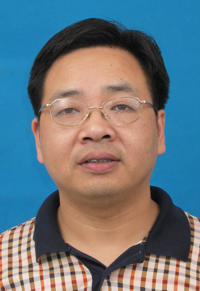 Zhaoming Ye (叶招明)合作论文数Department of Orthopaedic Surgery, the Second Affilited Hospital, College of Medical Science, Zhejiang University94
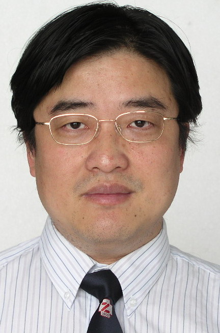 Huimin Tao (陶惠民)合作论文数The Second Affiliated Hospital, Zhejiang University School of Medicine70
 Weixu Li合作论文数48
 Renfu Quan (全仁夫)合作论文数Zhejiang Chinese Medicine University30Nong Lin合作论文数28Xudong Miao (苗旭东)合作论文数23ZhengMing Yang (杨正明)合作论文数19
 Shunwu Fan (范顺武)合作论文数Sir Run Run Shaw Hospital, Zhejiang University School of Medicine18Rongxue Shao合作论文数14
 Weiqi Yan (严伟琪)合作论文数The Second Affiliated Hospital, Zhejiang University School of Medicine12Weibo Pan合作论文数11
 Jie Feng (冯杰)合作论文数College of Animal Sciences, Zhejiang University11Wei Li (李伟)合作论文数10Jinwei Xu合作论文数9
 Xiaochun Wu (吴晓春)合作论文数School of Materials Science and Engineering, Shanghai University9Xiaobo Yan合作论文数8ZhongMing Huang (黄忠名)合作论文数8Meng Liu (柳萌)合作论文数8Zhang Liang (张亮)合作论文数8
 Wenjian Weng (翁文剑)合作论文数School of Materials Science and Engineering, Zhejiang University8Binggang Guan (官丙刚)合作论文数8
 Xicheng Wei (韦习成)合作论文数School of Materials Science and Engineering, Shanghai University8
 Shigui Yan (严世贵)合作论文数The Second Affiliated Hospital, School of Medicine, Zhejiang University8Wei ZHUANG (庄伟)合作论文数7WeiBin Du (杜伟斌)合作论文数7Guo-Ping Cao合作论文数6ShangJu Xie (谢尚举)合作论文数6
 Xin Huang (黄昕)合作论文数The Second Affiliated Hospital, School of Medicine, Zhejiang University6Xie Xian-Kuan (解先宽)合作论文数6Yanghua Tang (汤样华)合作论文数6Jinhong Miao合作论文数5Xuan Zheng (郑宣)合作论文数5ShiChao Xu (许世超)合作论文数5YongPing Wu (吴永平)合作论文数5
 Rongzhen Xu (徐荣臻)合作论文数Zhejiang University Cancer Center;Department of Hematology, The Second Affiliated Hospital, Zhejiang University School of Medicine;Hematology Institute, Zhejiang University5Zhongli Shi合作论文数5GaoShun Li (李高舜)合作论文数4
 Xiang Gao (高翔)合作论文数School of Physics & Information Technology, Shaanxi Normal University4
 Zongbao Yang (杨宗保)合作论文数School of Medicine, Xiamen University4Hongbin Wang合作论文数4Qiang Li (李强)合作论文数3Chen Rongliang合作论文数3Libin Jin合作论文数3Meng-Xuan Du合作论文数3RongKun Lv (吕荣坤)合作论文数3XianKuan Jie (解先宽)合作论文数3YiJin Wang (王以进)合作论文数3JianZhong Xu (许建中)合作论文数3




