BACKGROUND:G protein-coupled receptors (GPCRs) are the largest class of membrane-bound receptors and are emerging as targets for the effective treatment of cancer. The role of orphan GPCR GPR52 in cancer has not been characterized. Low mRNA expression of GPR52 in breast tumours correlates with reduced overall survival, leading to the hypothesis that loss of GPR52 supports breast cancer progression. METHODS:CRISPR-Cas9 was used to knock out GPR52 in the human triple-negative breast cancer cell lines MDA-MB-468 and MDA-MB-231. 2D and 3D in vitro studies, electron microscopy, and a zebrafish xenograft model were used to assess the morphology and behaviour of GPR52 KO cells. RESULTS:Loss of GPR52 was associated with elevated levels of cAMP, increased cell-cell interaction in 2D cultures, more spindle-like morphology on collagen, altered 3D spheroid morphology, and increased propensity to organize and invade collectively. Zebrafish injected with GPR52 KO cells developed a greater total cancer area than control. RNA sequencing and proteomic analyses of GPR52-null cells revealed an increased cAMP signalling signature. Re-expression of GPR52 and inhibition of cAMP production rescued some GPR52 KO phenotypes. CONCLUSIONS:GPR52 loss is a potential mechanism by which breast cancer progression may occur and supports the investigation of GPR52 agonism as a therapeutic option for breast cancer. STATEMENT OF SIGNIFICANCE:Loss of the orphan GPCR GPR52 in human breast cell lines leads to increased cell clustering, hybrid/partial EMT, and increased tumour burden in zebrafish, further expanding our understanding of mechanisms driving cancer progression and opening the door to novel therapeutic approaches.
Bromodomain and extraterminal domain (BET) family proteins are ubiquitous transcriptional co-activators that function broadly in cellular differentiation, proliferation, and stress responses. Pharmacological inhibition of BET proteins with small molecules that disrupt bromodomain engagement with acetyllysine residues (such as JQ1) or drive their degradation through the ubiquitin–proteasome system (such as dBET6) ameliorates pathological gene expression in a range of systems and shows promise as a potential therapeutic strategy. Understanding the cell-type and signaling pathway requirements that dictate BET dependence in a particular cellular context remains incomplete. We previously demonstrated that, in neonatal rat cardiomyocytes, GPCR-induced hypertrophy responses depended strongly on the BET protein Brd4 when signaling was coupled to Gαs, but not Gαq. Here, we tested whether Brd4 was differentially responsive to G protein isoforms in HEK 293 cells by expressing Gαs- or Gαq-coupled Designer Receptors Exclusively Activated by Designer Drugs (DREADDs). Gαq induced the expression of a group of early response genes and inflammatory genes in a manner largely insensitive to pharmacological BET inhibition, consistent with our previous data in cardiomyocytes. Gαs activated a small subset of the Gαq-induced genes, but this effect was largely reversed by dBET6. Our data further suggest that there may be general signaling requirements to activate Brd4 across cell types.
Statement of Significance:We showed that loss of the orphan G protein-coupled receptor GPR52 in human breast cell lines leads to increased cell clustering, hybrid/partial EMT, and increased tumor burden in zebrafish. Background:G protein-coupled receptors (GPCRs) are the largest class of membrane-bound receptors that transmit critical signals from extracellular to intracellular spaces. Transcriptomic data of resected breast tumors show that low mRNA expression of orphan GPCR GPR52 correlates with reduced overall survival in patients with breast cancer, leading to the hypothesis that loss of GPR52 supports breast cancer progression. Methods:CRISPR-Cas9 was used to knockout GPR52 in the human triple-negative breast cancer (TNBC) cell lines MDA-MB-468 and MDA-MB-231, and in the non-cancerous breast epithelial cell line MCF10A. 2D and 3D in vitro studies, electron microscopy, Matrigel culture, and a zebrafish xenograft model were used to assess the morphology and behavior of GPR52 KO cells. RNA-sequencing and proteomic analyses were also conducted on these cell lines, and transcriptomic data from The Cancer Genome Atlas (TCGA) database were used to compare GPR52-null and wild-type (WT) signatures in breast cancer. Results:Loss of GPR52 was found to be associated with increased cell-cell interaction in 2D cultures, altered 3D spheroid morphology, and increased propensity to organize and invade collectively in Matrigel. Furthermore, GPR52 loss was associated with features of EMT in MDA-MB-468 cells, and zebrafish injected with GPR52 KO cells developed a greater total cancer area than those injected with control cells. RNA sequencing and proteomic analyses of GPR52-null breast cancer cells revealed an increased cAMP signaling signature. Consistently, we found that treatment of wild-type (WT) cells with forskolin, which stimulates the production of cAMP, induces phenotypic changes associated with GPR52 loss, and inhibition of cAMP production rescued some GPR52 KO phenotypes. Conclusion:GPR52 is an orphan GPCR and its role in cancer progression has not been previously characterized. We found that GPR52 loss in breast cancer cells can lead to increased cell clustering, collective invasion, and EMT in vitro . These are features of increased cancer aggression. Our results reveal that GPR52 loss is a potential mechanism by which breast cancer progression may occur and support the investigation of GPR52 agonism as a therapeutic option for breast cancer. Abstract Figure:
Despite the observation of synergistic interactions between the urotensinergic and angiotensinergic systems, the interplay between the urotensin II receptor (hUT) and the angiotensin II type 1 receptor (hAT1R) in regulating cellular signaling remains incompletely understood. Notably, the putative interaction between hUT and hAT1R could engender reciprocal allosteric modulation of their signaling signatures, defining a unique role for these complexes in cardiovascular physiology and pathophysiology. Using a combination of co-immunoprecipitation, bioluminescence resonance energy transfer (BRET) and FlAsH BRET-based conformational biosensors, we first demonstrated the physical interaction between hUT and hAT1R. Next, to analyze how this functional interaction regulated proximal and distal hUT- and hAT1R-associated signaling pathways, we used BRET-based signaling biosensors and western blots to profile pathway-specific signaling in HEK 293 cells expressing hUT, hAT1R or both. We observed that hUT-hAT1R heterodimers triggered distinct signaling outcomes compared to their respective parent receptors alone. Notably, co-transfection of hUT and hAT1R has no impact on hUII-induced Gq activation but significantly reduced the potency and efficacy of Ang II to mediate Gq activation. Interestingly, URP, the second hUT endogenous ligand, produce a distinct signaling signature compared to hUII at hUT-hAT1R. Our results therefore suggest that assembly of hUT with hAT1R might be important for allosteric modulation of outcomes associated with specific hardwired signaling complexes in healthy and disease states. Altogether, our work, which potentially explains the interplay observed in native cells and tissues, validates such complexes as potential targets to promote the design of compounds that can modulate heterodimer function selectively.
We explored the functional redundancy of three structurally related KCTD (Potassium Channel Tetramerization Domain) proteins, KCTD2, KCTD5, and KCTD17, by progressively knocking them out in HEK 293 cells using CRISPR/Cas9 genome editing. After validating the knockout, we assessed the effects of progressive knockout on cell growth and gene expression. We noted that the progressive effects of knockout of KCTD isoforms on cell growth were most pervasive when all three isoforms were deleted, suggesting some functions were conserved between them. This was also reflected in progressive changes in gene expression. Our previous work indicated that Gβ1 was involved in the transcriptional control of gene expression, so we compared the gene expression patterns between GNB1 and KCTD KO. Knockout of GNB1 led to numerous changes in the expression levels of other G protein subunit genes, while knockout of KCTD isoforms had the opposite effect, presumably because of their role in regulating levels of Gβ1. Our work demonstrates a unique relationship between KCTD proteins and Gβ1 and a global role for this subfamily of KCTD proteins in maintaining the ability of cells to survive and proliferate.
Heterotrimeric G proteins can be regulated by posttranslational modifications, including ubiquitylation. KCTD5, a pentameric substrate receptor protein consisting of an N-terminal BTB domain and a C-terminal domain, engages CUL3 to form the central scaffold of a cullin-RING E3 ligase complex (CRL3KCTD5) that ubiquitylates Gβγ and reduces Gβγ protein levels in cells. The cryo-EM structure of a 5:5:5 KCTD5/CUL3NTD/Gβ1γ2 assembly reveals a highly dynamic complex with rotations of over 60° between the KCTD5BTB/CUL3NTD and KCTD5CTD/Gβγ moieties of the structure. CRL3KCTD5 engages the E3 ligase ARIH1 to ubiquitylate Gβγ in an E3-E3 superassembly, and extension of the structure to include full-length CUL3 with RBX1 and an ARIH1~ubiquitin conjugate reveals that some conformational states position the ARIH1~ubiquitin thioester bond to within 10 Å of lysine-23 of Gβ and likely represent priming complexes. Most previously described CRL/substrate structures have consisted of monovalent complexes and have involved flexible peptide substrates. The structure of the KCTD5/CUL3NTD/Gβγ complex shows that the oligomerization of a substrate receptor can generate a polyvalent E3 ligase complex and that the internal dynamics of the substrate receptor can position a structured target for ubiquitylation in a CRL3 complex.
BACKGROUND AND AIMS:Inflammatory bowel diseases (IBD) result in chronic inflammation of the gastrointestinal tract. Genetic studies have shown that the GPR65 gene, as well as its missense coding variant, GPR65*Ile231Leu, is associated with IBD. We aimed to define the signalling and biological pathways downstream of GPR65 activation and evaluate the impact of GPR65*231Leu on these. METHODS:We used HEK 293 cells stably expressing GPR65 and deficient for either Gαs, Gαq/11 or Gα12/13, to define GPR65 signalling pathways, IBD patient biopsies and a panel of human tissues, primary immune cells and cell lines to determine biologic context, and genetic modulation of human THP-1-derived macrophages to examine the impact of GPR65 in bacterial phagocytosis and NLRP3 inflammasome activation. RESULTS:We confirmed that GPR65 signals via the Gαs pathway, leading to cAMP accumulation. GPR65 can also signal via the Gα12/13 pathway leading to formation of stress fibers, actin remodeling and RhoA activation; all impaired by the IBD-associated GPR65*231Leu allele. Gene expression profiling revealed greater expression of GPR65 in biopsies from inflamed compared to non-inflamed tissues from IBD patients or control individuals, potentially explained by infiltration of inflammatory immune cells. Decreased GPR65 expression in THP-1-derived macrophages leads to impaired bacterial phagocytosis, increased NLRP3 inflammasome activation and IL-1β secretion in response to an inflammatory stimulus. CONCLUSIONS:We demonstrate that GPR65 exerts its effects through Gαs- and Gα12/13-mediated pathways, that the IBD-associated GPR65*231Leu allele has compromised interactions with Gα12/13 and that KD of GPR65 leads to impaired bacterial phagocytosis and increased inflammatory signalling via the NLRP3 inflammasome. This work identifies a target for development of small molecule therapies.
Agonist-induced phosphorylation of G protein-coupled receptors (GPCRs) is a primary determinant of β-arrestin (βarr) recruitment and trafficking. For several GPCRs such as the vasopressin receptor subtype 2 (V 2 R), agonist-stimulation first drives the translocation of βarrs to the plasma membrane, followed by endosomal trafficking, which is generally considered to be orchestrated by multiple phosphorylation sites. We have previously shown that mutation of a single phosphorylation site in the V 2 R (i.e., V 2 R T360A ) results in near-complete loss of βarr translocation to endosomes despite robust recruitment to the plasma membrane, and compromised ERK1/2 activation. Here, we discover that a synthetic intrabody (Ib30), which selectively recognizes activated βarr1, efficiently rescues the endosomal trafficking of βarr1 and ERK1/2 activation for V 2 R T360A . Molecular dynamics simulations reveal that Ib30 enriches active-like βarr1 conformation with respect to the inter-domain rotation, and cellular assays demonstrate that it also enhances βarr1-β 2 -adaptin interaction. Our data provide an experimental framework to positively modulate the receptor-transducer-effector axis for GPCRs using intrabodies, which can be potentially integrated in the paradigm of GPCR-targeted drug discovery.
Protein ubiquitination is a common posttranslational modification with central roles in eukaryotic cellular physiology. The selection of targets for modification is largely determined by the E3 ubiquitin ligases, which catalyze the transfer of ubiquitin by positioning substrates next to activated E2~ubiquitin conjugates. Of the over 600 known E3 ubiquitin ligases, the largest subclass are the Cullin3-Ring-Ligases (CRL3) with over 70 members. CRL3s are modular assemblies that involve multiple components, including BTB domain substrate binders. These binders usually combine a N-terminal BTB Cul3-binding domain and a C-terminal substrate-binding domain within a single polypeptide. Notably, BTB domains can self-associate into stable dimers, pentamers and oligomers and thus drive the multimerization of CRL3 complexes. We have identified interactions between KCTD5, a pentameric CRL3 BTB adaptor protein, and several G-protein heterodimers. This raises the possibility that members of the CRL3 E3 ligase family regulate G-protein signalling by targeted ubiquitination. We demonstrate the direct, non- exclusive binding of both Gßγ heterodimers and the Cul3 N terminal domain with KCTD5 and determined the cryo-EM s tructure of a 560 kDa 5:5:5 KCTD5:Gßγ:Cul3 complex to a resolution of 3.0 Å resolution. The 15-chain assembly has pseudo-C5 symmetry with large scale dynamics involving rotations of over 40° between the KCTD5/Gßγ and KCTD5/Cul3 moieties of the complex. Mod eling a full- length Cul3/Rbx1/E2~ubiquitin assembly into the complex reveals that one particular rotamer positions Gßγ within ~5 Å of the E2~Ub thioester bond. Previously described E3/substrate structures were monovalent and involved flexible peptide subst rates. The KCTD5/Gßγ/Cul3 complex presented here demonstrates the role of multivalency in the CRL3 ligases and reveals how the architecture of an E3 ligase can position a structured target for ubiquitination.
The hormone oxytocin (OT) has pleiotropic activities both in the central nervous system as well as in peripheral tissues, including uterotonic effects on the myometrium during parturition. OT effects are mediated by a single transmembrane receptor, belonging to the GPCR (G protein-coupled receptor) superfamily and coupled primarily to Gq- and Gi-containing heterotrimeric G proteins. Upon receptor stimulation, one well-studied downstream effect is activation of the ERK1/2 MAP (mitogen-activated protein) kinase, and studies have shown that induction of COX-2 by OT in the myometrium required ERK1/2 activity. Many studies investigating the role of ERK1/2 in myometrial tissue were based on the use of chemical inhibitors that, to varying degrees, also inhibited ERK5/MAPK7. Here we report that OT activates ERK5 in a human myometrial cell line in a dose- and time-dependent manner through the activation of Gi/o heterotrimers. Using complementary approaches, we demonstrate that OT-induced COX-2 induction and the concomitant release of PGF2α into the media are primarily ERK5-dependent and to a much lesser extent ERK1/2-dependent. Moreover, in contrast to ERK1/2 activation, ERK5 activation is downstream of Gi/o activation. Here, we also found that ERK5 impacted both basal and to a lesser extent, OT-mediated myometrial cell contraction in vitro. Finally, tracking both ERK1/2 and ERK5 activity during different stages of gestation in rat myometrium, we showed that they followed distinct patterns starting at the onset of labor corresponding to the highest COX-2 expression levels. Overall, our results reveal an important, hitherto unrecognized role for ERK5 in myometrial cell contraction involving induction of COX-2. This novel pathway is likely to play an important role in supporting uterine contractions during parturition.
Gonadotropin-releasing hormone (GnRH) is the primary neuropeptide controlling reproduction in vertebrates. GnRH stimulates follicle-stimulating hormone (FSH) and luteinizing hormone (LH) synthesis via a G-protein-coupled receptor, GnRHR, in the pituitary gland. In mammals, GnRHR lacks a C-terminal cytosolic tail (Ctail) and does not exhibit homologous desensitization. This might be an evolutionary adaptation that enables LH surge generation and ovulation. To test this idea, we fused the chicken GnRHR Ctail to the endogenous murine GnRHR in a transgenic model. The LH surge was blunted, but not blocked in these mice. In contrast, they showed reductions in FSH production, ovarian follicle development, and fertility. Addition of the Ctail altered the nature of agonist-induced calcium signaling required for normal FSH production. The loss of the GnRHR Ctail during mammalian evolution is unlikely to have conferred a selective advantage by enabling the LH surge. The adaptive significance of this specialization remains to be determined.
The authors would like to point out that the wrong version of Fig. 7 has appeared in the published article. The figure currently appearing as Fig. 7 was, as part of the review process, incorporated into Fig. 6 and a new Fig. 7 was included. The submitted manuscript for review was correct, but unfortunately in the final uploads and proofing an error must have occurred and went undetected. The authors and journal apologise for this error. The correct Fig. 7 is below: [Figure presented]
As the largest family of cell surface receptors, G protein-coupled receptors (GPCRs) represent an important strategic class of therapeutic targets. Attaining a clearer perspective of how such signaling complexes set molecular events in motion could have significant impact on our understanding and treatment of human diseases. As such, many experimental approaches have set out to better understand signaling networks associated with individual receptors to understand signaling architectures and their relationship to signaling outcomes. However, designing in vitro assays aimed at addressing signaling events downstream of single GPCRs must also take into account their propensity to form homo- and heterooligomeric complexes. In the context of GPCR oligomers, physical interactions with a partner protein can have a number of potential consequences, which we will explore in this review. We will also discuss methods used to identify putative dimer partners as well as the various techniques used to study the functional consequences of such complex formation. Since the full functional significance and physiological relevance of GPCR oligomers remains incompletely understood, owing in part to technical limitations, new tools to elucidate molecular mechanisms underlying allosteric co-regulation occurring between two GPCRs are required. Accordingly, using the example of the FP/AT1R heterodimer, we discuss the potential of the FlAsH-BRET approach as a simple tool to reveal how allosteric information is transmitted via conformational rearrangements within putative GPCR complexes and as a means to deorphanize receptors.
A comprehensive understanding of signalling downstream of GPCRs requires a broad approach to capture novel signalling modalities in addition to established pathways. Here, using an array of sixteen validated BRET-based biosensors, we analyzed the ability of seven different β-adrenergic ligands to engage five distinct signalling pathways downstream of the β1-adrenergic receptor (β1AR). In addition to generating signalling signatures and capturing functional selectivity for the different ligands toward these pathways, we also revealed coupling to signalling pathways that have not previously been ascribed to the βAR. These include coupling to Gz and G12 pathways. The signalling cascade linking the β1AR to calcium mobilization was also characterized using a combination of BRET-based biosensors and CRISPR-engineered HEK 293 cells lacking the Gαs subunit or with pharmacological or genetically engineered pathway inhibitors. We show that both Gs and G12 are required for the full calcium response. Our work highlights the power of combining signal profiling with genome editing approaches to capture the full complement of GPCR signalling activities in a given cell type and to probe their underlying mechanisms.
Initially identified as monomers, G protein-coupled receptors (GPCRs) can also form functional homo- and heterodimers that act as distinct signaling hubs for cellular signal integration. We previously found that the angiotensin II (Ang II) type 1 receptor (AT1R) and the prostaglandin F2α (PGF2α) receptor (FP), both important in the control of smooth muscle contractility, form such a functional heterodimeric complex in HEK 293 and vascular smooth muscle cells. Here, we hypothesize that both Ang II- and PGF2α-induced activation of the AT1R/FP dimer, or the parent receptors alone, differentially regulate signaling by distinct patterns of β-arrestin recruitment. Using BRET-based biosensors, we assessed the recruitment kinetics of β-arrestin1/2 to the AT1R/FP dimer, or the parent receptors alone, when stimulated by either Ang II or PGF2α. Using cell lines with CRISPR/Cas9-mediated gene deletion, we also examined the role of G proteins in such recruitment. We observed that Ang II induced a rapid, robust, and sustained recruitment of β-arrestin1/2 to AT1R and, to a lesser extent, the heterodimer, as expected, since AT1R is a strong recruiter of both β-arrestin subtypes. However, PGF2α did not induce such recruitment to FP alone, although it did when the AT1R is present as a heterodimer. β-arrestins were likely recruited to the AT1R partner of the dimer. Gαq, Gα11, Gα12, and Gα13 were all involved to some extent in PGF2α-induced β-arrestin1/2 recruitment to the dimer as their combined absence abrogated the response, and their separate re-expression was sufficient to partially restore it. Taken together, our data sheds light on a new mechanism whereby PGF2α specifically recruits and signals through β-arrestin but only in the context of the AT1R/FP dimer, suggesting that this may be a new allosteric signaling entity.
Cardiovascular homeostasis is tightly regulated by numerous neurohormonal mediators such as the renin-angiotensin system which plays an important role in the maintenance of blood pressure. Central to this system is the peptide hormone angiotensin II (Ang II) whose signals are transduced via the AT1 receptor (AT1R), an important member of the superfamily of G protein-coupled receptors (GPCRs). Ang II binding results in receptor activation characterized by structural re-arrangements within the receptor structure and the subsequent activation of its cognate G protein partners. GPCRs are allosteric in nature and their biological activity is highly dependent on the cell context in which they are expressed1. Changes in the cellular background such as the differential availability of G proteins and effector molecules including putative dimer partners can affect receptor conformation and function. As such, we are interested in understanding how AT1R conformation and signaling are modulated by the cell context in which it is expressed1. In the past, studies that aimed at understanding signaling downstream of GPCRs mostly relied on heterologous expression systems such as HEK 293 cells because of their ease of culture. Such studies led to a ‘one size fits all’ notion that our findings could be reasonably extrapolated to guide drug discovery platforms relevant for human disease. However, it is clear that with the high rate of drug attrition, we need more physiologically relevant cellular models for studies of molecular signal transduction events to be translatable. With this in mind, we are generating iPSCs that stably express a panel of conformation-sensitive biosensors that reliably report on the conformational changes in the AT1R1,2. Our biosensors use resonance energy transfer between a bioluminescent donor and a fluorescent acceptor (FlAsH) where agonist-mediated conformational changes can be recorded1. Here, we will investigate how the conformation of the AT1R changes when expressed in AT1R-relevant cell types such as iPSC-derived cardiomyocytes and vascular smooth muscle cells. We will investigate how our conformational profiles differ in different iPSC-derived cell types in response to AT1R-specific agonists. Our goal is to gain a better mechanistic understanding of how cells are differentially wired leading to cell-specific conformational and signaling responses. We hope our results can guide rational drug design to better target the AT1R and other GPCRs. 1Devost D., et al (2017). Journal of Biological Chemistry, jbc-M116. 2Pei Y., et al (2015). Scientific reports, 5, 9205.
Background: The pituitary adenylate cyclase-activating polypeptide (PACAP) type 1 receptor (PAC1), a class B G protein-coupled receptor (GPCR), has emerged as a promising target for treating neurodegenerative conditions. Unfortunately, despite years of research, no PAC1-specific agonist has been discovered, as activity on two other GPCRs, VPAC1 and VPAC2, is retained with current analogs. Cell signaling is related to structural modifications in the intracellular loops (ICLs) of GPCRs. Thus, we hypothesized that peptides derived from the ICLs (called pepducins) of PAC1 might initiate, as allosteric ligands, signaling cascades after recognition of the parent receptor and modulation of its conformational landscape. Methods: Three pepducins were synthesized and evaluated for their ability to 1) promote cell survival; 2) stimulate various signaling pathways associated with PAC1 activation; 3) modulate selectively PAC1, VPAC1 or VPAC2 activation; and 4) sustain mobility and prevent death of dopaminergic neurons in a zebrafish model of neurodegeneration. Results: Assays demonstrated that these molecules promote SH-SY5Y cell survival, a human neuroblastoma cell line expressing PAC1, and activate signaling via Ga-s and Ga-q, with distinct potencies and efficacies. Also, PAC1-Pep1 and PAC1-Pep2 activated selectively PAC1-mediated Ga-s stimulation. Finally, experiments, using a zebrafish neurodegeneration model, showed a neuroprotective action with all three pepducins and in particular, revealed the ability of PAC1-Pep1 and PAC1-Pep3 to preserve fish mobility and tyrosine hydroxylase expression in the brain. Conclusion: We have developed the first neuroprotective pepducins derived from PAC1, a class B GPCR. General significance: PAC1-derived pepducins represent attractive templates for the development of innovative neuroprotecting molecules.
Initially identified as monomers, G protein-coupled receptors (GPCRs) can also form functional dimers that act as distinct signalling hubs for the integration of cellular signalling. We previously found that the angiotensin II (Ang II) type 1 receptor (AT1R) and the prostaglandin F2a (PGF2a) receptor (FP), both important in the control of smooth muscle contractility, form such a functional heterodimeric complex in HEK 293 and vascular smooth muscle cells (Goupil et al., J Biol Chem 290:3137-3148, 2015; Sleno et al., J Biol Chem 292:12139-12152, 2017). In addition to canonical G protein coupling, GPCRs recruit and engage beta-arrestin-dependent pathways. Using BRET-based biosensors, we demonstrate how to assess recruitment of beta-arrestin-1 and -2 to AT1R and the AT1R/FP dimer in response to Ang II. Surprisingly, beta-arrestin-1 and -2 were recruited to the dimer, in response to PGF2a as well, even though FP alone cannot recruit either beta-arrestin-1 and -2.
 Terence E. Hebert合作论文数40Pétrin Darlaine合作论文数16
 Hans H Zingg合作论文数16Sleno Rory合作论文数From the Department of Pharmacology and Therapeutics, McGill University13
 Asuka Inoue合作论文数12
 Stéphane A. Laporte合作论文数Division of Endocrinology and Metabolism, Department of Medicine, Faculty of Medicine and Health Sciences, McGill University8
 Kyla Bourque合作论文数7
 Alice Zhang合作论文数5Jacinta N Conroy合作论文数5Liz Johnstone合作论文数5
 Shannon O'Brien合作论文数Centre of Membrane Proteins and Receptors (COMPARE),, Universities of Birmingham and Nottingham5
 Mohammed Akli Ayoub合作论文数Institute of Functional Genomics, Universities Montpellier 1 and 2 Montpellier5
 Reichelt Melissa E合作论文数Department of Physiology, University of Melbourne5
 Brooke W Purdue合作论文数5
 Wally Thomas合作论文数School of Biomedical Sciences, Faculty of Medicine, The University of Queensland5
 Satoru Eguchi合作论文数Department of Cardiovascular Sciences, Lewis Katz School of Medicine, Temple University;Center for Metabolic Disease Research, Lewis Katz School of Medicine, Temple University5
 Michel Bouvier合作论文数Institut De Recherche En Immunologie Et En Cancérologie;Department of Biochemistry and Molecular Medicine, Faculty of Medicine, Université de Montréal5
 Kevin D. G. Pfleger合作论文数University of Western Australia Molecular Endocrinology Research Group/7TM Receptor Laboratory, Western Australian Institute for Medical Research (WAIMR),5
 Tatsuo Kawai (河合達男)合作论文数Cardiovascular Research Center, Lewis Katz School of Medicine, Temple University5Christian Le Gouill合作论文数4Gérard Morel合作论文数4
 Yoon Namkung合作论文数McGill Univ, Ctr Hlth, Res Inst, Dept Med, Montreal, PQ H4A 3J1, Canada4
 Junken Aoki (青木淳賢)合作论文数Graduate School of Pharmaceutical Sciences, Faculty of Pharmaceutical Sciences, The University of Tokyo4
 Smallwood Sébastien A合作论文数Epigenet Programme, Babraham Inst4Goupil Eugénie合作论文数4Shinjo Yuji合作论文数4Jean-Marie Boutin合作论文数4Hichem Mertani合作论文数Cancer Research Centre of Lyon, University Claude Bernard Lyon I4Mireille Raccurt合作论文数Laboratoire d'Histologie-Embryologie4Dong Wha Jun合作论文数3Paulina K Wrzal合作论文数3
 Dany Fillion合作论文数3Chihiro Sakurai合作论文数3Ml Barreiro合作论文数3
 Edith Sánchez-Jaramillo合作论文数hungarian academy of sciences3
 Patricia de Gortari合作论文数Lab Neurofisiol Mol, Inst Nacl Psiquiatria Ramon Fuente Muniz3
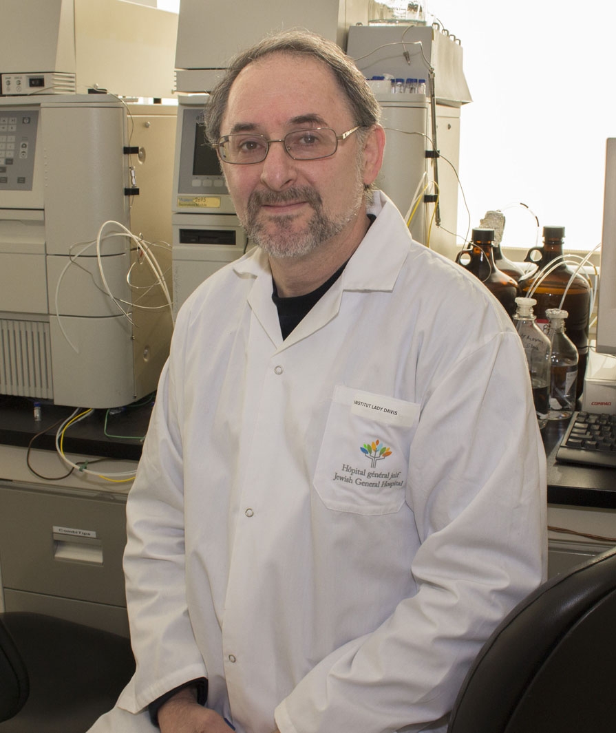 Uri Saragovi合作论文数Department of Pharmacology & Therapeutics, Faculty of Medicine and Health Sciences, McGill University3
 Kilarski Witold W合作论文数Institute of Bioengineering and Swiss Institute of Experimental, Cancer Research (ISREC),, École Polytechnique Fédérale de Lausanne3Balderas Israela合作论文数3
 Paolo Nicolas Catalano合作论文数3Bonaventura Maria Marta合作论文数3
 Manuel Tena-Sempere合作论文数Research Centre of Integrative Physiology and Pharmacology, Institute of Biomedicine, University of Turku;Instituto Maimonides de Investigación Biomedica de Cordoba;Universidad de Córdoba Facultad de Medicina y Enfermería3
 Werner a Scherbaum合作论文数Universitätsklinikum Düsseldorf3
 Bernhard Bettler合作论文数Therapeutic Area Nervous System, Novartis Pharma AG3Minoru Ohta合作论文数3
 Allan E Herbison合作论文数Department of Physiology, Development and Neuroscience, University of Cambridge3
 Kyoko Miyasaka (宮坂京子)合作论文数Department of Physiology and Nutrition, Tokyo Kasei University3Patricia Joseph-Bravo合作论文数Instituto de Biotecnología, Universidad Nacional Autónoma de México3Victor Ramirez-Amaya合作论文数3




