Objective To prospectively evaluate safety and efficacy of brain-responsive neurostimulation in adults with medically intractable focal onset seizures (FOS) over 9 years. Methods Adults treated with brain-responsive neurostimulation in 2-year feasibility or randomized controlled trials were enrolled in a long-term prospective open label trial (LTT) to assess safety, efficacy, and quality of life (QOL) over an additional 7 years. Safety was assessed as adverse events (AEs), efficacy as median percent change in seizure frequency and responder rate, and QOL with the Quality of Life in Epilepsy (QOLIE-89) inventory. Results Of 256 patients treated in the initial trials, 230 participated in the LTT. At 9 years, the median percent reduction in seizure frequency was 75% (p < 0.0001, Wilcoxon signed rank), responder rate was 73%, and 35% had a ≥90% reduction in seizure frequency. We found that 18.4% (47 of 256) experienced ≥1 year of seizure freedom, with 62% (29 of 47) seizure-free at the last follow-up and an average seizure-free period of 3.2 years (range 1.04–9.6 years). Overall QOL and epilepsy-targeted and cognitive domains of QOLIE-89 remained significantly improved (p < 0.05). There were no serious AEs related to stimulation, and the sudden unexplained death in epilepsy (SUDEP) rate was significantly lower than predefined comparators (p < 0.05, 1-tailed χ2). Conclusions Adjunctive brain-responsive neurostimulation provides significant and sustained reductions in the frequency of FOS with improved QOL. Stimulation was well tolerated; implantation-related AEs were typical of other neurostimulation devices; and SUDEP rates were low. ClinicalTrials.gov identifier NCT00572195. Classification of evidence This study provides Class IV evidence that brain-responsive neurostimulation significantly reduces focal seizures with acceptable safety over 9 years.
For each important type of drug toxic emergency, available information on predisposing risks, incidence, detection (clinical and laboratory), management (monitoring, specific therapy, modification of antiepileptic drug regimen), and outcome will be reviewed. Methods and timing of reinstitution of antiepileptic medications after overdose is a matter of some discussion. In 1987, 2577 cases of phenytoin (PHT) overdosage were reported in the United States, with 4 deaths. Patients routinely requiring high doses for seizure control are at particular risk. Iatrogenic poisoning occurs easily due to saturation pharmacokinetics of PHT metabolism. The earliest and most reliable indicator of developing clinical toxicity from PHT overdose is accurate reporting by the patient of symptoms. A specific association of valproate (VPA) therapy and idiosyncratic isolated hepatic failure, in the absence of systemic hypersensitivity, has been studied and reviewed recently by Dreifuss et al. Acute hemorrhagic pancreatitis rarely has been associated with VPA therapy.
Background. Accurate prediction of clinical impairment in upper-extremity motor function following therapy in chronic stroke patients is a difficult task for clinicians but is key in prescribing appropriate therapeutic strategies. Machine learning is a highly promising avenue with which to improve prediction accuracy in clinical practice. Objectives. The objective was to evaluate the performance of 5 machine learning methods in predicting postintervention upper-extremity motor impairment in chronic stroke patients using demographic, clinical, neurophysiological, and imaging input variables. Methods. A total of 102 patients (female: 31%, age 61 ± 11 years) were included. The upper-extremity Fugl-Meyer Assessment (UE-FMA) was used to assess motor impairment of the upper limb before and after intervention. Elastic net (EN), support vector machines, artificial neural networks, classification and regression trees, and random forest were used to predict postintervention UE-FMA. The performances of methods were compared using cross-validated R2. Results. EN performed significantly better than other methods in predicting postintervention UE-FMA using demographic and baseline clinical data (median REN2=0.91,RRF2=0.88,RANN2=0.83,RSVM2=0.79,RCART2=0.70;P < .05). Preintervention UE-FMA and the difference in motor threshold (MT) between the affected and unaffected hemispheres were the strongest predictors. The difference in MT had greater importance than the absence or presence of a motor-evoked potential (MEP) in the affected hemisphere. Conclusion. Machine learning methods may enable clinicians to accurately predict a chronic stroke patient's postintervention UE-FMA. Interhemispheric difference in the MT is an important predictor of chronic stroke patients' response to therapy and, therefore, could be included in prospective studies.
Pediatric vagal nerve stimulation (VNS) studies have featured various epilepsy syndromes, follow-up durations and treatment protocols. Only 19 partial onset seizure patients between 12 and 18 years old were enrolled in the above acute-phase double-blind controlled trials. VNS is an appealing 'add-on' antiepileptic therapy because it offers fewer seizures without the common additional medication side effects, such as poor concentration and fatigue. While reports on pediatric patients receiving VNS have tended to include short- and long-term results, in adults, the long-term results have been presented separately. The following unique observations have been made concerning VNS therapy for epilepsy. Large clinical trials indicate that vagal nerve stimulation reduces partial-onset seizures in adults by -30% in the first 3 months of therapy. A few recent single case reports of unique adverse effects of VNS warrant mention. In patients with epilepsy, VNS also has been associated with improved mood, and memory.
PURPOSE:To assess the potential biologic significance of variations in burst-suppression patterns (BSPs) after cardiac arrest in relation to recovery of consciousness. In the context of recent theoretical models of BSP, bursting frequency may be representative of underlying network dynamics; discontinuous activation of membrane potential during impaired cellular energetics may promote neuronal rescue. METHODS:We reviewed a database of 73 comatose post-cardiac arrest patients who underwent therapeutic hypothermia to assess for the presence of BSP and clinical outcomes. In a subsample of patients with BSP (n = 14), spectral content of burst and suppression periods were quantified using multitaper method. RESULTS:Burst-suppression pattern was seen in 45/73 (61%) patients. Comparable numbers of patients with (31.1%) and without (35.7%) BSP regained consciousness by the time of hospital discharge. In addition, in two unique cases, BSP initially resolved and then spontaneously reemerged after completion of therapeutic hypothermia and cessation of sedative medications. Both patients recovered consciousness. Spectral analysis of bursts in all patients regaining consciousness (n = 6) showed a prominent theta frequency (5-7 Hz) feature, but not in age-matched patients with induced BSP who did not recover consciousness (n = 8). CONCLUSIONS:The prognostic implications of BSP after hypoxic brain injury may vary based on the intrinsic properties of the underlying brain state itself. The presence of theta activity within bursts may index potential viability of neuronal networks underlying recovery of consciousness; emergence of spontaneous BSP in some cases may indicate an innate neuroprotective mechanism. This study highlights the need for better characterization of various BSP patterns after cardiac arrest.
Accurate predictions of motor improvement resulting from intensive therapy in chronic stroke patients is a difficult task for clinicians, but is key in prescribing appropriate therapeutic strategies. Statistical methods, including machine learning, are a highly promising avenue with which to improve prediction accuracy in clinical practice. The first main objective of this study was to use machine learning methods to predict a chronic stroke individual’s motor function improvement after 6 weeks of intervention using pre-intervention demographic, clinical, neurophysiological and imaging data. The second main objective was to identify which data elements were most important in predicting chronic stroke patients’ impairment after 6 weeks of intervention. Data from one hundred and two patients (Female: 31%, age 61±11 years) who suffered first ischemic stroke 3-12 months prior were included in this study. After enrollment, patients underwent 6 weeks of intensive motor and transcranial magnetic stimulation therapy. Age, gender, handedness, time since stroke, pre-intervention Fugl-Meyer Assessment, stroke lateralization, the difference in motor threshold between the unaffected and affected hemispheres, absence or presence of motor evoked potential in the affected hemisphere and various imaging metrics were used as predictors of post-intervention Fugl-Meyer Assessment. Five machine learning methods, including Elastic-Net, Support Vector Machines, Artificial Neural Networks, Classification and Regression Trees, and Random Forest, were used to predict post-intervention Fugl-Meyer Assessment based on either demographic, clinical and neurophysiological data alone or in combination with the imaging metrics. Cross-validated R-squared and root of mean squared error were used to assess the prediction accuracy and compare the performance of methods. Elastic-Net performed significantly better than the other methods for the model containing pre-intervention Fugl-Meyer Assessment, demographic, clinical and neurophysiological data as predictors of post-intervention Fugl-Meyer Assessment (). Pre-intervention Fugl-Meyer Assessment and difference in motor threshold between affected and unaffected hemispheres were commonly found as the strongest two predictors in the clinical model. The difference in motor threshold had greater importance than the absence or presence of motor evoked potential in the affected hemisphere. The various imaging metrics, including lesion overlap with the spinal cord, largely did not improve the model performance. The approach implemented here may enable clinicians to more accurately predict a chronic stroke patient’s individual response to intervention. The predictive models used in this study could assist clinicians in making treatment decisions and improve the accuracy of prognosis in chronic stroke patients.
Background and Purpose: Accurate predictions of motor improvement resulting from intensive therapy in chronic stroke patients is a difficult task for clinicians, but is key in prescribing appropriate therapeutic strategies. Statistical methods, including machine learning, are a highly promising avenue with which to improve prediction accuracy in clinical practice. The first main objective of this study was to use machine learning methods to predict a chronic stroke individual9s motor function improvement after 6 weeks of intervention using pre-intervention demographic, clinical, neurophysiological and imaging data. The second main objective was to identify which data elements were most important in predicting chronic stroke patients9 impairment after 6 weeks of intervention. Materials and methods: Data from one hundred and two patients (Female: 31%, age 61±11 years) who suffered first ischemic stroke 3-12 months prior were included in this study. After enrollment, patients underwent 6 weeks of the intensive motor and transcranial magnetic stimulation therapy. Age, gender, handedness, time since stroke, pre-intervention Fugl-Meyer Assessment, stroke lateralization, the difference in motor threshold between the unaffected and affected hemispheres, absence or presence of motor evoked potential in the affected hemisphere and various imaging metrics were used as predictors of post-intervention Fugl-Meyer Assessment. Five machine learning methods, including Elastic-Net (EN), Support Vector Machines (SVM), Artificial Neural Networks (ANN), Classification and Regression Trees (CART), and Random Forest (RF), were used to predict post-intervention Fugl-Meyer Assessment based on either demographic, clinical and neurophysiological data alone or in combination with the imaging metrics. Cross-validated R-squared and root of mean squared error were used to assess the prediction accuracy and compare the performance of methods. Results: EN performed significantly better than the other methods for the model containing pre-intervention Fugl-Meyer Assessment, demographic, clinical and neurophysiological data as predictors of post-intervention Fugl-Meyer Assessment (R-squared of EN=0.91, R-squared of RF=0.88, R-squared of ANN=0.83, R-squared of SVM=0.79, R-squared of CART=0.70, p
Purpose: The authors report a case of chronic oral-pharyngeal dystonia, and a resultant profound dysarthria, in a post-encephalitic young woman who demonstrated quantitative improvement during a two-week combined course of priming repetitive transcranial magnetic stimulation (rTMS) and speech therapy.
Evaluate the seizure‐reduction response and safety of brain‐responsive stimulation in adults with medically intractable partial‐onset seizures of neocortical origin.
SummaryObjectiveEvaluate the seizure‐reduction response and safety of mesial temporal lobe (MTL) brain‐responsive stimulation in adults with medically intractable partial‐onset seizures of mesial temporal lobe origin.MethodsSubjects with mesial temporal lobe epilepsy (MTLE) were identified from prospective clinical trials of a brain‐responsive neurostimulator (RNS System, NeuroPace). The seizure reduction over years 2–6 postimplantation was calculated by assessing the seizure frequency compared to a preimplantation baseline. Safety was assessed based on reported adverse events.ResultsThere were 111 subjects with MTLE; 72% of subjects had bilateral MTL onsets and 28% had unilateral onsets. Subjects had one to four leads placed; only two leads could be connected to the device. Seventy‐six subjects had depth leads only, 29 had both depth and strip leads, and 6 had only strip leads. The mean follow‐up was 6.1 ± (standard deviation) 2.2 years. The median percent seizure reduction was 70% (last observation carried forward). Twenty‐nine percent of subjects experienced at least one seizure‐free period of 6 months or longer, and 15% experienced at least one seizure‐free period of 1 year or longer. There was no difference in seizure reduction in subjects with and without mesial temporal sclerosis (MTS), bilateral MTL onsets, prior resection, prior intracranial monitoring, and prior vagus nerve stimulation. In addition, seizure reduction was not dependent on the location of depth leads relative to the hippocampus. The most frequent serious device‐related adverse event was soft tissue implant‐site infection (overall rate, including events categorized as device‐related, uncertain, or not device‐related: 0.03 per implant year, which is not greater than with other neurostimulation devices).SignificanceBrain‐responsive stimulation represents a safe and effective treatment option for patients with medically intractable epilepsy, including patients with unilateral or bilateral MTLE who are not candidates for temporal lobectomy or who have failed a prior MTL resection.
The aim of this study was to evaluate bihemispheric motor cortex excitability in relation to lesion location and the severity of motor impairment in patients with chronic post-stroke hemiparesis.
Purpose: The reliability of somatosensory evoked potentials (SSEPs) in predicting outcome in comatose survivors of cardiac arrest treated with therapeutic hypothermia (TH) has been questioned. We investigated whether the absence of cortical (N20) responses was a reliable predictor of a nonawakening in the setting of TH. Methods: A retrospective review was conducted in cardiac arrest survivors treated with TH admitted to a single tertiary care hospital from April, 2010 to March, 2013 who underwent SSEP testing at various time points after cardiac arrest. N20 responses were categorized as normal, present but abnormal, bilaterally absent, or inadequate for interpretation. Neurologic outcome was assessed at discharge by the Cerebral Performance Category Scale (CPC). Results: Ninety-three SSEP studies were performed in 73 patients. Fourteen patients had absent N20 responses; all had poor outcome (CPC 4–5). Eleven patients had absent N20 s during hypothermia, three of whom had follow-up SSEPs after rewarming and cortical responses remained absent. Fifty-seven patients had N20 peaks identified and had variable outcomes. Evaluation of 1 or more N20 peaks was limited or inadequate in 11.4% of SSEPs performed during the cooling because of artifact. Conclusions: Somatosensory evoked potentials remain a reliable prognostic indicator in patients undergoing TH. The limited sample size of patients who had SSEP performed during TH and repeated after normothermia added to the effect of self-fulfilling prophecy limit the interpretation of the reliability of this testing when performed during cooling. Further prospective, multicenter, large scale studies correlating cortical responses in SSEPs during and after TH are warranted. Technical challenges are commonplace during TH and caution is advised in the interpretation of suboptimal recordings.
Purpose: Continuous video EEG is a tool to assess brain function in injuries, including cardiac arrest (CA). In post-CA therapeutic hypothermia (TH) studies, some EEG features are linked to poor prognosis, but the evolvement of EEG characteristics during two temperature phases and its significance is unclear. We systematically analyzed EEG characteristics in cooled and rewarmed phases of post-CA therapeutic hypothermia patients and investigated their correlation to patient outcome. Methods: This is a retrospective study of EEG analyses, from a single academic center, of 20 patients who underwent CA and therapeutic hypothermia. For each patient, three 30-minute EEG segments in cooled and rewarmed phases were analyzed for continuity, frequency, interictal epileptiform discharges, and seizures. Mortality at the time of discharge was used as outcome. Results: Rewarming was associated with the emergence of interictal epileptiform discharges, 2.6 times as likely compared with the cooled period (P = 0.03), and was not affected by systemic factors. Continuity, frequency, and discrete seizures were unaffected by temperature and did not show variance within each temperature phase. There was a trend toward the emergence of interictal epileptiform discharges upon rewarming and mortality, but it was not statistically significant. Conclusions: Increased interictal epileptiform discharges with rewarming in post-CA therapeutic hypothermia patients may suggest poor prognosis, but a larger scale prospective study is needed.
Introduction: Existing strategies to enhance motor function following Spinal Cord Injury (SCI) are suboptimal leaving patients with considerable disability. Available evidence suggests that transcranial direct current stimulation (tDCS) has emerged as a promising method to improve motor dysfunction in other neurological disorders. However, little is known about the specific brain changes that result in improved motor performance following tDCS. Studying electroencephalography (EEG) responses to tDCS, has not been well-explored, and may provide us with valuable insight regarding the effects of neuromodulation techniques for individuals with SCI.
Our goal was to systematically analyze EEG characteristics in cooled and re-warmed phases of post-cardiac arrest (CA) therapeutic hypothermia (TH) patients and investigate their correlation to patient outcome.
Transcranial direct current stimulation (tDCS) is emerging as a promising method to treat motor and sensory impairment following neurological disorders such as spinal cord injury (SCI). However, the degree to which responses to tDCS are personalized from subject-to-subject, and how this affects physiological response, is largely unknown.
Upon its inception, repetitive transcranial magnetic stimulation (rTMS) was delivered at rest, without regard to the potential impact of activity occurring during or around the time of stimulation. rTMS was considered an experimental intervention imposed on the brain; therefore, the myriad features that might suppress or enhance its desired effects had not yet been explored. The field of rTMS has since grown substantially and therapeutic benefits have been reported, albeit with modest and inconsistent improvements. Work in this field accelerated following approval of a psychiatric application (depression), and it is now expanding to other applications and disciplines. In the last decade, experimental enquiry has sought new ways to improve the therapeutic benefits of rTMS, intended to enhance underlying brain reorganization and functional recovery by combining it with behavioral therapy. This concept is appealing, but poorly defined and requires clarity. We provide an overview of how combined rTMS and behavioral therapy has been delineated in the literature, highlighting the diversity of approaches. We outline a framework for study design and reporting such that the effects of this emerging method can be better understood.
Objective: The long-term efficacy and safety of responsive direct neurostimulation was assessed in adults with medically refractory partial onset seizures. Methods: All participants were treated with a cranially implanted responsive neurostimulator that delivers stimulation to 1 or 2 seizure foci via chronically implanted electrodes when specific electrocorticographic patterns are detected (RNS System). Participants had completed a 2-year primarily open-label safety study (n = 65) or a 2-year randomized blinded controlled safety and efficacy study (n = 191); 230 participants transitioned into an ongoing 7-year study to assess safety and efficacy. Results: The average participant was 34 (±11.4) years old with epilepsy for 19.6 (±11.4) years. The median preimplant frequency of disabling partial or generalized tonic-clonic seizures was 10.2 seizures a month. The median percent seizure reduction in the randomized blinded controlled trial was 44% at 1 year and 53% at 2 years (p < 0.0001, generalized estimating equation) and ranged from 48% to 66% over postimplant years 3 through 6 in the long-term study. Improvements in quality of life were maintained (p < 0.05). The most common serious device-related adverse events over the mean 5.4 years of follow-up were implant site infection (9.0%) involving soft tissue and neurostimulator explantation (4.7%). Conclusions: The RNS System is the first direct brain responsive neurostimulator. Acute and sustained efficacy and safety were demonstrated in adults with medically refractory partial onset seizures arising from 1 or 2 foci over a mean follow-up of 5.4 years. This experience supports the RNS System as a treatment option for refractory partial seizures. Classification of evidence: This study provides Class IV evidence that for adults with medically refractory partial onset seizures, responsive direct cortical stimulation reduces seizures and improves quality of life over a mean follow-up of 5.4 years.
Objective: To investigate the effects of anodal transcranial direct current stimulation (a-tDCS) intensity on corticospinal excitability and affected muscle activation in individuals with chronic spinal cord injury (SCI).Design: Single-blind, randomized, sham-controlled, crossover study.Setting: Medical research institute and rehabilitation hospital.Participants: Volunteers (N=9) with chronic SCI and motor dysfunction in wrist extensor muscles.Interventions: Three single session exposures to 20 minutes of a-tDCS (anode over the extensor carpi radialis [ECR] muscle representation on the left primary motor cortex, cathode over the right supraorbital area) using lmA, 2mA, or sham stimulation, delivered at rest, with at least 1 week between sessions.Main Outcome Measures: Corticospinal excitability was assessed with motor-evoked potentials (MEPs) from the ECR muscle using surface electromyography after transcranial magnetic stimulation. Changes in spinal excitability, sensory threshold, and muscle strength were also investigated.Results: Mean MEP amplitude significantly increased by approximately 40% immediately after 2mA a-tDCS (pre: 0.36 +/- 0.1mV; post: 0.47 +/- 0.11mV; P=.001), but not with lmA or sham. Maximal voluntary contraction measures remained unaltered across all conditions. Sensory threshold significantly decreased over time after lmA (P=.002) and 2mA (P=.039) a-tDCS and did not change with sham. F-wave persistence showed a nonsignificant trend for increase (pre: 32%+/- 112%; post: 41%+/- 110%; follow-up: 46%+/- 112%) after 2mA stimulation. No adverse effects were reported with any of the experimental conditions.Conclusions: The a-tDCS can transiently raise corticospinal excitability to affected muscles in patients with chronic SCI after 2mA stimulation. Sensory perception can improve with both 1 and 2mA stimulation. This study gives support to the safe and effective use of a-tDCS using small electrodes in patients with SCI and highlights the importance of stimulation intensity. (C) 2015 by the American Congress of Rehabilitation Medicine
OBJECTIVE: We aim to improve our understanding of contralateral cortical activity during stroke recovery as a target of TMS-based therapy. BACKGROUND: TMS is being widely investigated as a tool to improve outcomes after stroke. Inhibition of the unaffected hemisphere causes disinhibition of the affected hemisphere and improved functionality. However, there is wide variability between subjects. We seek to identify those who will benefit most. We start by investigating the hyperexcitability of the unaffected hemisphere following stroke and its relationship to lesion anatomy and clinical presentation. DESIGN/METHODS: Eight adults (5 females, 3 males, mean age=62.38) were enrolled with the following criteria: first clinical stroke within the past 3-12 months resulting in moderate hemiparesis involving the hand. Subjects were assessed neurophysiologically with navigated single pulse TMS and functionally with the Fugl-Meyer Assessment (FMA), a detailed measure of hand strength and coordination, and NIH Stroke Scale (NIHSS). TMS was used to map primary cortex corresponding to the Abductor Pollicis Brevis (APB) and the Extensor digitorum (EDC) muscles and to determine motor threshold. RESULTS: We show that motor threshold (low values being a marker of elevated excitability) of unaffected hemisphere M1 is not always excessively low, and may relate to severity of hemiparesis depending on whether the stroke lesion involves the cerebral cortex. We demonstrate that motor evoked potentials in the affected hemisphere are often small or absent. CONCLUSIONS: Our data suggest that lesion location may be indicative of whether contralesional M1 is hyper-excitable and therefore warrant targeted de-excitatory brain stimulation as a therapeutic strategy. These findings are important to clinical trials underway that presently do not assess contralesional excitability or stratify patients according to lesion location. We also suggest that transcallosal and intracortical excitability measures can be invalid in the affected hemisphere if evoked potentials are small or absent.
 Cynthia L. Harden合作论文数Northwell Health21
 Dylan J. Edwards合作论文数Human Motor Recovery Laboratory, Jefferson Moss Rehabilitation Research Institute;Ribeirao Preto Medical School, University of São Paulo;School of Medical and Health Sciences, Edith Cowan University;Department of Rehabilitation Medicine, Thomas Jefferson University11
 Blagovest Nikolov合作论文数11
 Martha Morrell合作论文数Department of Neurology, School of Medicine, Stanford University;NeuroPace9
 Gregory K. Bergey合作论文数Department of Neurology;Johns Hopkins Epilepsy Center;School of Medicine, Johns Hopkins University9
 Robert Duckrow合作论文数9
 Gregory A. Worrell合作论文数Department of Neurology, Mayo Clinic;Department of Physiology & Biomedical Engineering, Mayo Clinic9
 Anthony Murro合作论文数8Lawrence J. Hirsch合作论文数8
 Ryder Gwinn合作论文数Swedish Neuroscience Institute8Salanova Vicenta合作论文数7
 Robert E. Gross合作论文数Department of Neurosurgery, School of Medicine, Emory University7
 Robert Goodman合作论文数Institute of Psychiatry, Department of Child and Adolescent Psychiatry, King's College London7Nathan B. Fountain合作论文数7Gregory L. Barkley合作论文数7L. Ponticello合作论文数6Syed Hosain合作论文数6
 Michael R. Sperling合作论文数Department of Neurology, Sidney Kimmel Medical College, Thomas Jefferson University;Jefferson Comprehensive Epilepsy Center, Thomas Jefferson University Hospital;Clinical Neurophysiology Laboratory, Thomas Jefferson University Hospital6
 Theodore H. Schwartz合作论文数Minimally Invasive Surgery, Weill Cornell Medicine, Cornell University;Neurological Surgery, Weill Cornell Medicine, Cornell University6Michael C. Smith合作论文数6
 Padmaja Kandula合作论文数Department of Neurology, Weill Cornell Medicine, Cornell University6
 K Zoe Tsagaris合作论文数Burke Neurol Inst5S Hosain合作论文数5Carl W. Bazil合作论文数5Adrian Handforth合作论文数5
 Leslie Burton合作论文数5Richard Fraser合作论文数5
 Mar Cortes合作论文数Winifred Masterson Burke Medical Research Institute5
 Robert E. Wharen合作论文数Department of Neurosurgery (Reimer, Wharen),, Mayo Clinic Jacksonville5Eric Geller合作论文数4Lakshmi Nagarajan合作论文数4
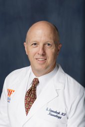 Stephan Eisenschenk合作论文数College of Medicine, University of Florida4David Spencer合作论文数Department of Neurology, Oregon Health & Science University4
 Nitin Sethi合作论文数Comprehensive Epilepsy Center, NYP-Weill Cornell Medical Center4Schaul Neil合作论文数4Srinivasan Shraddha合作论文数4
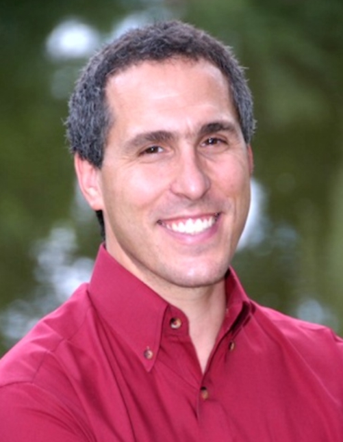 Sydney Cash合作论文数Division of Epilepsy, Department of Neurology, Mass General Brigham;Department of Neurology, Massachusetts General Hospital;Harvard Medical School, Harvard University4
 Dileep Nair合作论文数Epilepsy Center, Cleveland Clinic Foundation4Cole Andrew J合作论文数4Heck Christianne N合作论文数4
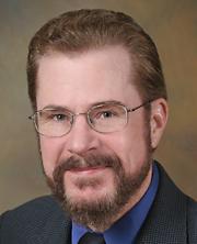 Thomas Reid Henry合作论文数Department of Neurology, University of Minnesota Medical School4
 Charles Liu合作论文数Clinical Neurological Surgery;Director, USC Neurorestoration Center4
 Alica Goldman合作论文数Baylor College of Medicine, Baylor College of Medicine4Yong D. Park合作论文数4
 Amy Kuceyeski合作论文数Department of Radiology, Weill Cornell Medicine;Department of Computational Biology, Cornell University4Thomas C. Witt合作论文数4Paul Rutecki合作论文数4David R. Chabolla合作论文数4




