Thalamic stimulation has emerged as a promising neuromodulation target for treating disorders of consciousness. Impaired consciousness, a debilitating outcome in temporal lobe epilepsy (TLE) remains a central problem for patients whose seizures cannot be treated pharmacologically and cannot be stopped with conventional surgery or responsive hippocampal stimulation. Although prior studies suggest an essential role of the thalamic intralaminar central lateral (CL) nucleus in arousal and sleep, evidence for a direct effect of thalamic intralaminar stimulation on human arousal has been limited. To address these gaps, the START (stimulation of the thalamus for arousal restoral in TLE) clinical trial investigated the efficacy of bilateral CL thalamic stimulation to restore consciousness during human sleep and TLE seizures. Five patients with medically refractory mesial temporal lobe epilepsy were implanted with an investigational neurostimulator, the Medtronic Summit RC+STM. Optimal CL stimulation parameters were obtained through individualized titration in slow wave sleep, evaluated through analysis of patient movement from video recordings and electrophysiology from simultaneously recorded scalp and hippocampal electroencephalography (EEG). We found that bilateral CL stimulation led to robust arousal from sleep characterized by increased body movements and decreased low frequency power (2-15 Hz) in both cortical and hippocampal EEG during 5 minute epochs of stimulation compared to baseline slow wave sleep. We evaluated impaired consciousness during seizures using verbal and non-verbal behavioral tests administered automatically by smartwatch, and found significant behavioral impairment in three of five patients during seizures with hippocampal stimulation. Administering CL stimulation in patients with impaired consciousness during TLE seizures showed significant improvement in behavioral outcomes in two of three patients, with one patient reaching their baseline performance comparable to non-seizure times. Overall, we found that stimulation of the thalamic CL in TLE patients increased arousal during both sleep and seizures. These findings demonstrate the potential of CL as a therapeutic target for mitigating impaired consciousness in TLE and can serve as a foundation for additional studies to test generalizability of CL stimulation effects on other disorders of consciousness. ### Competing Interest Statement The authors have declared no competing interest. ### Clinical Trial NCT04897776 ### Funding Statement This work was supported by NIH/NINDS UG3/UH3 NS112826. ### Author Declarations I confirm all relevant ethical guidelines have been followed, and any necessary IRB and/or ethics committee approvals have been obtained. Yes The details of the IRB/oversight body that provided approval or exemption for the research described are given below: Ethics committee/IRB of Yale University School of Medicine, Mayo Clinic, and Dartmouth-Hitchcock medical center gave ethical approval for this work. I confirm that all necessary patient/participant consent has been obtained and the appropriate institutional forms have been archived, and that any patient/participant/sample identifiers included were not known to anyone (e.g., hospital staff, patients or participants themselves) outside the research group so cannot be used to identify individuals. Yes I understand that all clinical trials and any other prospective interventional studies must be registered with an ICMJE-approved registry, such as ClinicalTrials.gov. I confirm that any such study reported in the manuscript has been registered and the trial registration ID is provided (note: if posting a prospective study registered retrospectively, please provide a statement in the trial ID field explaining why the study was not registered in advance). Yes I have followed all appropriate research reporting guidelines, such as any relevant EQUATOR Network research reporting checklist(s) and other pertinent material, if applicable. Yes All data generated in the present study will be made available from the corresponding author upon reasonable request.
Objective To identify the causative gene in a large unsolved family with genetic epilepsy with febrile seizures plus (GEFS+), we sequenced the genomes of family members, and then determined the contribution of the identified gene to the pathogenicity of epilepsies by examining sequencing data from 2,772 additional patients. Methods We performed whole genome sequencing of 3 members of a GEFS+ family. Subsequently, whole exome sequencing data from 1,165 patients with epilepsy from the Epi4K dataset and 1,329 Australian patients with epilepsy from the Epi25 dataset were interrogated. Targeted resequencing was performed on 278 patients with febrile seizures or GEFS+ phenotypes. Variants were validated and familial segregation examined by Sanger sequencing. Results Eight previously unreported missense variants were identified in SLC32A1, coding for the vesicular inhibitory amino acid cotransporter VGAT. Two variants cosegregated with the phenotype in 2 large GEFS+ families containing 8 and 10 affected individuals, respectively. Six further variants were identified in smaller families with GEFS+ or idiopathic generalized epilepsy (IGE). Conclusion Missense variants in SLC32A1 cause GEFS+ and IGE. These variants are predicted to alter gamma-aminobutyric acid (GABA) transport into synaptic vesicles, leading to altered neuronal inhibition. Examination of further epilepsy cohorts will determine the full genotype-phenotype spectrum associated with SLC32A1 variants.
PURPOSE:Posterior quadrant disconnection (PQD) is a surgical procedure for medically refractory epilepsy (MRE) involving diffuse regions of the temporo-parieto-occipital lobes. We sought to compare factors and efficacy according to PQD extent. METHODS:We performed a systematic review of the literature reporting the use of PQD since 2004. We analyzed various characteristics of pooled cases, including the role of preoperative studies in patient selection, intraoperative techniques, and outcomes. RESULTS:Our review of 137 patients from nine studies revealed 66% undergoing total PQD and 34% undergoing partial PQD. Interictal electroencephalography (EEG) findings were predominantly characterized as lateralized for total PQD (56%) and localized within the ipsilateral posterior quadrant in patients undergoing partial PQD (53%). Metabolic functional studies [positron emission tomography (PET) or ictal single-photon emission computed tomography (SPECT)] played a role in surgical decision-making in 42% of patients who underwent total PQD. Wada and/or functional magnetic resonance imaging (fMRI) was more often utilized for partial PQD (22%) than total PQD (3%) as was intracranial electroencephalography (icEEG) (30% versus 13%, respectively). Overall, 75% of total PQD patients achieved seizure freedom [defined as Engel I or International League Against Epilepsy (ILAE) Class 1 outcome] in comparison to 63% of partial PQD patients (p = .078). New visual field deficits were seen in 12% and new or worsened hemiparesis in 6%. For patients in either cohort, concordance of interictal and ictal EEG findings was found to be predictive of seizure freedom (p = .048). CONCLUSION:Both total and partial PQD represent effective alternatives for managing patients with MRE whose seizure onset zone (SOZ) involves a diffuse region within the posterior quadrant. While PET and/or SPECT frequently aided in the decision to proceed with total PQD, patients who underwent a tailored, partial multilobar resection were more likely to undergo Wada and/or fMRI testing as well as stage I icEEG studies.
Drug-resistant focal epilepsy (DRFE) in children can impair cognition and behavior, and lead to premature death. Increased pediatric epilepsy surgery numbers reflect the improvements in seizure control and long-term developmental outcomes. Yet, many children with DRFE are not candidates for surgical resection due to overlap of the seizure network with eloquent cortex or multiple seizure-onset zones, making surgery dangerous or ineffective. In adults, responsive neurostimulation (RNS System) therapy is safe and effective treatment for DRFE with one or two seizure foci, especially when the seizure focus is in eloquent cortex. We present six pediatric patients with DRFE who underwent RNS implantation. Our outcomes demonstrate safety, decreased clinical seizure frequency, as well as improved functional status and quality of life. Changes in the clinical seizure semiology and frequency occurred in conjunction with adjustments to the stimulation parameters, supporting the efficacy of responsive neuromodulation in children.
Objective To prospectively evaluate safety and efficacy of brain-responsive neurostimulation in adults with medically intractable focal onset seizures (FOS) over 9 years. Methods Adults treated with brain-responsive neurostimulation in 2-year feasibility or randomized controlled trials were enrolled in a long-term prospective open label trial (LTT) to assess safety, efficacy, and quality of life (QOL) over an additional 7 years. Safety was assessed as adverse events (AEs), efficacy as median percent change in seizure frequency and responder rate, and QOL with the Quality of Life in Epilepsy (QOLIE-89) inventory. Results Of 256 patients treated in the initial trials, 230 participated in the LTT. At 9 years, the median percent reduction in seizure frequency was 75% (p < 0.0001, Wilcoxon signed rank), responder rate was 73%, and 35% had a ≥90% reduction in seizure frequency. We found that 18.4% (47 of 256) experienced ≥1 year of seizure freedom, with 62% (29 of 47) seizure-free at the last follow-up and an average seizure-free period of 3.2 years (range 1.04–9.6 years). Overall QOL and epilepsy-targeted and cognitive domains of QOLIE-89 remained significantly improved (p < 0.05). There were no serious AEs related to stimulation, and the sudden unexplained death in epilepsy (SUDEP) rate was significantly lower than predefined comparators (p < 0.05, 1-tailed χ2). Conclusions Adjunctive brain-responsive neurostimulation provides significant and sustained reductions in the frequency of FOS with improved QOL. Stimulation was well tolerated; implantation-related AEs were typical of other neurostimulation devices; and SUDEP rates were low. ClinicalTrials.gov identifier NCT00572195. Classification of evidence This study provides Class IV evidence that brain-responsive neurostimulation significantly reduces focal seizures with acceptable safety over 9 years.
Objective To describe seizure outcomes in patients with medically refractory epilepsy who had evidence of bilateral mesial temporal lobe (MTL) seizure onsets and underwent MTL resection based on chronic ambulatory intracranial EEG (ICEEG) data from a direct brain-responsive neurostimulator (RNS) system. Methods We retrospectively identified all patients at 17 epilepsy centers with MTL epilepsy who were treated with the RNS System using bilateral MTL leads, and in whom an MTL resection was subsequently performed. Presumed lateralization based on routine presurgical approaches was compared to lateralization determined by RNS System chronic ambulatory ICEEG recordings. The primary outcome was frequency of disabling seizures at last 3-month follow-up after MTL resection compared to seizure frequency 3 months before MTL resection. Results We identified 157 patients treated with the RNS System with bilateral MTL leads due to presumed bitemporal epilepsy. Twenty-five patients (16%) subsequently had an MTL resection informed by chronic ambulatory ICEEG (mean = 42 months ICEEG); follow-up was available for 24 patients. After MTL resection, the median reduction in disabling seizures at last follow-up was 100% (mean: 94%; range: 50%-100%). Nine patients (38%) had exclusively unilateral electrographic seizures recorded by chronic ambulatory ICEEG and all were seizure-free at last follow-up after MTL resection; eight of nine continued RNS System treatment. Fifteen patients (62%) had bilateral MTL electrographic seizures, had an MTL resection on the more active side, continued RNS System treatment, and achieved a median clinical seizure reduction of 100% (mean: 90%; range: 50%-100%) at last follow-up, with eight of fifteen seizure-free. For those with more than 1 year of follow-up (N = 21), 15 patients (71%) were seizure-free during the most recent year, including all eight patients with unilateral onsets and 7 of 13 patients (54%) with bilateral onsets. Significance Chronic ambulatory ICEEG data provide information about lateralization of MTL seizures and can identify additional patients who may benefit from MTL resection.
OBJECTIVE:The RNS System is a direct brain-responsive neurostimulation system that is US Food and Drug Administration-approved for adults with medically intractable focal onset seizures based on safety and effectiveness data from controlled clinical trials. The purpose of this study was to retrospectively evaluate the real-world safety and effectiveness of the RNS System. METHODS:Eight comprehensive epilepsy centers conducted a chart review of patients treated with the RNS System for at least 1 year, in accordance with the indication for use. Data included device-related serious adverse events and the median percent change in disabling seizure frequency from baseline at years 1, 2, and 3 of treatment and at the most recent follow-up. RESULTS:One hundred fifty patients met the criteria for analysis. The median reduction in seizures was 67% (interquartile range [IQR] = 33%-93%, n = 149) at 1 year, 75% (IQR = 50%-94%, n = 93) at 2 years, 82% (IQR = 50%-96%, n = 38) at ≥3 years, and 74% (IQR = 50%-96%, n = 150) at last follow-up (mean = 2.3 years). Thirty-five percent of patients had a ≥90% seizure frequency reduction, and 18% of patients reported being clinically seizure-free at last follow-up. Seizure frequency reductions were similar regardless of patient age, age at epilepsy onset, duration of epilepsy, seizure onset in mesial temporal or neocortical foci, magnetic resonance imaging findings, prior intracranial monitoring, prior epilepsy surgery, or prior vagus nerve stimulation treatment. The infection rate per procedure was 2.9% (6/150 patients); five of the six patients had an implant site infection, and one had osteomyelitis. Lead revisions were required in 2.7% (4/150), and 2.0% (3/150) of patients had a subdural hemorrhage, none of which had long-lasting neurological consequences. SIGNIFICANCE:In this real-world experience, safety was similar and clinical seizure outcomes exceeded those of the prospective clinical trials, corroborating effectiveness of this therapy and suggesting that clinical experience has informed more effective programming.
Objective: Autism spectrum disorder (ASD) is a neurodevelopmental disorder frequently associated with epilepsy and epilepsy is a leading cause of death in ASD patients. Despite growing interest in genetic, neurophysiological and clinical overlaps, data on ictal electroencephalographic (EEG) recordings in ASD are lacking since behavioral disorders often make it difficult to obtain EEG recordings. We examined ictal EEG features in a consecutive series of patients with ASD and epilepsy. Methods: We retrospectively identified 400 consecutive patients with ASD and epilepsy at our Level 4 Epilepsy center between 2015 and 2019; 45 had at least one EEG-recorded seizure captured. Demographics, age of nonfebrile seizure onset, age of ASD diagnosis, language, magnetic resonance imagining findings, genetic testing and EEG studies were reviewed. Seizures were classified by semiologic and electrographic features. Ictal findings were analyzed. Results: A total of 497 seizures were captured in 45 patients: 20 patients with focal onset epilepsy had 126 seizures (median: 1, range: 1-30), 17 patients with generalized onset epilepsy had 88 seizures (median: 2, range: 1-15), 7 patients with Lennox-Gastaut syndrome had 270 seizures (median: 12, range: 1-74) and one patient had both right hemisphere focal and generalized onsets (12 focal, 1 generalized). Significance: Our study is the first to analyze a large set of ictal data in patients with autism spectrum disorder, a population traditionally difficult to obtain ictal recordings. Our results confirm the diverse spectrum of seizure types and provide clinical-EEG correlates of seizures in ASD patients. Both focal-onset and generalized-onset seizures were recorded, confirming that ASD patients have higher rates of both focal and generalized epilepsy syndromes. Among patients with focal epilepsy, temporal and frontal onsets were frequent, suggesting the possibility of epilepsy surgery or brain stimulation. EEG to classify seizures and epilepsies is critical to determine therapeutic options and effort should be made to obtain EEGs in this heterogenous population.
Objective Classification of epilepsy into types and subtypes is important for both clinical care and research into underlying disease mechanisms. A quantitative, data-driven approach may augment traditional electroclinical classification and shed new light on existing classification frameworks. Methods We used latent class analysis, a statistical method that assigns subjects into groups called latent classes based on phenotypic elements, to classify individuals with common familial epilepsies from the Epi4K Multiplex Families study. Phenotypic elements included seizure types, seizure symptoms, and other elements of the medical history. We compared class assignments to traditional electroclinical classifications and assessed familial aggregation of latent classes. Results A total of 1120 subjects with epilepsy were assigned to five latent classes. Classes 1 and 2 contained subjects with generalized epilepsy, largely reflecting the distinction between absence epilepsies and younger onset (class 1) versus myoclonic epilepsies and older onset (class 2). Classes 3 and 4 contained subjects with focal epilepsies, and in contrast to classes 1 and 2, these did not adhere as closely to clinically defined focal epilepsy subtypes. Class 5 contained nearly all subjects with febrile seizures plus or unknown epilepsy type, as well as a few subjects with generalized epilepsy and a few with focal epilepsy. Family concordance of latent classes was similar to or greater than concordance of clinically defined epilepsy types. Significance Quantitative classification of epilepsy has the potential to augment traditional electroclinical classification by (1) combining some syndromes into a single class, (2) splitting some syndromes into different classes, (3) helping to classify subjects who could not be classified clinically, and (4) defining the boundaries of clinically defined classifications. This approach can guide future research, including molecular genetic studies, by identifying homogeneous sets of individuals that may share underlying disease mechanisms.
Decisions under uncertainty distinguish between those made under risk (known probabilities) and those made under ambiguity (unknown probabilities). Despite widespread interest in decisions under uncertainty and the successful documentation that these distinct psychological constructs profoundly-and differentially-impact behavior, research has not been able to systematically converge on which brain regions are functionally involved in processing risk and ambiguity. We merge a lesion approach with computational modeling and simultaneous measurement of the arousal response to investigate the impact the medial prefrontal cortex (mPFC), lateral prefrontal cortex (lPFC), and amygdala have on decisions under uncertainty. Results reveal that the lPFC acts as a unitary system for processing uncertainty: Lesions to this region disrupted the relationship between arousal and choice, broadly increasing both risk and ambiguity seeking. In contrast, the mPFC and amygdala appeared to play no role in processing risk, and the mPFC only had a tenuous relationship with ambiguous uncertainty. Together, these findings reveal that only the lPFC plays a global role in processing the highly aversive nature of uncertainty.
The epilepsies affect around 65 million people worldwide and have a substantial missing heritability component. We report a genome-wide mega-analysis involving 15,212 individuals with epilepsy and 29,677 controls, which reveals 16 genome-wide significant loci, of which 11 are novel. Using various prioritization criteria, we pinpoint the 21 most likely epilepsy genes at these loci, with the majority in genetic generalized epilepsies. These genes have diverse biological functions, including coding for ion-channel subunits, transcription factors and a vitamin-B6 metabolism enzyme. Converging evidence shows that the common variants associated with epilepsy play a role in epigenetic regulation of gene expression in the brain. The results show an enrichment for monogenic epilepsy genes as well as known targets of antiepileptic drugs. Using SNP-based heritability analyses we disentangle both the unique and overlapping genetic basis to seven different epilepsy subtypes. Together, these findings provide leads for epilepsy therapies based on underlying pathophysiology.
For drug-resistant epilepsy, nonpharmacologic treatments should be considered early rather than late. Of the nondrug treatments, only resective surgery can be curative. Neurostimulation is palliative, i.e., not expected to achieve a seizure-free outcome. While resective surgery is the goal, other options are necessary because the majority of patients with drug-resistant epilepsy are not surgical candidates, and others have seizures that fail to improve with surgery or have only partial improvement but not seizure freedom. Neurostimulation modalities include vagus nerve stimulation (VNS), responsive neurostimulation (RNS), and deep brain stimulation (DBS), each with its own advantages, disadvantages, and side effects. In most scenarios, determined by noninvasive evaluation, especially EEG and MRI, several strategies are reasonable. For focal epilepsies, the choices are between resective surgery, with or without intracranial EEG, and all three modalities of neurostimulation. In situations where resective surgery is likely to result in seizure freedom, such as mesiotemporal lobe epilepsy or lesional focal epilepsy, resection (standard, laser, or radiofrequency) is preferred. For difficult cases like extratemporal nonlesional epilepsies, neurostimulation offers a less invasive option than resective surgery. For generalized and multifocal epilepsies, VNS is an option, RNS is not, and DBS has only limited evidence. "This article is part of the Supplement issue Neurostimulation for Epilepsy."
The responsive neurostimulator (RNS ®, NeuroPace Inc.) has been available clinically since 2013 for the treatment of medically refractory partial epilepsy. Using intracranial electrodes and a cranially implanted device, RNS ® provides on-demand electrical cortical stimulation to reduce seizures. A randomized, multicenter, double-blind clinical trial demonstrated seizure reduction compared with sham stimulation. Seizure reduction was improved and sustained over years in a long-term treatment trial. The RNS ® provides chronic ambulatory electrographic monitoring over years giving unprecedented insight into epilepsy dynamics. Studies to date have looked at the length of time to detecting bilateral seizure onsets in mesial temporal lobe epilepsy (MTLE), demonstrated biorhythms in interictal epileptiform activity over varied time scales, and shown promise in early detection of benefits of adding a new antiepileptic drug. Questions remain as to the boundaries of patient selection and lead placement.
De novo mutations of the TRIM8 gene, which codes for a tripartite motif protein, have been identified using whole exome sequencing (WES) in two patients with epileptic encephalopathy (EE), but these reports were not sufficient to conclude that TRIM8 was a novel gene responsible for EE. Here we report four additional patients presenting with EE and de novo truncating mutations of TRIM8 detected by WES, and give further details of the patient previously reported by the Epi4K consortium. Epilepsy of variable severity was diagnosed in children aged 2 months to 3.5 years of age. All patients had developmental delay of variable severity with no or very limited language, often associated with behavioral anomalies and unspecific facial features or MRI brain abnormalities. The phenotypic variability observed in these patients appeared related to the severity of the epilepsy. One patient presented pharmacoresistant EE with regression, recurrent infections and nephrotic syndrome, compatible with the brain and kidney expression of TRIM8. Interestingly, all mutations were located at the highly conserved C-terminus section of TRIM8. This collaborative study confirms that TRIM8 is a novel gene responsible for EE, possibly associated with nephrotic syndrome. This report brings new evidence on the pathogenicity of TRIM8 mutations and highlights the value of data-sharing to delineate the phenotypic characteristics and biological basis of extremely rare disorders.
Evaluate the seizure‐reduction response and safety of brain‐responsive stimulation in adults with medically intractable partial‐onset seizures of neocortical origin.
SummaryObjectiveEvaluate the seizure‐reduction response and safety of mesial temporal lobe (MTL) brain‐responsive stimulation in adults with medically intractable partial‐onset seizures of mesial temporal lobe origin.MethodsSubjects with mesial temporal lobe epilepsy (MTLE) were identified from prospective clinical trials of a brain‐responsive neurostimulator (RNS System, NeuroPace). The seizure reduction over years 2–6 postimplantation was calculated by assessing the seizure frequency compared to a preimplantation baseline. Safety was assessed based on reported adverse events.ResultsThere were 111 subjects with MTLE; 72% of subjects had bilateral MTL onsets and 28% had unilateral onsets. Subjects had one to four leads placed; only two leads could be connected to the device. Seventy‐six subjects had depth leads only, 29 had both depth and strip leads, and 6 had only strip leads. The mean follow‐up was 6.1 ± (standard deviation) 2.2 years. The median percent seizure reduction was 70% (last observation carried forward). Twenty‐nine percent of subjects experienced at least one seizure‐free period of 6 months or longer, and 15% experienced at least one seizure‐free period of 1 year or longer. There was no difference in seizure reduction in subjects with and without mesial temporal sclerosis (MTS), bilateral MTL onsets, prior resection, prior intracranial monitoring, and prior vagus nerve stimulation. In addition, seizure reduction was not dependent on the location of depth leads relative to the hippocampus. The most frequent serious device‐related adverse event was soft tissue implant‐site infection (overall rate, including events categorized as device‐related, uncertain, or not device‐related: 0.03 per implant year, which is not greater than with other neurostimulation devices).SignificanceBrain‐responsive stimulation represents a safe and effective treatment option for patients with medically intractable epilepsy, including patients with unilateral or bilateral MTLE who are not candidates for temporal lobectomy or who have failed a prior MTL resection.
Gene identification in epilepsy has mainly been limited to large families segregating genes of major effect and de novo mutations in epileptic encephalopathies. Many families that present with common non-acquired focal epilepsies and genetic generalized epilepsies remain unexplained. We assembled a cohort of 'genetically enriched' common epilepsies by collecting and phenotyping families containing multiple individuals with unprovoked seizures. We aimed to determine if specific clinical epilepsy features aggregate within families, and whether this segregation of phenotypes may constitute distinct 'familial syndromes' that could inform genomic analyses. Families with three or more individuals with unprovoked seizures were studied across multiple international centres. Affected individuals were phenotyped and classified according to specific electroclinical syndromes. Families were categorized based on syndromic groupings of affected family members, examined for pedigree structure and phenotypic patterns and, where possible, assigned specific familial epilepsy syndromes. A total of 303 families were assembled and analysed, comprising 1120 affected phenotyped individuals. Of the 303 families, 117 exclusively segregated generalized epilepsy, 62 focal epilepsy, and 22 were classified as genetic epilepsy with febrile seizures plus. Over one-third (102 families) were observed to have mixed epilepsy phenotypes: 78 had both generalized and focal epilepsy features within the same individual (n = 39), or within first or second degree relatives (n = 39). Among the genetic generalized epilepsy families, absence epilepsies were found to cluster within families independently of juvenile myoclonic epilepsy, and significantly more females were affected than males. Of the 62 familial focal epilepsy families, two previously undescribed familial focal syndrome patterns were evident: 15 families had posterior quadrant epilepsies, including seven with occipito-temporal localization and seven with temporo-parietal foci, and four families displayed familial focal epilepsy of childhood with multiple affected siblings that was suggestive of recessive inheritance. The findings suggest (i) specific patterns of syndromic familial aggregation occur, including newly recognized forms of familial focal epilepsy; (ii) although syndrome-specificity usually occurs in multiplex families, the one-third of families with features of both focal and generalized epilepsy is suggestive of shared genetic determinants; and (iii) patterns of features observed across families including pedigree structure, sex, and age of onset may hold clues for future gene identification. Such detailed phenotypic information will be invaluable in the conditioning and interpretation of forthcoming sequencing data to understand the genetic architecture and inter-relationships of the common epilepsy syndromes.
Background Despite progress in understanding the genetics of rare epilepsies, the more common epilepsies have proven less amenable to traditional gene-discovery analyses. We aimed to assess the contribution of ultra-rare genetic variation to common epilepsies.Methods We did a case-control sequencing study with exome sequence data from unrelated individuals clinically evaluated for one of the two most common epilepsy syndromes: familial genetic generalised epilepsy, or familial or sporadic non-acquired focal epilepsy. Individuals of any age were recruited between Nov 26, 2007, and Aug 2, 2013, through the multicentre Epilepsy Phenome/Genome Project and Epi4K collaborations, and samples were sequenced at the Institute for Genomic Medicine (New York, USA) between Feb 6, 2013, and Aug 18, 2015. To identify epilepsy risk signals, we tested all protein-coding genes for an excess of ultra-rare genetic variation among the cases, compared with control samples with no known epilepsy or epilepsy comorbidity sequenced through unrelated studies.Findings We separately compared the sequence data from 640 individuals with familial genetic generalised epilepsy and 525 individuals with familial non-acquired focal epilepsy to the same group of 3877 controls, and found significantly higher rates of ultra-rare deleterious variation in genes established as causative for dominant epilepsy disorders (familial genetic generalised epilepsy: odd ratio [OR] 2.3, 95% CI 1.7-3.2, p=9.1 x 10(-8); familial non acquired focal epilepsy 3.6, 2.7-4.9, p=1.1 x 10(17)). Comparison of an additional cohort of 662 individuals with sporadic non-acquired focal epilepsy to controls did not identify study-wide significant signals. For the individuals with familial non-acquired focal epilepsy, we found that five known epilepsy genes ranked as the top five genes enriched for ultra-rare deleterious variation. After accounting for the control carrier rate, we estimate that these five genes contribute to the risk of epilepsy in approximately 8% of individuals with familial non-acquired focal epilepsy. Our analyses showed that no individual gene was significantly associated with familial genetic generalised epilepsy; however, known epilepsy genes had lower p values relative to the rest of the protein-coding genes (p=5.8 x 10(-8)) that were lower than expected from a random sampling of genes.Interpretation We identified excess ultra-rare variation in known epilepsy genes, which establishes a clear connection I between the genetics of common and rare, severe epilepsies, and shows that the variants responsible for epilepsy risk are exceptionally rare in the general population. Our results suggest that the emerging paradigm of targeting of treatments to the genetic cause in rare devastating epilepsies might also extend to a proportion of common epilepsies. These findings might allow clinicians to broadly explain the cause of these syndromes to patients, and lay the foundation for possible precision treatments in the future.
(The American Journal of Human Genetics 95, 360–370; October 2, 2014) In the list of consortium members for the Epilepsy Phenome/Genome Project, member Dina Amrom’s name was misspelled as Amron. The authors regret the error. De Novo Mutations in Synaptic Transmission Genes Including DNM1 Cause Epileptic EncephalopathiesAppenzeller et al.The American Journal of Human GeneticsSeptember 25, 2014In BriefEmerging evidence indicates that epileptic encephalopathies are genetically highly heterogeneous, underscoring the need for large cohorts of well-characterized individuals to further define the genetic landscape. Through a collaboration between two consortia (EuroEPINOMICS and Epi4K/EPGP), we analyzed exome-sequencing data of 356 trios with the “classical” epileptic encephalopathies, infantile spasms and Lennox Gastaut syndrome, including 264 trios previously analyzed by the Epi4K/EPGP consortium. Full-Text PDF Open Archive
 Orrin Devinsky合作论文数Department of Neurology, Grossman School of Medicine, New York University;Department of Psychiatry, Grossman School of Medicine, New York University20
 Nathan B. Fountain合作论文数13Peter Widdess-Walsh合作论文数Beaumont Hosp Epilepsy Programme10
 Werner K. Doyle合作论文数Department of Neurosurgery, Grossman School of Medicine, New York University9Heck Christianne N合作论文数9Michael C. Smith合作论文数9
 Ruben Kuzniecky合作论文数Donald & Barbara Zucker School of Medicine, Hofstra University;School of Medicine, New York University;Northwell Health9
 Ryder Gwinn合作论文数Swedish Neuroscience Institute9
 Erin Heinzen合作论文数Division of Pharmacotherapy and Experimental Therapeutics, Eshelman School of Pharmacy, The University of North Carolina at Chapel Hil9Salanova Vicenta合作论文数8
 Dileep Nair合作论文数Epilepsy Center, Cleveland Clinic Foundation8
 Petrovski Slavé合作论文数Ctr Human Genome Variat, Duke Univ8
 Martha Morrell合作论文数Department of Neurology, School of Medicine, Stanford University;NeuroPace8
 Sam Berkovic合作论文数Department of Medicine, University of Melbourne;Epilepsy Research Centre, Austin Health8Lawrence J. Hirsch合作论文数8
 Ingrid Scheffer合作论文数Department of Medicine, The University of Melbourne;Faculty of Medicine, Dentistry and Health Sciences, The University of Melbourne;Florey Institute of Neuroscience and Mental Health;Department of Neurology, Royal Children's Hospital Melbourne;Austin Health8
 Daniel Lowenstein合作论文数Department of Neurology, School of Medicine, University of California, San Francisco8Barbara C. Jobst合作论文数Department of Medicine, Dartmouth Medical School8
 Sheryl R. Haut合作论文数Department of Neurology and the Comprehensive Epilepsy Management Center8Jonathan C. Edwards合作论文数7
 Stephan Eisenschenk合作论文数College of Medicine, University of Florida7David Spencer合作论文数Department of Neurology, Oregon Health & Science University7Dennis J. Dlugos合作论文数Department of Neurology, Perelman School of Medicine, University of Pennsylvania7
 Kossoff Eric H合作论文数Johns Hopkins University School of Medicine7
 Liu Lin Thio合作论文数7
 Robert E. Gross合作论文数Department of Neurosurgery, School of Medicine, Emory University7
 Judith L Z Weisenberg合作论文数7
 Tracy A. Glauser合作论文数Department of Pediatrics, College of Medicine, University of Cincinnati;Comprehensive Epilepsy Center, Cincinnati Children's Hospital Medical Center7
 Tony Marson合作论文数Institute of Systems, Molecular & Integrative Biology, Faculty of Health and Life Sciences, University of Liverpool7Cole Andrew J合作论文数7Knowlton Robert C合作论文数7
 Alica Goldman合作论文数Baylor College of Medicine, Baylor College of Medicine7
 Gregory D. Cascino合作论文数Department of Neurology, Mayo Clinic7Eileen P. Vining合作论文数7
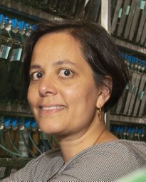 Annapurna Poduri合作论文数National Institute of Neurological Disorders and Stroke, National Institutes of Health;Eunice Kennedy Shriver National Institute of Child Health and Human Development, National Institutes of Health;Broad Institute of MIT and Harvard7
 Jerry J. Shih合作论文数7Norman Delanty合作论文数7
 Heidi Elyse Kirsch合作论文数Department of Neurology, University of California7
 Gregory K. Bergey合作论文数Department of Neurology;Johns Hopkins Epilepsy Center;School of Medicine, Johns Hopkins University7Paul Rutecki合作论文数7
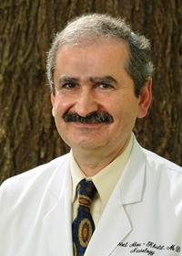 Bassel W. Abou Khalil合作论文数Department of Neurology, School of Medicine, Vanderbilt University7
 Robert E. Wharen合作论文数Department of Neurosurgery (Reimer, Wharen),, Mayo Clinic Jacksonville7
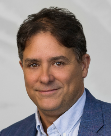 David B. Goldstein合作论文数Actio BioSciences, Inc.;Praxis Precision Medicines, Inc.7
 Gregory A. Worrell合作论文数Department of Neurology, Mayo Clinic;Department of Physiology & Biomedical Engineering, Mayo Clinic7Gregory L. Barkley合作论文数7Paul Motika合作论文数6Anu Venkat合作论文数6
 Paul C. Van Ness合作论文数6Simon Glynn合作论文数6




