OBJECTIVE: We aimed at explaining the mechanism of therapeutic effect of Umbilical Cord Mesenchymal Stem Cells (UC-MSC) in subjects with COVID-19 Acute Respiratory Distress Syndrome (ARDS). Patients with COVID-19 ARDS present with a hyperinflammatory response characterized by high levels of circulating pro-inflammatory mediators, including tumor necrosis factor α and β (TNFα and TNFβ). Inflammatory functions of these TNFs can be inhibited by soluble TNF Receptor 2 (sTNFR2). In patients with COVID-19 ARDS, UC-MSC appear to impart a robust anti-inflammatory effect, and treatment is associated with remarkable clinical improvements. We investigated the levels of TNFα, TNFβ and sTNFR2 in blood plasma samples collected from subjects with COVID-19 ARDS enrolled in our trial of UC-MSC treatment. PATIENTS AND METHODS: We analyzed plasma samples from subjects with COVID-19 ARDS (n=24) enrolled in a Phase 1/2a randomized controlled trial of UC-MSC treatment. Plasma samples were obtained at Day 0 (baseline, before UC-MSC or control infusion), and Day 6 post infusion. Plasma concentrations of sTNFR2, TNFα, and TNFβ were evaluated using a quantitative multiplex protein array. RESULTS: Our data indicate that at Day 6 after infusion, UC-MSC recipients develop significantly increased levels of plasma sTNFR2 and significantly decreased levels of TNFα and TNFβ, compared to controls. CONCLUSIONS: These observations suggest that sTNFR2 plays a mechanistic role in mediating UC-MSC effect on TNFα and TNFβ plasma levels, determining a decrease in inflammation in COVID-19 ARDS.
Background & Aim: Mesenchymal stem cells (MSCs) have been shown to modulate hyperinflammation, promote tissue repair and secrete antimicrobial factors. MSCs have been studied in clinical trials of autoimmune diseases, inflammatory disorders, refractory GvHD and acute respiratory distress syndrome (ARDS). MSCs can be isolated and expanded from multiple tissues, including umbilical cord (UC). A number of clinical studies demonstrated safety and feasibility of UCMSCs therapy for the treatment of COVID-19 ARDS. UC-derived MSCs are easily available and can be quickly expanded to relevant numbers. UC-MSCs have an extended population doubling capacity and express low levels of class I and class II leukocyte antigen, which may reduce alloreactivity. To meet clinical manufacture demands, UC-MSC production requires an innovative, scaled-up manufacturing platform. We describe the manufacturing strategy developed in support of a double-blind, randomized, controlled UC- MSC clinical trial in subjects with COVID-19 ARDS. Methods, Results & Conclusion: UC-MSC Final Product was manufactured from the master cell bank (MCB) derived from subepithelial lining of a UC from a healthy term delivery, in cGMP conditions. Utilizing a 2D culture xenogeneic protein-free process, UC-MSC MCB was culture-expanded during 3 expansion cycles, in tissue culture treated vessels with increased surface area for each expansion, in commercially available tissue culture media supplemented with platelet lysate. Cells were harvested during log phase, at 75-80% confluence. The manufacturing process yielded ~ 300x increase in total viable cells at the end of the last expansion cycle. The Final Product was cryopreserved using a controlled rate freezer. Each subject in the treatment group received two doses of 100×106 UC-MSCs. A single UC-MSC Final Product batch was sufficient to treat all subjects randomized to the treatment group and complete the trial. The final product was tested for identity (label verification), effectiveness by viable cell dose and cell viability (>80%), safety by assessment of endotoxin ( 90%, CD34/CD45 <10%). UC-MSC cell doses prepared for infusion produced similar results to UC-MSC Final Product when tested to confirm product identity, effectiveness, safety and purity. The developed 2D culture and expansion process can be successfully scaled up without compromising integrity of the final UC-MSC product.
This commentary describes how diabetes research using human pancreatic islets has been drastically altered over the past six months, due to COVID is based on the IIDP experience and on interviews conducted with IIC staff members and IIDP recipient investigators.
BACKGROUND: Coronavirus Disease 2019 (COVID-19) caused by SARS-CoV-2 coronavirus is a worldwide epidemic. Estimates of the infection vary by country and region, and US reports over a quarter of the total COVID-19 cases, reported worldwide. COVID-19 has made a significant impact on organ transplantation, in general, and islet cell transplantation, in particular. Islet cell transplantation has been proven a viable cell replacement strategy for treatment of patients with impaired awareness of hypoglycemia and severe hypoglycemia and is now approved as standard of care in Canada, Europe, Japan and Australia. Clinical success of an islet transplant is largely dependent on the quality of a deceased donor pancreas. Hence, careful selection and testing of potential organ donors are of critical importance. The threat of COVID-19 transmission has either significantly slowed down or completely shut down islet transplant programs in most US transplant centers. MATERIALS AND METHODS: Literature regarding COVID-19 infection rates and mitigation strategies, National Institutes of Health, American Society of Transplantation and UNOS (United Network for Organ Sharing) recommendations regarding donor organ testing for SARS-CoV-2 and resource allocation were reviewed. CONCLUSIONS: Impact of local COVID-19 transmission and changing epidemiology of the disease, availability of resources that include protective equipment, donor procurement teams and adequate donor testing, impact of immunosuppression regiments on COVID-19 infection, as well as local regulations, are issues that should be critically assessed prior to reopening islet transplant programs.
The coronavirus SARS-CoV-2 is cause of a global pandemic of a pneumonia-like disease termed Coronavirus Disease 2019 (COVID-19). COVID-19 presents a high mortality rate, estimated at 3.4%. More than 1 out of 4 hospitalized COVID-19 patients require admission to an Intensive Care Unit (ICU) for respiratory support, and a large proportion of these ICU-COVID-19 patients, between 17% and 46%, have died. In these patients COVID-19 infection causes an inflammatory response in the lungs that can progress to inflammation with cytokine storm, Acute Lung Injury (ALI), Acute Respiratory Distress Syndrome (ARDS), thromboembolic events, disseminated intravascular coagulation, organ failure, and death. Mesenchymal Stem Cells (MSCs) are potent immunomodulatory cells that recognize sites of injury, limit effector T cell reactions, and positively modulate regulatory cell populations. MSCs also stimulate local tissue regeneration via paracrine effects inducing angiogenic, anti-fibrotic and remodeling responses. MSCs can be derived in large number from the Umbilical Cord (UC). UC-MSCs, utilized in the allogeneic setting, have demonstrated safety and efficacy in clinical trials for a number of disease conditions including inflammatory and immune-based diseases. UC-MSCs have been shown to inhibit inflammation and fibrosis in the lungs and have been utilized to treat patients with severe COVID-19 in pilot, uncontrolled clinical trials, that reported promising results. UC-MSCs processed at our facility have been authorized by the FDA for clinical trials in patients with an Alzheimer's Disease, and in patients with Type 1 Diabetes (T1D). We hypothesize that UC-MSC will also exert beneficial therapeutic effects in COVID-19 patients with cytokine storm and ARDS. We propose an early phase controlled, randomized clinical trial in COVID-19 patients with ALI/ARDS. Subjects in the treatment group will be treated with two doses of UC-MSC (l00 × 106 cells). The first dose will be infused within 24 hours following study enrollment. A second dose will be administered 72 ± 6 hours after the first infusion. Subject in the control group will receive infusion of vehicle (DPBS supplemented with 1% HSA and 70 U/kg unfractionated Heparin, delivered IV) following the same timeline. Subjects will be evaluated daily during the first 6 days, then at 14, 28, 60, and 90 days following enrollment (see Schedule of Assessment for time window details). Safety will be determined by adverse events (AEs) and serious adverse events (SAEs) during the follow-up period. Efficacy will be defined by clinical outcomes, as well as a variety of pulmonary, biochemical and immunological tests. Success of the current study will provide a framework for larger controlled, randomized clinical trials and a means of accelerating a possible solution for this urgent but unmet medical need. The proposed early phase clinical trial will be performed at the University of Miami (UM), in the facilities of the Diabetes Research Institute (DRI), UHealth Intensive Care Unit (ICU) and the Clinical Translational Research Site (CTRS) at the University of Miami Miller School of Medicine and at the Jackson Memorial Hospital (JMH).
Cell Transplant Center Diabetes Research Institute University of Miami School of Medicine th 1450 NW 10 Avenue (R134) Miami, FL 33136 SOP: Cellular Composition and Fractional β-Cell Viability Assay Page 1 of 12 Original Version: 05/2005 Updated Version: 07/2014 CellR4 2014; 2 (4): e1121 – www.cellr4.org – ISSN: 2329-7042 Original Version: 05/2005 Attachments: N/A Cellular Composition and Fractional β-Cell Viability Assay PURPOSE: To outline the procedure for assessment of cellular composition and fractional beta cell viability for purified islets of Langerhans.
The NIH CIT Consortium Chemistry Manufacturing Controls Monitoring Committee: J. Ansite, A.N. Balamurugan, B. Barbaro, J. Battle, D. Brandhorst, J. Cano, X. Chen, S. Deng, D. Feddersen, A. Friberg, T. Gilmore, J.S. Goldstein, E. Holbrook, A. Khan, T. Kin, J. Lei, E. Linetsky, C. Liu, X. Luo, K. McElvaney, Z. Min, J. Moreno, D. O’Gorman, K.K. Papas, G. Putz, C. Ricordi, G. Szot, T. Templeton, L. Wang, J.J. Wilhelm, J. Willits, T. Wilson, X. Zhang
ackground: T cell depleting strategies have become an integral part of immunosuppressive regimens widely used for induction in solid organ transplantation. Alemtuzumab (AL) is a recombinant humanized monoclonal antibody against human CD52, a cell surface antigen expressed on B and T cells, monocytes, and natural killer (NK) cells. Although the depletion of lymphocytes would be expected to result in an increased risk of infections, some studies reported not to increase the incidence of infections. We observed early large granulocyte reconstitution in liver transplantation (LT) with AL. However, information is limited regarding the phenotype and function of liver NK cells in T cell depletion using AL. Methods: Absolute counts and proportion of NK and T cells were measured after LT with AL. We collected the mononuclear cells from the liver (LMC), peripheral blood (PBMC), spleen (SP), and lymph nodes (LN) from the donor in LT patients. Phenotype and functional differences were examined by flow cytometry and in vitro cytotoxicity assays. Anti- CD3, CD16, TRAIL, NKp30, NKp44, NKp46, NKG2D, CD52, CD56, CD94, CD117, and CD158b mAbs are used for phenotyping lymphocyte and NK cells. Results: Absolute counts of T cells remained low for 6 months. In contrast, those of NK cells had emerged with larger number since early days after LT (Fig 1A). LMC (n=7) contained high percentage of CD52- CD56+ NK cells (54.2±9.0 % especially CD56 bright 81.2±5.7 %, Fig 1B). It was statistically significant higher percentage than that of PBMC, SP, and LN (25.1%, 28.4%, and 9.3 % respectively, P< 0.05). The phenotype of almost liver NK cells showed mature type (stage5; CD94+CD117-: 99.1%). The expressions of other markers had not any differences between CD52-CD56+ and CD52+CD56+ NK cells. On the other hand, PBMC contained high percentage of CD52+CD56+ NK cells (99.3%). Donor liver NK cells had stronger cytotoxicity after IL-2 stimulation in the presence of AL. These data indicated that NK cells in PBMC were depleted but NK cells in LMC remained alive and active in LT patient with AL induction.[Figure 1]Conclusions: The liver contains high percentage of CD52-CD56+ NK cells which are mature type. The function of CD52-CD56+ NK cells had stronger cytotoxicity even in the presence of AL. These results suggest that the functionally maintained CD52-CD56+ NK cells contribute to protect the recipients from severe infections even after T cell depletion therapy such as AL induction.
At the present time, transplantation of pancreatic islet cells is considered an experimental therapy for a selected cohort of patients with type 1 diabetes, and is conducted under an Investigational New Drug (IND) application. Encouraging results of the Edmonton Protocol published in the year 2000 sparked a renewed interest in clinical transplantation of allogeneic islets, triggering a large number of IND applications for phase I clinical trials. Promising results reported by a number of centers since then prompted the Food and Drug Administration (FDA) to consider the possibility of licensing allogeneic islets as a therapeutic treatment for patients with type 1 diabetes. However, prior to licensure, issues such as safety, purity, efficacy, and potency of the islet product must be addressed. This is complicated by the intricate nature of pancreatic islets and limited characterization prior to transplantation. In this context, control of the manufacturing process plays a critical role in the definition of the final product. Despite significant progress made in standardization of the donor organ preservation methods, reagents used, and characterization assays performed to qualify an islet cell product, control of the isolation process remains a challenge. Within the scope of the FDA regulations, islet cells meet the definition of a biologic product, somatic cell therapy, and a drug. In addition, AABB standards that address cellular therapy products apply to manufacturing facilities accredited by this organization. Control of the source material, isolation process, and final product are critical issues that must be addressed in the context of FDA and other relevant regulations applicable to islet cell products.
Transplantation of islets of Langerhans in patients with type 1 diabetes allows for improved metabolic control and insulin independence. The need for chronic immunosuppression limits this procedure to selected patients with brittle diabetes. Definition of therapeutic strategies allowing permanent engraftment without the need for chronic immunosuppression could overcome such limitations. We tested the effect of the use of protoporphyrins (CoPP and FePP), powerful inducers of the cytoprotective protein heme-oxygenase 1 (HO-1), on allogeneic islet graft survival. Chemically induced diabetic C57BL/6 mice received DBA/2 islets. Treatment consisted in peritransplant administration of CoPP or saline. Islets were either cultured in the presence of FePP or vehicle before implant. Short-course administration of CoPP led to long-term islet allograft survival in a sizable proportion of recipients. Long-term graft-bearing animals rejected third-party islets while accepting a second set donor-specific graft permanently, without additional treatment. Preconditioning of islets with FePP by itself led to improved graft survival in untreated recipients, and provided additional advantage in CoPP-treated recipients, resulting in an increased proportion of long-term surviving grafts. Preconditioning of the graft with protoporphyrins prior to implant resulted in reduction of class II expression. Administration of protoporphyrins to the recipients of allogeneic islets also resulted in transient powerful immunosuppression with reduced lymphocyte proliferative responses, increased proportion of regulatory cells (CD4+CD25+), decreased mononuclear cell infiltrating the graft, paralleled by a systemic upregulation of HO-1 expression. All these mechanisms may have contributed to the induction of donor-specific hyporesponsiveness in a proportion of the protoporphyrin-treated animals.
Background. Transplantation tolerance, defined as acceptance of a graft by an otherwise fully immunocompetent host, has been an elusive goal. Although robust tolerance has been achieved by the induction of stable hematopoietic chimerism after bone marrow transplantation, lethal or sublethal radiation conditioning used to induce long-term chimerism precludes its clinical use. We studied whether targeted delivery of radiation to bone marrow could allow for bone marrow cell (BMC) engraftment, chimerism, and donor-specific tolerance in the absence of the side effects associated with external irradiation. Methods. We administered a radioactive bone-seeking compound (153Samarium-Lexidronam, Quadramet, Berlex Laboratories, Wayne, NJ) together with transient T-cell costimulatory blockade to recipient mice. Allogeneic BMCs were given 7 or 14 days after preconditioning. Costimulatory blockade was obtained by the use of an anti-CD154 antibody for 4 weeks. Chimerism was assessed by flow cytometry. Mice then received donor-specific and third-party skin grafts. Graft survival was analyzed with mechanisms of donor-specific hyporesponsiveness. Results. High levels of stable chimerism across an allogeneic barrier were achieved in mice by a single administration of 153Samarium-Lexidronam, transient T-cell costimulatory blockade, and BMC transplantation. A large percentage of chimeric animals retained donor-derived skin grafts for more than 120 days without requiring additional immunosuppression, suggesting that harsh cytotoxic preconditioning is not necessary to achieve stable chimerism and donor specific hyporesponsiveness. Analysis of the T-cell repertoire in chimeras indicates T-cell deletional mechanisms. Conclusions. These data broaden the potential use of BMC transplantation for tolerance induction and argue for its potential in treating autoimmune diseases.
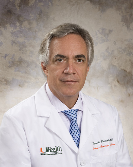 Camillo Ricordi合作论文数Division of Cellular Transplantation, DeWitt Daughtry Family Department of Surgery, Miller School of Medicine, University of Miami27
 Gennaro Selvaggi合作论文数10
 Rita Bottino合作论文数Rangos Research Center;Department of Pediatrics;Hospital of Pittsburgh;University of Pittsburgh;School of Medicine;School of Medicine, University of Pittsburgh10Rodolfo Alejandro合作论文数9S S Kong合作论文数7D. Feddersen合作论文数6A. Friberg合作论文数6
 Tatsuya Kin合作论文数6B. Barbaro合作论文数6
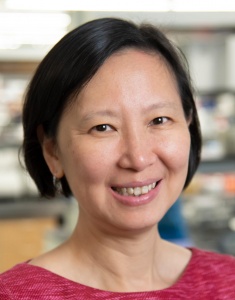 Xunrong Luo合作论文数Department of Medicine, School of Medicine, Duke University6
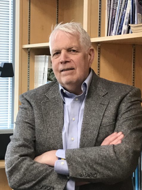 Thomas D. Gilmore合作论文数Dept of Pharmacology & Experimental Therapeutics, Boston University;The Gilmore Lab, Biology Department, Boston University;Cancer Center of the BU-Boston Medical Center;Genome Science Institute, Boston University6Lei Ji合作论文数6Luca Inverardi合作论文数6
 Eric H. Holbrook合作论文数Division of Rhinology, Massachusetts Eye and Ear, Harvard Medical School, Harvard University;Department of Otolaryngology Head and Neck Surgery, Harvard Medical School, Harvard University6
 Daniel Brandhorst合作论文数Nuffield Department of Surgical Sciences, University of Oxford6Min Zha合作论文数6Gregory L. Szot合作论文数6Norma S. Kenyon合作论文数Diabetes Research Institute;University of Miami;School of Medicine6J. Wilhelm合作论文数5J Ansite合作论文数5s carpentier合作论文数5
 A.N Balamurugan合作论文数5T Qian合作论文数4Schroeder A合作论文数4R D Molano合作论文数3S. Deng合作论文数3
 Luis Alberto Fernandez合作论文数3
 Hirohito Ichii合作论文数3P Ruiz合作论文数3
 Roger Lehmann合作论文数3
 O'Gorman Doug合作论文数Clinical Islet Transplant Program;Department of Transplant Services;Alberta Health Services;Edmonton;Department of Transplant Services, Alberta Health Services3H. Li合作论文数3
 Giacomo Lanzoni合作论文数Interuniversity Consortium I.N.B.B.3Antonello Pileggi合作论文数3D. Bhardwaj合作论文数2S Messinger Cayetano合作论文数2D Baidal合作论文数2Rosichuk S合作论文数2
 Xiaoming Xu合作论文数Laboratory of Spinal Cord Regenerative Medicine, School of Medicine, Indiana University2Andreas G Tzakis合作论文数2Malcolm C. Paterson合作论文数2
 Andreas Tzakis合作论文数Transplant Center, Department of Surgery, Cleveland Clinic2Klearchos K. Papas合作论文数Department of Surgery, University of Minnesota Medical School2
 Diego Correa合作论文数Skeletal Research Center, Case Western Reserve University2Roseline Godbout合作论文数Department of Oncology;University of Alberta;Cross Cancer Institute;Cross Cancer Institute, University of Alberta2Parastoo Dinyari合作论文数2
 Amit J. Patel合作论文数The Orthopaedic Department, Iris Hospital;Shri Krishna Hospital;Pramukhswami Medical College, Bhaikaka University2
 Rena Pawlick合作论文数2
 Nicole A Turgeon合作论文数2




