Radiotherapy-induced cardiac toxicity and consequent diseases still represent potential severe late complications for many cancer survivors who undergo therapeutic thoracic irradiation. We aimed to assess the phenotypic and paracrine features of resident cardiac mesenchymal stromal cells (CMSCs) at early follow-up after the end of thoracic irradiation of the heart as an early sign and/or mechanism of cardiac toxicity anticipating late organ dysfunction. Resident CMSCs were isolated from a rat model of fractionated thoracic irradiation with accurate and clinically relevant heart dosimetry that developed delayed dose-dependent cardiac dysfunction after 1 year. Cells were isolated 6 and 12 weeks after the end of radiotherapy and fully characterized at the transcriptional, paracrine, and functional levels. CMSCs displayed several altered features in a dose- and time-dependent trend, with the most impaired characteristics observed in those exposed in situ to the highest radiation dose with time. In particular, altered features included impaired cell migration and 3D growth and a and significant association of transcriptomic data with GO terms related to altered cytokine and growth factor signaling. Indeed, the altered paracrine profile of CMSCs derived from the group at the highest dose at the 12-week follow-up gave significantly reduced angiogenic support to endothelial cells and polarized macrophages toward a pro-inflammatory profile. Data collected in a clinically relevant rat model of heart irradiation simulating thoracic radiotherapy suggest that early paracrine and transcriptional alterations of the cardiac stroma may represent a dose- and time-dependent biological substrate for the delayed cardiac dysfunction phenotype observed in vivo.
Background: Post-COVID-19 Interstitial Lung Disease (PC-ILD) is characterized by fibrotic-like signs at high-resolution computed tomography (HRCT) and pulmonary function tests (PFTs) abnormalities after SARS-CoV-2 infection. It is still not clear how frequent these tests should be performed to rule out long-term consequences of COVID-19 pneumonia. Objectives: The aims of our study were to evaluate the incidence and risk factors of PC-ILD and possibly to propose a long-term follow-up program. Method: One-hundred patients, hospitalized in our ward for moderate to critical COVID-19, underwent two follow-up visits at three and 15 months in which PFTs and HRCT were performed. Results: At the 15-month follow-up, 8% of patients showed residual radiological and functional signs consistent with PC-ILD. All but one of these patients had already demonstrated PFTs and HRCT alterations at first follow-up visit, and the last 1 patient showed worsening of lung function during follow-up. These findings highlight the negative predictive value of PFTs at 3-month follow-up for the development of PC-ILD. Aging, severity of COVID-19, and degree of pulmonary involvement during acute infection proved to be significant risk factors for developing PC-ILD. Conclusions: Our study highlights the importance of PFTs in the long-term follow-up of patients affected by moderate to critical COVID-19 pneumonia. Further studies are needed to confirm our hypothesis that HRCT should be performed only in patients with PFTs abnormalities.
Methacryloyl gelatin (GelMA) was blended with dextran methacrylate (DexMA) to produce polymer networks with interconnected macropores via cryostructuring and radical crosslinking of the polymers at subzero tem-peratures. The experimental set-up was optimized to allow the formation of monolithic networks characterized by highly uniform structure with interconnected macropores. The total polymers mass, the amount of the gel -forming reagents and particularly the rheological properties of GelMA resulted the most critical factors for the fabrication of homogeneous and not collapsed scaffolds. Indeed, only the use of GelMA with very low gelation temperature resulted in the formation of uniform monolithic cryogels. However, blending with DexMA produced general worsening of the mechanical properties of the scaffolds, due to DexMA interference with secondary structuring of GelMA during the cryogelation process. DexMA also had negative effect on the ability of the cryogel to support growth and proliferation of HaCat cells, bringing to slower cell adhesion to the scaffold.
Background: Conversion of cardiac stromal cells into myofibroblasts is typically associated with hypoxia conditions, metabolic insults, and/or inflammation, all of which are predisposing factors to cardiac fibrosis and heart failure. We hypothesized that this conversion could be also mediated by response of these cells to mechanical cues through activation of the Hippo transcriptional pathway. The objective of the present study was to assess the role of cellular/nuclear straining forces acting in myofibroblast differentiation of cardiac stromal cells under the control of YAP (yes-associated protein) transcription factor and to validate this finding using a pharmacological agent that interferes with the interactions of the YAP/TAZ (transcriptional coactivator with PDZ-binding motif) complex with their cognate transcription factors TEADs (TEA domain transcription factors), under high-strain and profibrotic stimulation. Methods: We employed high content imaging, 2-dimensional/3-dimensional culture, atomic force microscopy mapping, and molecular methods to prove the role of cell/nuclear straining in YAP-dependent fibrotic programming in a mouse model of ischemia-dependent cardiac fibrosis and in human-derived primitive cardiac stromal cells. We also tested treatment of cells with Verteporfin, a drug known to prevent the association of the YAP/TAZ complex with their cognate transcription factors TEADs. Results: Our experiments suggested that pharmacologically targeting the YAP-dependent pathway overrides the profibrotic activation of cardiac stromal cells by mechanical cues in vitro, and that this occurs even in the presence of profibrotic signaling mediated by TGF-β1 (transforming growth factor beta-1). In vivo administration of Verteporfin in mice with permanent cardiac ischemia reduced significantly fibrosis and morphometric remodeling but did not improve cardiac performance. Conclusions: Our study indicates that preventing molecular translation of mechanical cues in cardiac stromal cells reduces the impact of cardiac maladaptive remodeling with a positive effect on fibrosis.
The contact between integrated microelectronics circuits and cellular membrane represented a great issue which research tried to solve in the last years. This achievement could open a huge field of device and systems, bringing the sensitivity and intelligence of microelectronics toward the biological systems. We describe an innovative approach to gain the electric sensing of the membrane by covering the chip contact surface with a carpet of nanowires, in particular silicon and zinc oxide nanowires. Both material ensures low invasiveness toward the biological samples. The nanowires growth is with ICs, allowing on-site amplification of bioelectric signals. We report our preliminary results showing biocompatibility and neutrality of silicon and zinc oxide nanowires used as seeding substrate for cells in culture.
Long COVID-19 is defined as persistency of symptoms, such as exertional dyspnea, twelve weeks after recovery from SARS-CoV-2 infection; its pathophysiology still needs to be fully understood. We investigated exercise tolerance and ventilatory efficiency using cardiopulmonary exercise testing (CPET) in patients with long COVID-19. Methods. One hundred patients admitted to our hospital from March to August 2020 for a moderate to critical COVID-19 were enrolled in our long COVID-19 program. Medical history, physical examination and chest HRCT were obtained at hospitalization (T0), at 3 (T3) and 15 months (T15). All HRCTs were revised using a semi-quantitative CT severity score (Pan, F. et al. Radiology 2020; 295(3):715–721). Pulmonary function tests (PFTs) were obtained at T3 and T15. CPET was performed at T15 in twenty patients (10 male/10 female; mean age 62 years) with residual respiratory symptoms (e.g., exertional dyspnea) and/or an impairment in PFTs, DLCO and/or KCO. Results. At CPET, peak oxygen uptake (VO2-peak) and ventilatory efficiency (VE/VCO2 slope) were 95.9±18.4 SD %pred and 31.4±3.9 SD, respectively. Of notice, significant correlations between VE/V’CO2 slope and CT score (T0) (r=0.403; p=0.039), CT score (T3) (r=0.453; p=0.022) and DLCO (T3) (r=-0.465; p=0.019) were observed. Conclusions. At fifteen-months from COVID-19 pneumonia, a significant number of subjects (20%) still complains of exertional dyspnea. At CPET this may be explained by reduced ventilatory efficiency (i.e., increase in VE/VCO2), possibly related to the degree of lung parenchymal involvement in the COVID-19 acute phase, likely reflecting a damage in the interstitial/pulmonary capillary structure.
Nano- or microdevices, enabling simultaneous, long-term, multisite, cellular recording and stimulation from many excitable cells, are expected to make a strategic turn in basic and applied cardiology (particularly tissue engineering) and neuroscience. We propose an innovative approach aiming to elicit bioelectrical information from the cell membrane using an integrated circuit (IC) bearing a coating of nanowires on the chip surface. Nanowires grow directly on the backend of the ICs, thus allowing on-site amplification of bioelectric signals with uniform and controlled morphology and growth of the NWs on templates. To implement this technology, we evaluated the biocompatibility of silicon and zinc oxide nanowires (NWs), used as a seeding substrate for cells in culture, on two different primary cell lines. Human cardiac stromal cells were used to evaluate the effects of ZnO NWs of different lengths on cell behavior, morphology and growth, while BV-2 microglial-like cells and GH4-C1 neuroendocrine-like cell lines were used to evaluate cell membrane–NW interaction and contact when cultured on Si NWs. As the optimization of the contact between integrated microelectronics circuits and cellular membranes represents a long-standing issue, our technological approach may lay the basis for a new era of devices exploiting the microelectronics’ sensitivity and “smartness” to both improve investigation of biological systems and to develop suitable NW-based systems available for tissue engineering and regenerative medicine.
Adipose-derived mesenchymal stem cells (ASCs) represent a valid therapeutic option for clinical application in several diseases, due to their ability to repair damaged tissues and to mitigate the inflammatory/immune response. A better understanding of the underlying mechanisms regulating ASC biology might represent the chance to modulate their in vitro characteristics and differentiation potential for regenerative medicine purposes. Herein, we investigated the effects of the demethylating agent 5-azacytidine (5-aza) on proliferation, clonogenicity, migration, adipogenic differentiation and senescence of ASCs, to identify the molecular pathways involved. Through functional assays, we observed a detrimental effect of 5-aza on ASC self-renewal capacity and migration, accompanied by actin cytoskeleton reorganization, with decreased stress fibers. Conversely, 5-aza treatment enhanced ASC adipogenic differentiation, as assessed by lipid accumulation and expression of lineage-specific markers. We analyzed the involvement of the Akt/mTOR, MAPK and Wnt/β-catenin pathways in these processes. Our results indicated impairment of Akt and ERK phosphorylation, potentially explaining the reduced cell proliferation and migration. We observed a 5-aza-mediated inhibition of the Wnt signaling pathway, this potentially explaining the pro-adipogenic effect of the drug. Finally, 5-aza treatment significantly induced ASC senescence, through upregulation of the p53/p21 axis. Our data may have important translational implications, by helping in clarifying the potential risks and advantages of using epigenetic treatment to improve ASC characteristics for cell-based clinical approaches.
Ex vivo modelling systems for cardiovascular research are becoming increasingly important in reducing lab animal use and boosting personalized medicine approaches. Integrating multiple cell types in complex setups adds a higher level of significance to the models, simulating the intricate intercellular communication of the microenvironment in vivo. Cardiac fibrosis represents a key pathogenetic step in multiple cardiovascular diseases, such as ischemic and diabetic cardiomyopathies. Indeed, allowing inter-cellular interactions between cardiac stromal cells, endothelial cells, cardiomyocytes, and/or immune cells in dedicated systems could make ex vivo models of cardiac fibrosis even more relevant. Moreover, culture systems with 3D architectures further enrich the physiological significance of such in vitro models. In this review, we provide a summary of the multicellular 3D models for the study of cardiac fibrosis described in the literature, such as spontaneous microtissues, bioprinted constructs, engineered tissues, and organs-on-chip, discussing their advantages and limitations. Important discoveries on the physiopathology of cardiac fibrosis, as well as the screening of novel potential therapeutic molecules, have been reported thanks to these systems. Future developments will certainly increase their translational impact for understanding and modulating mechanisms of cardiac fibrosis even further.
Here we present a novel technique to sense bioelectric signals enabling high space-time resolution and minimizing invasiveness based on low-temperature (200°C) growth of silicon nanowires (SiNWs). Such SiNWs are IC-compatible and allow in situ amplification of bioelectrical phenomena. In this paper we show our initial results in SiNWs biocompatibility and neutrality suggesting their use as seeding material for cell culturing. Using this technology our goal is to put together an innovative and compact device that allows synchronous, on-site recording of large amounts of biological signals from excitable cells networks with high SNR. Moreover, we aim to record signals from cell membrane distinct subdomains, providing very highly resolved measurements of electric waveforms propagation within living cells and networks.
We describe an innovative approach to sensing bioelectric signals at high space-time resolution with low invasiveness based on growing small Silicon Nano Wires (SiNW) at low-temperature (200 °C). The resulting SiNWs are compatible with ICs, allowing on-site amplification of bioelectric signals. We report our preliminary results showing biocompatibility and neutrality of SiNWs used as seeding substrate for cells in culture. With this technology, we aim to produce a compact device allowing on-site, synched and high signal/noise recordings of a large amounts of biological signals from networks of excitable cells (e.g. neurons) or distinct subdomains of the cell membrane, thus providing super-resolved descriptions of the propagation of electric waveforms within living cells and networks.
During embryonic morphogenesis, the heart undergoes a complex series of cellular phenotypic maturations (e.g., transition of myocytes from proliferative to quiescent or maturation of the contractile apparatus), and this involves stiffening of the extracellular matrix (ECM) acting in concert with morphogenetic signals. The maladaptive remodeling of the myocardium, one of the processes involved in determination of heart failure, also involves mechanical cues, with a progressive stiffening of the tissue that produces cellular mechanical damage, inflammation, and ultimately myocardial fibrosis. The assessment of the biomechanical dependence of the molecular machinery (in myocardial and non-myocardial cells) is therefore essential to contextualize the maturation of the cardiac tissue at early stages and understand its pathologic evolution in aging. Because systems to perform multiscale modeling of cellular and tissue mechanics have been developed, it appears particularly novel to design integrated mechano-molecular models of heart development and disease to be tested in ex vivo reconstituted cells/tissue-mimicking conditions. In the present contribution, we will discuss the latest implication of mechanosensing in heart development and pathology, describe the most recent models of cell/tissue mechanics, and delineate novel strategies to target the consequences of heart failure with personalized approaches based on tissue engineering and induced pluripotent stem cell (iPSC) technologies.
Cardiac adverse remodeling is characterized by biological changes that affect the composition and architecture of the extracellular matrix (ECM). The consequently disrupted signaling can interfere with the balance between cardiogenic and pro-fibrotic phenotype of resident cardiac stromal primitive cells (CPCs). The latter are important players in cardiac homeostasis and can be exploited as therapeutic cells in regenerative medicine. Our aim was to compare the effects of human decellularized native ECM from normal (dECM-NH) or failing hearts (dECM-PH) on human CPCs. CPCs were cultured on dECM sections and characterized for gene expression, immunofluorescence, and paracrine profiles. When cultured on dECM-NH, CPCs significantly upregulated cardiac commitment markers (CX43, NKX2.5), cardioprotective cytokines (bFGF, HGF), and the angiogenesis mediator, NO. When seeded on dECM-PH, instead, CPCs upregulated pro-remodeling cytokines (IGF-2, PDGF-AA, TGF-β) and the oxidative stress molecule H2O2. Interestingly, culture on dECM-PH was associated with impaired paracrine support to angiogenesis, and increased expression of the vascular endothelial growth factor (VEGF)-sequestering decoy isoform of the KDR/VEGFR2 receptor. Our results suggest that resident CPCs exposed to the pathological microenvironment of remodeling ECM partially lose their paracrine angiogenic properties and release more pro-fibrotic cytokines. These observations shed novel insights on the crosstalk between ECM and stromal CPCs, suggesting also a cautious use of non-healthy decellularized myocardium for cardiac tissue engineering approaches.
The increased knowledge in cell signals and stem cell differentiation, together with the development of new technologies, such as 3D bioprinting, has made the generation of artificial tissues more feasible for in vitro studies and in vivo applications. In the human body, cell fate, function, and survival are determined by the microenvironment, a rich and complex network composed of extracellular matrix (ECM), different cell types, and soluble factors. They all interconnect and communicate, receiving and sending signals, modulating and responding to cues. In the cardiovascular field, the culture of stem cells in vitro and their differentiation into cardiac phenotypes is well established, although differentiated cardiomyocytes often lack the functional maturation and structural organization typical of the adult myocardium. The recreation of an artificial microenvironment as similar as possible to the native tissue, though, has been shown to partly overcome these limitations, and can be obtained through the proper combination of ECM molecules, different cell types, bioavailability of growth factors (GFs), as well as appropriate mechanical and geometrical stimuli. This review will focus on the role of the ECM in the regulation of cardiac differentiation, will provide new insights on the role of supporting cells in the generation of 3D artificial tissues, and will also present a selection of the latest approaches to recreate a cardiac microenvironment in vitro through 3D bioprinting approaches.
The ‘cardiosphere’ is a 3D cluster of cardiac progenitor cells recapitulating a stem cell niche-like microenvironment with a potential for disease and regeneration modelling of the failing human myocardium. In this multicellular 3D context, it is extremely important to decrypt the spatial distribution of cell markers for dissecting the evolution of cellular phenotypes by direct quantification of fluorescent signals in confocal microscopy. In this study, we present a fully automated method, named CARE (‘CARdiosphere Evaluation’), for the segmentation of membranes and cell nuclei in human-derived cardiospheres. The proposed method is tested on twenty 3D-stacks of cardiospheres, for a total of 1160 images. Automatic results are compared with manual annotations and two open-source software designed for fluorescence microscopy. CARE performance was excellent in cardiospheres membrane segmentation and, in cell nuclei detection, the algorithm achieved the same performance as two expert operators. To the best of our knowledge, CARE is the first fully automated algorithm for segmentation inside in vitro 3D cell spheroids, including cardiospheres. The proposed approach will provide, in the future, automated quantitative analysis of markers distribution within the cardiac niche-like environment, enabling predictive associations between cell mechanical stresses and dynamic phenotypic changes.
Abstract Background Cardiac stromal cells are the most abundant cell type present in the heart, where they have an important homeostatic function in matrix renewal and electromechanical coupling. We have shown that YAP transcription factor (a component of the Hippo pathway) is important for the proliferation of these cells and, potentially, pathologic cardiac matrix stiffening in heart failure. Purpose Given the role of Hippo pathway in cell mechanosensation, we assessed the conditions for activation of the YAP-dependent transcriptional pathway related to geometry/strain sensing using human “cardiosphere”-derived cells. Methods Human cardiospheres (C-Sp) and C-Sp-derived cells (CDCs) were obtained from fragments of atrial appendages of patients admitted for AoC bypass. C-Sps and CDCs were cultured using published methods. To analyze cell/nuclear geometry in 2D/3D culture, we employed an adapting thresholding algorithm allowing segmentation of cell membrane/nuclei shape and the relative abundance of nuclear/cytoplasmic fluorescence intensities from z-stacked confocal immunofluorescence images. Inhibitors of signaling cascades converging onto actin cytoskeleton polymerization or of YAP/TEADs interaction were used to assess YAP target genes transcription. Results Previous results showed stiffness sensitivity of YAP nuclear translocation process. Namely, CDCs plated onto stiff (E >50kPa) culture plates exhibited high ratios of nuclear/cytoplasmic YAP, as compared to cells plated on substrates with lower stiffness (E ∼ 10kPa). Analogously, cells with a stretched cytoplasm/nuclear shape in the border regions of the 3D organoids exhibited a higher YAP nuclear localization level and expression of fibrotic markers compared with cells with a more round geometry in the center of the spheres. To uncouple the cytoskeleton tensioning-dependent YAP nuclear shuttling, we treated CDCs plated onto stiff substrates with blebbistatin and Y27632. This showed a fully reversible YAP relocation from the nucleus to the cytoplasm and a reversion of canonical target genes (CYR61, ANKRD1 and CTGF) expression. In parallel, a “softening” process of the nucleus, likely due to release from cytoskeleton traction forces, was observed by confocal imaging along the cellular z-axis. Interestingly, use of Verteporfin, a drug directly interfering with the interaction of YAP/TAZ complex with TEADs DNA binding proteins showed reversion of pro-fibrotic YAP targets independent of high cytoskeleton tensioning. Conclusions These results establish, for the first time, a deterministic relationship between pro-pathologic evolution of human adult cardiac stromal cells and activation of mechanosensing-dependent pathways. They also provide quantitative criteria for interpreting fibrotic evolution of the heart based on modifications of myocardial architecture and mechanical compliance, as well as the ability of the cells to generate tension forces affecting the nuclear structure.
We propose a discrete in continuous mathematical model describing the in vitro growth process of biophsy-derived mammalian cardiac progenitor cells growing as clusters in the form of spheres (Cardiospheres). The approach is hybrid: discrete at cellular scale and continuous at molecular level. In the present model, cells are subject to the self-organizing collective dynamics mechanism and, additionally, they can proliferate and differentiate, also depending on stochastic processes. The two latter processes are triggered and regulated by chemical signals present in the environment. Numerical simulations show the structure and the development of the clustered progenitors and are in a good agreement with the results obtained from in vitro experiments.
Exercise represents an important lifestyle factor in all human ages when felt in harmony with other psycho-physical and environmental variables that affect individual life (e. g. quality of interest, affections, environment, diet and food). Consequently, in addition to the training level, the amount, intensity and modality of exercise (ana-/aerobic, isometric/isotonic), need to be personalized, considering the underlying diseases, which may benefit from it or worsening.Greek mythology gives us good examples of the exercise concept's evolution.From Discus-thrower to Spear-carrier the idea of physical activity is more effectively expressed. The Myron Discobolus displays the enduring pattern of athletic energy translated into the dynamic force given by the exercise. In Doryphoros instead, the physical activity is oriented to the achievement of the required psyco-physical harmony, who's the concept is aimed of being expressed by the sculpture.As outlined below, even in the field of arrhythmia, scientific evidence as well as clinical experience, supports the same concept: physical activity may be important while safely managed and personalized.
 Alessandro Giacomello合作论文数50
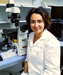 Isotta Chimenti合作论文数Pasteur Institute, University of Rome “Sapienza”,32Roberto Gaetani合作论文数Univ “La Sapienza”,31
 Giacomo Frati合作论文数Department of Medical-Surgical Sciences and Biotechnologies, Sapienza University of Rome22
 Francesco Angelini合作论文数21
 Lucio Barile合作论文数Pasteur Institute, University La Sapienza21
 Alberto Giacomello合作论文数Dipartimento di Ingegneria Meccanica e Aerospaziale, Università di Roma "La Sapienza;00184 Rome, Italy.;Dipartimento di Ingegneria Meccanica e Aerospaziale, Università di Roma "La Sapienza18
 Elvira Forte合作论文数Pasteur Institute, University of Rome “Sapienza”,16
 Eduardo Marbán合作论文数Department of physiology, University of California, Los Angeles12
 Rachel Smith合作论文数11L. Barile合作论文数9
 Vittoria Ionta合作论文数8
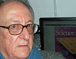 M Battaglia合作论文数CNR Institute of Neurobiology and Molecular Medicine7
 Francesca Pagano合作论文数7
 Roselle Abraham合作论文数Department of Medicine, University of California, San Francisco7
 Maurizio Pesce合作论文数Gene Expression Programme, EMBL7Hee Cho合作论文数6
 Vittorio Picchio合作论文数Dept Med Surg Sci & Biotechnol, Univ Rome Sapienza6
 Joost Sluijter合作论文数Department of Experimental Cardiology, Department of Cardiology, Division of Heart & Lungs, University Medical Center Utrecht6Fabio Miraldi合作论文数5Ottolenghi Sergio合作论文数5Michelle K. Leppo合作论文数5Maria Cristina Magli合作论文数5
 Umberto Morbiducci合作论文数Department of Mechanical and Aerospace Engineering, Politecnico di Torino5
 Francesco Cerisoli合作论文数ITB, CNR5I Chimenti合作论文数4Yuriy Derevyanchuk合作论文数4
 Pieter Doevendans合作论文数Division of Heart & Lungs, Department of Cardiology, University Medical Center Utrecht4
 Fabrizio Palma合作论文数Dipartimento di Elettronica, Università “La Sapienza”,4
 Fabrizio Rossi合作论文数Dipartimento di Informatica, Università di L’Aquila4Letizia Cassinelli合作论文数4F. Lupi合作论文数3Marcella Rocchetti合作论文数3C Salerno合作论文数3
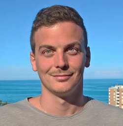 Massimo Salvi合作论文数Biolab, Department of Electronics and Telecommunications, Politecnico di Torino, Turin, Italy3Francesco Amadeo合作论文数3
 Georgina Ellison-Hughes合作论文数School of Basic & Medical Biosciences, Faculty of Life Sciences & Medicine, King’s College London3
 Diana Massai合作论文数3
 Tan Jun Jie合作论文数Advanced Medical and Dental Institute, Universiti Sains Malaysia3
 Dries Feyen合作论文数GSK3
 Daniel J. Stuckey合作论文数Centre for Advanced Biomedical Imaging, UCL3
 Mark Mercola合作论文数Department of Medicine, Stanford School of Medicine, Stanford University;Stanford Cardiovascular Institute, Stanford School of Medicine, Stanford University3
 Kieran Clarke合作论文数Department of Physiology, Anatomy and Genetics, Division of Medical Sciences, University of Oxford;TdeltaS3
 Giuseppe Biondi Zoccai合作论文数Dip. di Scienze e Biotecnologie Medico Chirurgiche, Sapienza University of Rome;Ospedale Santa Maria Goretti3
 Carolyn A. Carr合作论文数Department of Physiology, Anatomy & Genetics, University of Oxford3
 Filippo Molinari合作论文数Biolab, Politecnico di Torino3
 Elena De Falco合作论文数Laboratorio di Patologia Vascolare;Istituto Dermopatico dell ' Immacolata;Istituto di Ricovero e Cura a Carattere Scientifico;Laboratorio di Patologia Vascolare, Istituto di Ricovero e Cura a Carattere Scientifico3
 Antonio Zaza合作论文数3
 Valeria Panebianco合作论文数Academic Urology Unit, University of Sheffield;Department of Radiological, Oncological and Anatomopathological Sciences, Policlinico Umberto I Hospital, “Sapienza” Rome University3




