Movement-related gamma activity (> 60 Hz) in cortico-basal ganglia networks reflects pro-kinetic synchronization dynamics. While in the cortex these temporal dynamics are known to unfold spatially across topographically distributed networks, it remains unclear whether a similar spatial propagation occurs within the basal ganglia, and how such spatial encoding may contribute to both physiological and disease-related mechanisms. The subthalamic nucleus (STN) is a key integrative hub for motor processing within the basal ganglia-cortical circuitry. At rest, STN activity is topographically distributed according to its spectral frequency components. To assess whether this spectral topography is dynamic and underlies movement encoding, we dissected the spatiotemporal properties of STN local field potentials recorded intraoperatively at rest and during movement across 63 hemispheres from patients with Parkinson's disease. Using multi-contact deep brain stimulation leads, we captured high-resolution anatomical signal dynamics and contrasted a broad frequency spectrum (60-400 Hz), including high-gamma, fast-gamma, slow high-frequency oscillations and fast high-frequency oscillations. Moreover, we compared these signals to upper limb muscle activity and movement-related beta desynchronization, and examined their association with clinical impairment and levodopa responsiveness. All sub-bands exhibited significant movement-related synchronization in both the contralateral and ipsilateral STN, however, with distinct magnitude and temporal dynamics. The presence and degree of temporal locking to muscle activity and inverse relationship to movement-related beta desynchronization also varied by sub-band. Importantly, each sub-band exhibited spatially segregated hotspots located within the STN that propagate primarily along the inferior-superior axis, yet in band-specific directions. This spatial propagation evolved throughout the movement period but temporally decoupled from synchronization magnitude, indicating that spatial dynamics reflect a distinct property relevant for motor encoding. Notably, propagation of frequencies above 110 Hz inversely correlated with dopamine-related motor improvement, suggesting that exaggerated spatial dynamics may reflect compensatory mechanisms secondary to neurodegeneration. These findings demonstrated that synchronization within the basal ganglia is not a spatially static phenomenon but rather unfolds in space which expands on the current understanding of the basal ganglia mechanism. Propagation of movement-related activity may serve as a potential marker for motor impairment in Parkinson's disease, opening new avenues for spectro-behavioural research and spatially informed neuromodulation strategies.
Autonomic dysreflexia is a life-threatening medical condition characterized by episodes of uncontrolled hypertension that occur in response to sensory stimuli after spinal cord injury (SCI)1. The fragmented understanding of the mechanisms underlying autonomic dysreflexia hampers the development of therapeutic strategies to manage this condition, leaving people with SCI at daily risk of heart attack and stroke2-5. Here we expose the neuronal architecture that develops after SCI and causes autonomic dysreflexia. In parallel, we uncover a competing, yet overlapping neuronal architecture activated by epidural electrical stimulation of the spinal cord that safely regulates blood pressure after SCI. The discovery that these adversarial neuronal architectures converge onto a single neuronal subpopulation provided a blueprint for the design of a mechanism-based intervention that reversed autonomic dysreflexia in mice, rats and humans with SCI. These results establish a path towards essential pivotal device clinical trials that will establish the safety and efficacy of epidural electrical stimulation for the effective treatment of autonomic dysreflexia in people with SCI.
For several decades, deep brain stimulation has been a major breakthrough in the treatment of movement disorders, significantly improving the quality of life of patients with Parkinson's disease, but also with dystonia or tremor. Thanks to technological advances, this therapy continues to evolve: directional electrodes, adaptive stimulation, and neural recordings allow for more precise targeting and better clinical outcomes. At the same time, epidural electrical stimulation opens up new perspectives, notably for gait disorders, and represents a promising step forward in the field of therapeutic neuromodulation.
Autonomic dysreflexia is a life-threatening medical condition characterized by episodes of uncontrolled hypertension that occur in response to sensory stimuli after spinal cord injury (SCI)[1][1]–[7][2]. The fragmented understanding of the mechanisms underlying autonomic dysreflexia hampers the development of therapeutic strategies to manage this condition, leaving people with SCI at daily risk of heart attack and stroke[8][3]–[18][4]. Here, we expose the complete de novo neuronal architecture that develops after SCI and causes autonomic dysreflexia. In parallel, we uncover a competing, yet overlapping neuronal architecture activated by epidural electrical stimulation of the spinal cord that safely regulates blood pressure after SCI. The discovery that these adversarial neuronal architectures converge onto a single neuronal subpopulation provided a blueprint for the design of a mechanism-based intervention that reversed autonomic dysreflexia in mice, rats, and humans with SCI. These results establish a path for the effective treatment of autonomic dysreflexia in people with SCI.### Competing Interest StatementThe authors declare competing financial interests: G.C., A.A.P., J.W.S, J.B., R.D. and S.P.L. hold various patents in relation with the present work. G.C, A.A.P. and R.D. are consultants of ONWARD medical. G.C., A.A.P., J.B. and S.P.L. are minority shareholders of ONWARD, a company with direct relationships with the presented work. [1]: #ref-1 [2]: #ref-7 [3]: #ref-8 [4]: #ref-18
Cardinal motor symptoms in Parkinson's disease (PD) include bradykinesia, rest tremor and/or rigidity. This symptomatology can additionally encompass abnormal gait, balance and postural patterns at advanced stages of the disease. Besides pharmacological and surgical therapies, physical exercise represents an important strategy for the management of these advanced impairments. Traditionally, diagnosis and classification of such abnormalities have relied on partially subjective evaluations performed by neurologists during short and temporally scattered hospital appointments. Emerging sports medical methods, including wearable sensor-based movement assessment and computational-statistical analysis, are paving the way for more objective and systematic diagnoses in everyday life conditions. These approaches hold promise to facilitate customizing clinical trials to specific PD groups, as well as personalizing neuromodulation therapies and exercise prescriptions for each individual, remotely and regularly, according to disease progression or specific motor symptoms. We aim to summarize exercise benefits for PD with a specific emphasis on gait and balance deficits, and to provide an overview of recent advances in movement analysis approaches, notably from the sports science community, with value for diagnosis and prognosis. Although such techniques are becoming increasingly available, their standardization and optimization for clinical purposes is critically missing, especially in their translation to complex neurodegenerative disorders such as PD. We highlight the importance of integrating state-of-the-art gait and movement analysis approaches, in combination with other motor, electrophysiological or neural biomarkers, to improve the understanding of the diversity of PD phenotypes, their response to therapies and the dynamics of their disease progression.
We developed a real-time decoding framework that can accurately predict leg motor functions, as well as key aspects of walking, from local field potentials (LFP) recorded from the subthalamic nucleus of patients with Parkinson's disease. Concretely, we designed decoders that can predict locomotor states, gait events, modulations in force during obstacle avoidance, and freezing of gait episodes while participants walked freely in unconstrained conditions. Our algorithms employed the full spectrum of LFP recorded bilaterally, either through externalized deep brain stimulation (DBS) leads connected to an external, high-resolution amplifier (six bipolar channels, Fs = 8 kHz), or wirelessly using a last-generation implantable stimulator with sensing capabilities (Percept PC, Medtronic, two bipolar channels, Fs = 250 Hz). These results represent the first neural decoding of leg motor function operating in real-time from therapeutically implanted DBS electrodes. Considering the large number of patients treated worldwide with DBS implants, as well as the capabilities of newest commercial stimulators, our results pave the way for the design and widespread deployment of closed-loop neuromodulation therapies that address gait deficits with new closed-loop approaches.
People with late-stage Parkinson's disease (PD) often suffer from debilitating locomotor deficits that are resistant to currently available therapies. To alleviate these deficits, we developed a neuroprosthesis operating in closed loop that targets the dorsal root entry zones innervating lumbosacral segments to reproduce the natural spatiotemporal activation of the lumbosacral spinal cord during walking. We first developed this neuroprosthesis in a non-human primate model that replicates locomotor deficits due to PD. This neuroprosthesis not only alleviated locomotor deficits but also restored skilled walking in this model. We then implanted the neuroprosthesis in a 62-year-old male with a 30-year history of PD who presented with severe gait impairments and frequent falls that were medically refractory to currently available therapies. We found that the neuroprosthesis interacted synergistically with deep brain stimulation of the subthalamic nucleus and dopaminergic replacement therapies to alleviate asymmetry and promote longer steps, improve balance and reduce freezing of gait. This neuroprosthesis opens new perspectives to reduce the severity of locomotor deficits in people with PD.
Disruption of subthalamic nucleus dynamics in Parkinson’s disease leads to impairments during walking. Here, we aimed to uncover the principles through which the subthalamic nucleus encodes functional and dysfunctional walking in people with Parkinson’s disease. We conceived a neurorobotic platform embedding an isokinetic dynamometric chair that allowed us to deconstruct key components of walking under well-controlled conditions. We exploited this platform in 18 patients with Parkinson’s disease to demonstrate that the subthalamic nucleus encodes the initiation, termination, and amplitude of leg muscle activation. We found that the same fundamental principles determine the encoding of leg muscle synergies during standing and walking. We translated this understanding into a machine learning framework that decoded muscle activation, walking states, locomotor vigor, and freezing of gait. These results expose key principles through which subthalamic nucleus dynamics encode walking, opening the possibility to operate neuroprosthetic systems with these signals to improve walking in people with Parkinson’s disease.
ABSTRACTBackgroundREM sleep behaviour disorder (RBD) is a disabling, often overlooked sleep disorder affecting up to 70% of patients with Parkinson’s disease. Identifying and treating RBD is critical to prevent severe sleep-related injuries, both to patients and bedpartners. Current diagnosis relies on nocturnal video-polysomnography, which is an expensive and cumbersome exam requiring specific clinical expertise.ObjectivesTo design, optimise, and validate a novel home-screening tool, termed RBDAct, that automatically identifies RBD in Parkinson’s patients based on wrist actigraphy only.MethodsTwenty-six Parkinson’s patients underwent two-week home wrist actigraphy worn on their more affected arm, followed by two non-consecutive in-lab evaluations. Patients were classified as RBD versus non-RBD based on dream enactment history and video-polysomnography. We characterised patients’ movement patterns during sleep using raw tri-axial accelerometer signals from wrist actigraphy. Machine learning classification algorithms were then trained to discriminate between patients with or without RBD using actigraphic features that described patients’ movements. Classification performance was quantified with respect to clinical diagnosis, separately for in-lab and at-home recordings.ResultsClassification performance from in-lab actigraphic data reached an accuracy of 92.9±8.16% (sensitivity 94.9±7.4%, specificity 92.7±13.8%). When tested on home recordings, accuracy rose to 100% over the two-week window. Features showed robustness across tests and conditions.ConclusionsRBDAct provides reliable predictions of RBD in Parkinson’s patients based on home wrist actigraphy only. These results open new perspectives for faster, cheaper and more regular screening of sleep disorders, both for routine clinical practice and for clinical trials.
Objectives Rapid eye movement sleep behavior disorder (RBD) is a potentially harmful, often overlooked sleep disorder affecting up to 70% of Parkinson's disease patients. Current diagnosis relies on nocturnal video‐polysomnography, which is an expensive and cumbersome examination requiring specific clinical expertise. Here, we explored the use of wrist actigraphy to enable automatic RBD diagnoses in home settings. Methods A total of 26 Parkinson's disease patients underwent 2‐week home wrist actigraphy, followed by two in‐laboratory evaluations. Patients were classified as RBD versus non‐RBD based on dream enactment history and video‐polysomnography. We comprehensively characterized patients' movement patterns during sleep using actigraphic signals. We then trained machine learning classification algorithms to discriminate patients with or without RBD using the most relevant features. Classification performance was quantified with respect to clinical diagnosis, separately for in‐laboratory and at‐home recordings. Performance was further validated in a control group of non‐Parkinson's disease patients with other sleep conditions. Results To characterize RBD, actigraphic features extracted from both (1) individual movement episodes and (2) global nocturnal activity were critical. RBD patients were more active overall, and showed movements that were shorter, of higher magnitude, and more scattered in time. Using these features, our classification algorithms reached an accuracy of 92.9 ± 8.16% during in‐clinic tests. When validated on home recordings in Parkinson's disease patients, accuracy reached 100% over a 2‐week window, and was 94.4% in non‐Parkinson's disease control patients. Features showed robustness across tests and conditions. Interpretation These results open new perspectives for faster, cheaper, and more regular screening of sleep disorders, both for routine clinical practice and clinical trials. ANN NEUROL 2023;93:317–329
Objective . Technical advances in deep brain stimulation (DBS) are crucial to improve therapeutic efficacy and battery life. We report the potentialities and pitfalls of one of the first commercially available devices capable of recording brain local field potentials (LFPs) from the implanted DBS leads, chronically and during stimulation. The aim was to provide clinicians with well-grounded tips on how to maximize the capabilities of this novel device, both in everyday practice and for research purposes. Approach . We collected clinical and neurophysiological data of the first 20 patients (14 with Parkinson’s disease (PD), five with dystonia, one with chronic pain) that received the Percept™ PC in our centres. We also performed tests in a saline bath to validate the recordings quality. Main results . The Percept PC reliably recorded the LFP of the implanted site, wirelessly and in real time. We recorded the most promising clinically useful biomarkers for PD and dystonia (beta and theta oscillations) with and without stimulation. Furthermore, we provide an open-source code to facilitate export and analysis of data. Critical aspects of the system are presently related to contact selection, artefact detection, data loss, and synchronization with other devices. Significance . New technologies will soon allow closed-loop neuromodulation therapies, capable of adapting stimulation based on real-time symptom-specific and task-dependent input signals. However, technical aspects need to be considered to ensure reliable recordings. The critical use by a growing number of DBS experts will alert new users about the currently observed shortcomings and inform on how to overcome them.
ABSTRACT Background Technical advances in deep brain stimulation (DBS) are crucial to improve therapeutic efficacy and battery life. A prerogative of new devices is the recording and processing of a given input signal to instruct the delivery of stimulation. Objective We studied the advances and pitfalls of one of the first commercially available devices capable of recording brain local field potentials (LFP) from the implanted DBS leads, chronically and during stimulation. Methods We collected clinical and neurophysiological data of the first 20 patients (14 with Parkinson’s disease [PD], five with various types of dystonia, one with chronic pain) that received the Percept™ PC in our centers. We also performed tests in a saline bath to validate the recordings quality. Results The Percept PC reliably recorded the LFP of the implanted site, wirelessly and in real time. We recorded the most promising clinically useful biomarkers for PD and dystonia (beta and theta oscillations) with and without stimulation. Critical aspects of the system are presently related to contact selection, artefact detection, data loss, and synchronization with other devices. Furthermore, we provide an open-source code to facilitate export and analysis of data. Conclusion New technologies will soon allow closed-loop neuromodulation therapies, capable of adapting the stimulation based on real-time symptom-specific and task-dependent input signals. However, technical aspects need to be considered to ensure clean synchronized recordings. The critical use by a growing number of DBS experts will alert new users about the currently observed shortcomings and inform on how to overcome them.
INTRODUCTION:Motor complication management is one of the main unmet needs in Parkinson's disease patients.AREAS COVERED:Among the most promising emerging approaches for handling motor complications in Parkinson's disease, adaptive deep brain stimulation strategies operating in closed-loop have emerged as pivotal to deliver sustained, near-to-physiological inputs to dysfunctional basal ganglia-cortical circuits over time. Existing sensing systems that can provide feedback signals to close the loop include biochemical-, neurophysiological- or wearable-sensors. Biochemical sensing allows to directly monitor the pharmacokinetic and pharmacodynamic of antiparkinsonian drugs and metabolites. Neurophysiological sensing relies on neurotechnologies to sense cortical or subcortical brain activity and extract real-time correlates of symptom intensity or symptom control during DBS. A more direct representation of the symptom state, particularly the phenomenological differentiation and quantification of motor symptoms, can be realized via wearable sensor technology.EXPERT OPINION:Biochemical, neurophysiologic, and wearable-based biomarkers are promising technological tools that either individually or in combination could guide adaptive therapy for Parkinson's disease motor symptoms in the future.
Closed-loop strategies for deep brain stimulation (DBS) are paving the way for improving the efficacy of existing neuromodulation therapies across neurological disorders. Unlike continuous DBS, closed-loop DBS approaches (cl-DBS) optimize the delivery of stimulation in the temporal domain. However, clinical and neurophysiological manifestations exhibit highly diverse temporal properties and evolve over multiple time-constants. Moreover, throughout the day, patients are engaged in different activities such as walking, talking, or sleeping that may require specific therapeutic adjustments. This broad range of temporal properties, along with inter-dependencies affecting parallel manifestations, need to be integrated in the development of therapies to achieve a sustained, optimized control of multiple symptoms over time. This requires an extended view on future cl-DBS design. Here we propose a conceptual framework to guide the development of multi-objective therapies embedding parallel control loops. Its modular organization allows to optimize the personalization of cl-DBS therapies to heterogeneous patient profiles. We provide an overview of clinical states and symptoms, as well as putative electrophysiological biomarkers that may be integrated within this structure. This integrative framework may guide future developments and become an integral part of next-generation precision medicine instruments.
Spinal cord injury (SCI) induces haemodynamic instability that threatens survival1-3, impairs neurological recovery4,5, increases the risk of cardiovascular disease6,7, and reduces quality of life8,9. Haemodynamic instability in this context is due to the interruption of supraspinal efferent commands to sympathetic circuits located in the spinal cord10, which prevents the natural baroreflex from controlling these circuits to adjust peripheral vascular resistance. Epidural electrical stimulation (EES) of the spinal cord has been shown to compensate for interrupted supraspinal commands to motor circuits below the injury11, and restored walking after paralysis12. Here, we leveraged these concepts to develop EES protocols that restored haemodynamic stability after SCI. We established a preclinical model that enabled us to dissect the topology and dynamics of the sympathetic circuits, and to understand how EES can engage these circuits. We incorporated these spatial and temporal features into stimulation protocols to conceive a clinical-grade biomimetic haemodynamic regulator that operates in a closed loop. This 'neuroprosthetic baroreflex' controlled haemodynamics for extended periods of time in rodents, non-human primates and humans, after both acute and chronic SCI. We will now conduct clinical trials to turn the neuroprosthetic baroreflex into a commonly available therapy for people with SCI.
Parkinson's disease motor symptoms are treated with levodopa, but long‐term treatment leads to disabling dyskinesia. Altered synaptic transmission and maladaptive plasticity of corticostriatal glutamatergic projections play a critical role in the pathophysiology of dyskinesia. Because the noble gas xenon inhibits excitatory glutamatergic signaling, primarily through allosteric antagonism of the N‐methyl‐ d ‐aspartate receptors, we aimed to test its putative antidyskinetic capabilities. We first studied the direct effect of xenon gas exposure on corticostriatal plasticity in a murine model of levodopa‐induced dyskinesia We then studied the impact of xenon inhalation on behavioral dyskinetic manifestations in the gold‐standard rat and primate models of PD and levodopa‐induced dyskinesia. Last, we studied the effect of xenon inhalation on axial gait and posture deficits in a primate model of PD with levodopa‐induced dyskinesia. This study shows that xenon gas exposure (1) normalized synaptic transmission and reversed maladaptive plasticity of corticostriatal glutamatergic projections associated with levodopa‐induced dyskinesia, (2) ameliorated dyskinesia in rat and nonhuman primate models of PD and dyskinesia, and (3) improved gait performance in a nonhuman primate model of PD. These results pave the way for clinical testing of this unconventional but safe approach. © 2018 The Authors. Movement Disorders published by Wiley Periodicals, Inc. on behalf of International Parkinson and Movement Disorder Society.
 Grégoire Courtine合作论文数Interschool Institute Neuro-X, School of Life Sciences, Ecole Polytechnique Fédérale de Lausanne;ONWARD;Centre Hospitalier Universitaire Vaudois31
 Silvestro Micera合作论文数School of Engineering, Ecole Polytechnique Federale de Lausanne16
 Jocelyne Bloch合作论文数Department of Neurosurgery, University Hospital of Lausanne15
 Pavel Musienko合作论文数Pavlov Institute of Physiology, Russian Academy of Sciences12
 Marco Capogrosso合作论文数11
 Nikolaus Wenger合作论文数Center for Neuroprosthetics and Brain Mind Institute, School of Life Sciences, Swiss Federal Institute of Technology (EPFL), Lausanne, Switzerland11
 Quentin Barraud合作论文数10
 Stéphanie Lacour合作论文数Bertarelli Foundation, School of Engineering, Ecole Polytechnique Federale de Lausanne;Laboratory for Soft Bioelectronic Interfaces, Ecole Polytechnique Federale de Lausanne;Center for Neuroprosthetics, Ecole Polytechnique Federale de Lausanne;Neuro-X institute, Ecole Polytechnique Federale de Lausanne10
 Jack Digiovanna合作论文数Department of Biomedical Engineering, University of Florida10
 Julien Bally合作论文数Neurology Department, HUG-Geneva University Hospitals9
 Jerome Gandar合作论文数9
 Leonie Asboth合作论文数8Simone Duis合作论文数7
 Tomislav Milekovic合作论文数Bernstein Center Freiburg, University of Freiburg7
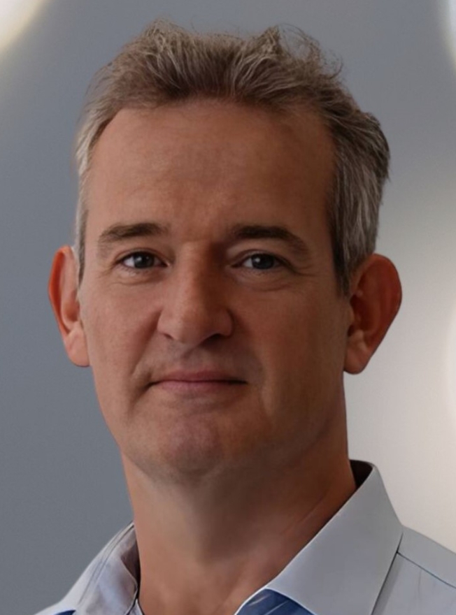 Erwan Bezard合作论文数Centre Broca Nouvelle-Aquitaine, Université de Bordeaux;SE-Therapeutics;Institut des Maladies Neurodégénératives CNRS;Motac Neuroscience Ltd;TreeFrog Therapeutics6Rubia Van Den Brand合作论文数6
 Ivan Minev合作论文数Department of Automatic Control and Systems Engineering, Faculty of Engineering, The University of Sheffield6Mayte Castro Jimenez合作论文数5
 Yohann Thenaisie合作论文数5
 Natalia Pavlova合作论文数Centre for Neuroprosthetics and Brain Mind Institute, EPFL;St. Petersburg, Pavlov Institute of Physiology;Centre for Neuroprosthetics and Brain Mind Institute, Pavlov Institute of Physiology5Jimmy Ravier合作论文数4Charlotte Moerman合作论文数4
 Arthur Hirsch合作论文数4
 Stefano Scafa合作论文数Institute of Digital Technologies for Personalized Healthcare (MedITech),, University of Applied Sciences and Arts of Southern Switzerland4
 Nicolas Vachicouras合作论文数Institute of Microengineering and Institute of Bioengineering, Ecole Polytechnique Fédérale de Lausanne (EPFL;Centre for Neuroprosthetics and Brain Mind Institute, EPFL;Institute of Microengineering and Institute of Bioengineering, EPFL4
 Jordan Squair合作论文数NeuroRestore Center, École Polytechnique Fédérale de Lausanne;Brain Mind Institute, School of Life Sciences, École Polytechnique Fédérale de Lausanne;Department of Clinical Neuroscience, Lausanne University Hospital4
 Qin Li合作论文数4
 Gerd Tinkhauser合作论文数Department of Neurology, Innsbruck Medical University4
 Joachim Von Zitzewitz合作论文数4
 Alessandro Puiatti合作论文数SUPSI4Andrea Galvez合作论文数3Kay Bartholdi合作论文数3
 Camille G. Le Goff合作论文数Center for Neuroprosthetics and Brain Mind Institute, Swiss Federal Institute of Technology Lausanne3Rebecca Charbonneau合作论文数3
 Achilleas Laskaratos合作论文数Med Sch Athens, Natl & Kapodistrian Univ Athens3
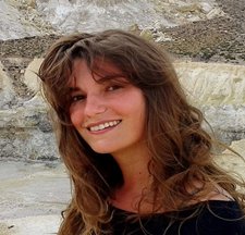 Chiara Palmisano合作论文数Department of Neurology, University Hospital of Würzburg and Julius-Maximilian-University Würzburg3Laetitia Baud合作论文数Swiss Fed Inst Technol EPFL, Ctr Neuroprosthet, Int Parapleg Fdn Chair Spinal Cord Repair, Lausanne, Switzerland3
 Robin Demesmaeker合作论文数Institute of Bioengineering/Center for Neuroprosthetics, École Polytechnique Fédérale de Lausanne, Lausanne, Switzerland3
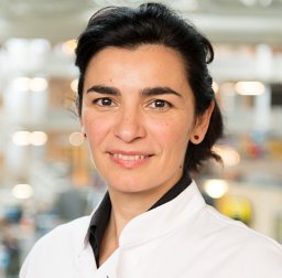 Maria Fiorella Contarino合作论文数Dept Neurol, Haga Teaching Hosp3
 Sophie M Wurth合作论文数Swiss Fed. Inst. of Technol. in Lausanne;c;Swiss Fed. Inst. of Technol. in Lausanne3
 David Borton合作论文数Center for Biomedical Engineering, Department of Molecular Pharmacology, Physiology, and Biotechnology, Brown University3
 Xiaoyang Kang (康晓洋)合作论文数Institute of Science and Technology for Brain-Inspired Intelligence, Fudan University3
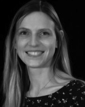 Kathe Claudia合作论文数Wolfson Centre for Age-Related Diseases, King's College London;claudia.kathe@kcl.ac.uk.;Wolfson Centre for Age-Related Diseases, King's College London3
 Qihan Liu合作论文数University of Science and Technology of China, Univ. of Sci. and Technol. of China3
 Jean-Baptiste Mignardot合作论文数3Wai Kin D. Ko合作论文数3Torres Rafael Fajardo合作论文数3
 Pirondini Elvira合作论文数3
 Marco Bonizzato合作论文数Center for Neuroprosthetics and Institute of Bioengineering, EPFL3




