Optogenetics has been a powerful scientific tool for two decades, yet its integration with non-human primate (NHP) electrophysiology has been limited due to several technical challenges. These include a lack of electrode arrays capable of supporting large-scale and long-term optical access, inaccessible viral vector delivery methods for transfection of large regions of cortex, a paucity of hardware designed for large-scale patterned cortical illumination, and limited designs for multi-modal experimentation. To address these gaps, we introduce a highly accessible platform integrating optogenetics and electrophysiology for behavioral and neural modulation with neurophysiological recording in NHPs. We employed this platform in two rhesus macaques and showcased its capability of optogenetically disrupting reaches, while simultaneously monitoring ongoing electrocorticography activity underlying the stimulation-induced behavioral changes. The platform exhibits long-term stability and functionality, thereby facilitating large-scale electrophysiology, optical imaging, and optogenetics over months, which is crucial for translationally relevant multi-modal studies of neurological and neuropsychiatric disorders.
1. We investigated the dimensions of output zones affecting specific combinations of forelimb muscles in the precentral "motor" cortex of macaque monkeys. Single-pulse intracortical microstimulation (S-ICMS) was used to evoke subthreshold effects in multiple wrist and finger muscles. S-ICMS consisted of 15 Hz stimulus trains at low intensity (5 μA) to avoid spread of effects due to temporal summation and to activate circumscribed foci of cortical cells in the bank of the precentral gyrus. 2. To detect subthreshold effects on active muscles, stimuli were delivered during wrist movements against elastic loads, and stimulus-triggered averages of rectified electromyographic (EMG) activity of 12 identified forelimb muscles were compiled. These averages detected statistical increases and decreases in averaged rectified EMG activity termed poststimulus facilitation and poststimulus suppression, respectively (7, 8). The "muscle profile" of a cortical output site was defined as the distribution and relative magnitude of these effects evoked in the recorded muscles. To document the spatial extent of a cortical output zone producing a particular "muscle profile," we delivered S-ICMS at successive sites along electrode tracks in the precentral bank, tangential to and near layer V cells. 3. In some cases the electrodes encountered cortical cells whose post-spike effects on muscles were also documented by spike-triggered averages of EMG activity. Near cells that had post-spike effects, S-ICMS evoked a similar profile of effects on these muscles, over distances of several hundred microns from the location of the cells with post-spike output effects. 4. The locations of tracks were marked by electrolytic lesions at specific depths and were identified in subsequent histological reconstructions. The physiological effects evoked from different sites were correlated with the locations of Nissl-stained cortical cells and corticospinal cells labeled by horseradish peroxidase (HRP) transported from the cervical spinal cord. 5. The "muscle profiles" of poststimulus effects elicited by 5-10 μA S-ICMS applied at successive sites were the same for multiple neighboring sites extending over tangential distances of about 1 mm. S-ICMS applied at 300 to 500 μm intervals evoked significant poststimulus facilitation or suppression effects over a range of 650-1250 μm. Assuming an effective excitation radius of 65 μm for 5-μA current pulses, the lower limit of the mean tangential dimension of cortical output zones on the precentral bank (n = 17) was estimated to be 800 μm. 6. A specific muscle could be affected, either singly or in combination with synergist muscles, from aggregate regions of output sites extending about 5 × 4 mm mediolateral x dorsoventral. These regions were comparable in size to output areas containing all the corticomotoneuronal (CM) cells that produced postspike facilitation in that muscle. 7. The cortical entry points of tracks with CM cells producing postspike facilitation in specific muscles were reconstructed for three monkeys. In two monkeys, the flexor and extensor CM cells were thoroughly intermingled; in the third, the flexor CM cells were preferentially located medially and showed less overlap with the region containing extensor CM cells. 8. These results indicate that each motor cortex site represents a different combination of muscles. The effects evoked from cortical sites separated by several hundred microns invariably involved different profiles of muscle activity. The muscle fields of remote CM cells were rarely identical, while the fields of neighboring CM cells were often similar. Given the number of unrecorded muscles, we conclude that primate motor cortex is a mosaic of output sites representing forelimb muscles in different combinations.
Objective.Closed-loop brain-computer interfaces can be used to bridge, modulate, or repair damaged connections within the brain to restore functional deficits. Towards this goal, we demonstrate that small artificial spiking neural networks can be bidirectionally interfaced with single neurons (SNs) in the neocortex of non-human primates (NHPs) to create artificial connections between the SNs to manipulate their activity in predictable ways.Approach.Spikes from a small group of SNs were recorded from primary motor cortex of two awake NHPs during rest. The SNs were then interfaced with a small network of integrate-and-fire units (IFUs) that were programmed on a custom clBCI. Spikes from the SNs evoked excitatory and/or inhibitory postsynaptic potentials in the IFUs, which themselves spiked when their membrane potentials exceeded a predetermined threshold. Spikes from the IFUs triggered single pulses of intracortical microstimulation (ICMS) to modulate the activity of the cortical SNs.Main results.We show that the altered closed-loop dynamics within the cortex depends on several factors including the connectivity between the SNs and IFUs, as well as the precise timing of the ICMS. We additionally show that the closed-loop dynamics can reliably be modeled from open-loop measurements.Significance.Our results demonstrate a new type of hybrid biological-artificial neural system based on a clBCI that interfaces SNs in the brain with artificial IFUs to modulate biological activity in the brain. Our model of the closed-loop dynamics may be leveraged in the future to develop training algorithms that shape the closed-loop dynamics of networks in the brain to correct aberrant neural activity and rehabilitate damaged neural circuits.
BACKGROUND:Computational models that predict effects of neural stimulation can serve as a preliminary tool to inform in-vivo research, reducing costs, time, and ethical considerations. However, current models do not support the diverse neural stimulation techniques used in-vivo, including the expanding selection of electrodes, stimulation modalities, and stimulation protocols. NEW METHOD:We developed several extensions to The Virtual Electrode Recording Tool for EXtracellular Potentials (VERTEX), the MATLAB-based neural stimulation tool. VERTEX simulates input currents in a large population of multi-compartment neurons within a small cortical slice to model electric field stimulation, while recording local field potentials (LFPs) and spiking activity. Our extensions enhance this framework with support for multiple pairs of parametrically defined electrodes and biphasic, bipolar stimulation delivered at programmable delays. To support the growing use of optogenetic approaches for targeted neural stimulation, we introduced a feature that models optogenetic stimulation through an additional VERTEX input function that converts irradiance to currents at optogenetically responsive neurons. Finally, we added extensions to allow complex stimulation protocols including paired-pulse, spatiotemporal patterned, and closed-loop stimulation. RESULTS:We demonstrated these novel features using VERTEX's built-in functionalities, with results consistent with other models and experimental work. COMPARISON WITH EXISTING METHODS:Unlike other tools, our extensions enable both electric field and optogenetic stimulation, provide a range of open- and closed-loop protocols, and offer flexible settings within a large-scale cortical network of neurons with realistic biophysical properties. CONCLUSIONS:Our extensions provide an all-in-one platform to efficiently and systematically test diverse, targeted, and individualized stimulation patterns.
To investigate intracortical microcircuits within primate motor cortical areas, we documented pairs of neurons whose synaptic interactions were identified by spike-triggered averaging (STA) of intracellularly recorded membrane potentials and whose relative location was histologically identified. Average synchronous excitation potentials (ASEPs) were the most commonly observed feature in STAs between neuron pairs in all cortical layers (about 70%). This synchrony spread broadly for distances of more than 4 mm, gradually decreasing in amplitude and probability with cell separation. Excitatory postsynaptic potentials (EPSPs) were observed among 9% of the neuron pairs, whose separation extended for distances of more than 4 mm. The peak amplitudes of EPSPs were inversely correlated with cell separation. Most source neurons were located in layer II-III or layer V, while the postsynaptic target neurons had wide laminar distribution. The probability of finding excitatory connections was not uniform within the cortical space. The connectivity between neuron pairs was relatively dense within 1.0 mm, and became sparse for distances between 1.0 and 2.0 mm, and showed a second peak for distance between 2.0 and 4.0 mm. Inhibitory postsynaptic potentials (IPSPs) and/or average synchronous inhibitory potentials (ASIPs) were observed for 10% of neuron pairs. Most of these were separated by less than 1.0 mm, suggesting that their influence was restricted within their own and neighboring columns. The major source neurons that provide these inhibitory effects were located within layer II-III. We conclude that excitatory synaptic effects (ASEPs and EPSPs) are widely distributed and omni-directional within primate motor cortex, while inhibitory connections (ASIPs and IPSPs) are restricted within columnar dimensions, and are predominantly directed from superficial to deeper layers. The appearance of independent zones that had no functional connectivity with neighboring columns indicates that primate motor cortical areas may not be organized in a uniform way. This would support the sparse coding theory and multiple representations of cortical output in the motor cortex. ### Competing Interest Statement The authors have declared no competing interest. NIH, , NS12542, RR00166
While neurofeedback represents a promising tool for neuroscience and a brain self-regulation approach to psychological rehabilitation, the field faces several problems and challenges. Current research has shown great variability and even failure among human participants in learning to self-regulate target features of brain activity with neurofeedback. A better understanding of cognitive mechanisms, psychological factors and neural substrates underlying self-regulation might help improve neurofeedback's scientific and clinical practices. This article reviews the current understanding of the neural mechanisms of brain self-regulation by drawing on findings from human and animal studies in neurofeedback, brain-computer/machine interfaces and neuroprosthetics. In this article, we look closer at the following topics: cognitive processes and psychophysiological factors affecting self-regulation, theoretical models and neural substrates underlying self-regulation, and finally, we provide an outlook on the outstanding gaps in knowledge and technical challenges. This article is part of the theme issue 'Neurofeedback: new territories and neurocognitive mechanisms of endogenous neuromodulation'.
The activity and connectivity of neurons in the primate brain underlying behavior cannot yet be completely specified, but neural networks provide complete models of the connectivity and activity that performs specific tasks and provide insight into the neural computations performed by the primate brain (Fetz and Shupe 2003). Studies of neurons in the monkey cortex have shown that short-term memory of sensory events may be mediated by sustained neural activity. Short-term memory tasks have been modeled with dynamic neural networks using a single continuous variable and a gate input to create a sample-and-hold (SAH) function (Zipser 1991; Maier 2003). Networks trained to perform these short-term memory tasks develop hidden unit activity which resembles that of cortical neurons in monkeys performing memory tasks. We here extend the investigation of single-input SAH networks to networks computing SAH for two continuous-variable inputs that have varying degrees of common mode signal. Results provide insights into computational mechanisms of associative short-term memory of sensory signals with common mode components, such as visual inputs to the two eyes, auditory inputs to the ears and proprioceptive input from multiple muscle spindle afferents. We also examined the attractor states that these SAH networks eventually reach after sufficiently long delay periods and found that these were determined by the shapes of the input-output functions of the hidden units rather than network architecture.
Motor intention is a high-level brain function related to planning for movement. Although studies have shown that motor intentions can be decoded from brain signals before movement execution, it is unclear whether intentions relating to mental imagery of movement can be decoded. Here, we investigated whether differences in spatial and temporal patterns of brain activation were elicited by intentions to perform different types of motor imagery and whether the patterns could be used by a multivariate pattern classifier to detect such differential intentions. The results showed that it is possible to decode intentions before the onset of different types of motor imagery from functional MR signals obtained from fronto-parietal brain regions, such as the premotor cortex and posterior parietal cortex, while controlling for eye movements and for muscular activity of the hands. These results highlight the critical role played by the aforementioned brain regions in covert motor intentions. Moreover, they have substantial implications for rehabilitating patients with motor disabilities.
Different sleep stages have been shown to be vital for a variety of brain functions, including learning, memory, and skill consolidation. However, our understanding of neural dynamics during sleep and the role of prominent LFP frequency bands remain incomplete. To elucidate such dynamics and differences between behavioral states we collected multichannel LFP and spike data in primary motor cortex of unconstrained macaques for up to 24 h using a head-fixed brain-computer interface (Neurochip3). Each 8-s bin of time was classified into awake-moving (Move), awake-resting (Rest), REM sleep (REM), or non-REM sleep (NREM) by using dimensionality reduction and clustering on the average spectral density and the acceleration of the head. LFP power showed high delta during NREM, high theta during REM, and high beta when the animal was awake. Cross-frequency phase-amplitude coupling typically showed higher coupling during NREM between all pairs of frequency bands. Two notable exceptions were high delta-high gamma and theta-high gamma coupling during Move, and high theta-beta coupling during REM. Single units showed decreased firing rate during NREM, though with increased short ISIs compared to other states. Spike-LFP synchrony showed high delta synchrony during Move, and higher coupling with all other frequency bands during NREM. These results altogether reveal potential roles and functions of different LFP bands that have previously been unexplored.
The initial-dip is a transient decrease frequently observed in functional neuroimaging signals, immediately after stimulus onset, believed to originate from a rise in deoxy-hemoglobin (HbR) caused by local neural activity. It has been shown to be more spatially specific than the hemodynamic response, and is believed to represent focal neuronal activity. However, despite being observed in various neuroimaging modalities (such as fMRI, fNIRS, etc), its origins are disputed, and its precise neuronal correlates are unknown. Here we show that the initial-dip is dominated by a decrease in total-hemoglobin (HbT). We also find a biphasic response in deoxy-Hb (HbR), with an early decrease and later rebound. Both the HbT-dip and HbR-rebound were strongly correlated to highly localized spiking activity. However, HbT decreases were always large enough to counter the spiking-induced increase in HbR. We find that the HbT-dip counters spiking induced HbR increases, imposing an upper-limit to HbR concentration in the capillaries. Building on our results, we explore the possibility of active venule dilation (purging) as a possible mechanism for the HbT dip.
Intracortical microstimulation (ICMS) is commonly used in many experimental and clinical paradigms; however, its effects on the activation of neurons are still not completely understood. To document the responses of cortical neurons in awake nonhuman primates to stimulation, we recorded single-unit activity while delivering single-pulse stimulation via Utah arrays implanted in primary motor cortex (M1) of three macaque monkeys. Stimuli between 5 and 50 μA delivered to single channels reliably evoked spikes in neurons recorded throughout the array with delays of up to 12 ms. ICMS pulses also induced a period of inhibition lasting up to 150 ms that typically followed the initial excitatory response. Higher current amplitudes led to a greater probability of evoking a spike and extended the duration of inhibition. The likelihood of evoking a spike in a neuron was dependent on the spontaneous firing rate as well as the delay between its most recent spike time and stimulus onset. Tonic repetitive stimulation between 2 and 20 Hz often modulated both the probability of evoking spikes and the duration of inhibition; high-frequency stimulation was more likely to change both responses. On a trial-by-trial basis, whether a stimulus evoked a spike did not affect the subsequent inhibitory response; however, their changes over time were often positively or negatively correlated. Our results document the complex dynamics of cortical neural responses to electrical stimulation that need to be considered when using ICMS for scientific and clinical applications.
Abstract Synchrony of oscillatory brain activity has been postulated to be a binding mechanism for cognitive and motor functions. Spectral analysis of human electrocorticogram (ECoG) in sensorimotor cortex has shown that power density of gamma band activity (30-60 Hz) increased and that of alpha-beta band activity (10-20 Hz) decreased during performance of manipulative visuomotor tasks, indicating that amplitude modulation of the gamma band activity occurred in relation to the task performance. Amplitude modulation may provide evidence for synchrony of local neuronal assembly. However, it does not implement the binding mechanisms for distributed networks that are necessary for cognitive and motor functions. To prove that oscillatory activity mediates a binding mechanism, phase modulation of oscillatory activity in a wide range area should be shown. We performed coherence analysis of the ECoG signals in sensorimotor cortex to study if synchrony of the gamma band activity between these areas occurs in relation to manipulative task performance. The ECoGs were recorded from 14 sites in sensorimotor cortex including hand-arm areas with subdural grid electrodes in four subjects. Coherence estimates in all pair-wise sites were calculated in different frequency bands with 10 Hz widths from 10 to 80 Hz. In all subjects, coherence estimates increased in the lower gamma band (20-50 Hz) during the performance of the manipulative tasks. But coherence in the alpha-beta band (10-20 Hz) also increased even though amplitude modulation did not occur in this frequency band. Coherence estimates increased in site pairs within and between sensory and motor areas, many separated by intervening sites. This interregional synchrony of the alpha-beta and the lower gamma activities may play a role in integration of sensorimotor information. Task-dependent increases in coherence estimates, i.e., greater increases during performance of the manipulative tasks than during the simple tasks, suggest another role of synchrony in attention mechanism. Time-series coherence analysis showed that phase modulation occurred in different timings for activities in the alpha-beta and the lower gamma bands. For the activity in higher gamma band (50-80 Hz), power density increased but coherence estimates decreased. Thus, only amplitude modulation occurred in this frequency band. Altogether these results suggest that oscillatory activities in different frequency bands may reflect different functional roles by modulating neural activity in different ways.
AbstractSynchrony of oscillatory brain activity has been postulated to be a binding mechanism for cognitive and motor functions. Spectral analysis of human electrocorticogram (ECoG) in sensorimotor cortex has shown that power density of gamma band activity (30-60 Hz) increased and that of alpha-beta band activity (10-20 Hz) decreased during performance of manipulative visuomotor tasks, indicating that amplitude modulation of the gamma band activity occurred in relation to the task performance. Amplitude modulation may provide evidence for synchrony of local neuronal assembly. However, it does not implement the binding mechanisms for distributed networks that are necessary for cognitive and motor functions. To prove that oscillatory activity mediates a binding mechanism, phase modulation of oscillatory activity in a wide range area should be shown. We performed coherence analysis of the ECoG signals in sensorimotor cortex to study if synchrony of the gamma band activity between these areas occurs in relation to manipulative task performance. The ECoGs were recorded from 14 sites in sensorimotor cortex including hand-arm areas with subdural grid electrodes in four subjects. Coherence estimates in all pair-wise sites were calculated in different frequency bands with 10 Hz widths from 10 to 80 Hz. In all subjects, coherence estimates increased in the lower gamma band (20-50 Hz) during the performance of the manipulative tasks. But coherence in the alpha-beta band (10-20 Hz) also increased even though amplitude modulation did not occur in this frequency band. Coherence estimates increased in site pairs within and between sensory and motor areas, many separated by intervening sites. This interregional synchrony of the alpha-beta and the lower gamma activities may play a role in integration of sensorimotor information. Task-dependent increases in coherence estimates, i.e., greater increases during performance of the manipulative tasks than during the simple tasks, suggest another role of synchrony in attention mechanism. Time-series coherence analysis showed that phase modulation occurred in different timings for activities in the alpha-beta and the lower gamma bands. For the activity in higher gamma band (50-80 Hz), power density increased but coherence estimates decreased. Thus, only amplitude modulation occurred in this frequency band. Altogether these results suggest that oscillatory activities in different frequency bands may reflect different functional roles by modulating neural activity in different ways.
Correlated activity of neurons can lead to long-term strengthening or weakening of the connections between them. In addition, the behavioral context, imparted by execution of physical movements or the presence of a reward, can modulate the plasticity induced by Hebbian mechanisms. In the present study, we have combined behavior and induced neuronal correlations to strengthen connections in the motor cortex of adult behaving monkeys. Correlated activity was induced using an electrical-conditioning protocol in which stimuli gated by voluntary movements were used to produce coactivation of neurons at motor-cortical sites involved in those movements. Delivery of movement-dependent stimulation resulted in small increases in the strength of associated cortical connections immediately after conditioning. Remarkably, when paired with further repetition of the movements that gated the conditioning stimuli, there were substantially larger gains in the strength of cortical connections, which occurred in a use-dependent manner, without delivery of additional conditioning stimulation. In the absence of such movements, little change was observed in the strength of motor-cortical connections. Performance of the motor behavior in the absence of conditioning also did not produce any changes in connectivity. Our results show that combining movement-gated stimulation with further natural use of the “conditioned” pathways after stimulation ends can produce use-dependent strengthening of connections in adult primates, highlighting an important role for behavior in cortical plasticity. Our data also provide strong support for combining movement-gated stimulation with use-dependent physical rehabilitation for strengthening connections weakened by a stroke or spinal cord injury.
Spike-timing dependent plasticity (STDP) is an extensively studied topic. Previous studies have demonstrated stimulus induced targeted STDP both in vitro and in vivo , but a more consistent and robust method is required. We hypothesized there were two reasons contributing to the inconsistent results previously reported: 1. the measure of connectivity is poorly understood, and 2. the timing of stimulation is static or has low temporal specificity. To test our hypotheses, we applied paired stimulation to the primary motor cortex of awake primates. Single unit responses to stimulation were used as measures of connectivity, and we applied inter-stimulus intervals (ISIs) from ±0.1 to ±50 ms with sub-millisecond intervals. The excitatory single unit response resulted in very consistent changes after conditioning that was dependent on the ISI. Negative ISIs resulted in depression similar to classic STDP, but positive ISI also often resulted in depression. Normalizing the ISIs to the timing of the excitatory response revealed that potentiation only occurred if the second stimulus arrived before the response. Stimuli occurring around the time of the response often resulted in depression as strong as negative ISIs. We additionally tracked the changes in cortico-cortical evoked potentials (CCEPs), a commonly used measure of connectivity in plasticity experiments. CCEP changes showed a similar but more variable dependence to ISI. These results show that the classic STDP curve may be more difficult to induce due to interactions between excitatory and inhibitory circuitry, and that CCEPs may not be the ideal measure of changes in strength of connectivity.
Vagus nerve stimulation (VNS) is tested as therapy for several brain disorders and as a means to modulate brain plasticity. Cortical effects of VNS, manifesting as vagal-evoked potentials (VEPs), are thought to arise from activation of ascending cholinergic and noradrenergic systems. However, it is unknown whether those effects are dependent on oscillatory brain activity underling different brain states. In 2 freely behaving macaque monkeys, we delivered trains of left cervical VNS, at different pulsing frequencies (5-300 Hz), while recording local field potentials (LFP) from sites in contralateral prefrontal, sensorimotor and parietal cortical areas, continuously over 11-16 hours. Different brain states were inferred from oscillatory components of LFPs and the presence of overt movement: active awake, resting awake, REM sleep and NREM sleep. VNS elicited VEPs comprising early (<70 ms), intermediate (70-250 ms) and late (>250 ms) components in all sampled cortical areas. The magnitude of only the intermediate and late components was modulated by brain state and pulsing frequency. These findings have implications for the role of ongoing brain activity in shaping cortical responses to peripheral stimuli, for the modulation of vagal interoceptive signaling by cortical states, and for the calibration of VNS therapies.
Vagus nerve stimulation (VNS) has been tested as therapy for several brain disorders and as a means to modulate cortical excitability and brain plasticity. Cortical effects of VNS, manifesting as vagal-evoked potentials (VEPs), are thought to arise from activation of ascending cholinergic and noradrenergic systems. However, it is unknown whether those effects are modulated by brain state at the time of stimulation. In 2 freely behaving macaque monkeys, we delivered short trains of 5 pulses to the left cervical vagus nerve at different frequencies (5-300 Hz) while recording local field potentials (LFPs) from sites in contralateral prefrontal, sensorimotor and parietal cortical areas. Brain states were inferred from spectral components of LFPs and the presence of overt movement: active awake, resting awake, REM sleep and NREM sleep. VNS elicited VEPs in all sampled cortical areas. VEPs comprised early (<70 ms), intermediate (70-250 ms) and late (>250 ms) components. The magnitude of the intermediate and late components was largest during NREM sleep and smallest during wakefulness, whereas that of the early component was not modulated by brain state. VEPs during NREM were larger for stimuli delivered at the depolarized phase of ongoing delta oscillations. Higher pulsing frequencies generated larger VEPs. These short VNS trains did not affect brain state transitions during wakefulness or sleep. Our findings suggest that ongoing brain state modulates the evoked effects of VNS on cortical activity. This has implications for the role of ongoing cortical activity and brain state in shaping cortical responses to peripheral stimuli, for the modulation of vagal interoceptive signaling by cortical activity, and for the dose calibration of VNS therapies.
We describe an integrate-and-fire (IF) spiking neural network that incorporates spike-timing-dependent plasticity (STDP) and simulates the experimental outcomes of four different conditioning protocols that produce cortical plasticity. The original conditioning experiments were performed in freely moving non-human primates (NHPs) with an autonomous head-fixed bidirectional brain-computer interface (BCI). Three protocols involved closed-loop stimulation triggered from (1) spike activity of single cortical neurons, (2) electromyographic (EMG) activity from forearm muscles, and (3) cycles of spontaneous cortical beta activity. A fourth protocol involved open-loop delivery of pairs of stimuli at neighboring cortical sites. The IF network that replicates the experimental results consists of 360 units with simulated membrane potentials produced by synaptic inputs and triggering a spike when reaching threshold. The 240 cortical units produce either excitatory or inhibitory postsynaptic potentials (PSPs) in their target units. In addition to the experimentally observed conditioning effects, the model also allows computation of underlying network behavior not originally documented. Furthermore, the model makes predictions about outcomes from protocols not yet investigated, including spike-triggered inhibition, γ-triggered stimulation and disynaptic conditioning. The success of the simulations suggests that a simple voltage-based IF model incorporating STDP can capture the essential mechanisms mediating targeted plasticity with closed-loop stimulation.
Toward addressing many neuroprosthetic applications, the Neurochip3 (NC3) is a multichannel bidirectional brain-computer interface that operates autonomously and can support closed-loop activity-dependent stimulation. It consists of four circuit boards populated with off-the-shelf components and is sufficiently compact to be carried on the head of a non-human primate (NHP). NC3 has six main components: (1) an analog front-end with an Intan biophysical signal amplifier (16 differential or 32 single-ended channels) and a 3-axis accelerometer, (2) a digital control system comprised of a Cyclone V FPGA and Atmel SAM4 MCU, (3) a micro SD Card for 128 GB or more storage, (4) a 6-channel differential stimulator with ±60 V compliance, (5) a rechargeable battery pack supporting autonomous operation for up to 24 h and, (6) infrared transceiver and serial ports for communication. The NC3 and earlier versions have been successfully deployed in many closed-loop operations to induce synaptic plasticity and bridge lost biological connections, as well as deliver activity-dependent intracranial reinforcement. These paradigms to strengthen or replace impaired connections have many applications in neuroprosthetics and neurorehabilitation.
 Steve I. Perlmutter合作论文数28
 Larry E. Shupe合作论文数14Paul D. Cheney合作论文数14
 Stavros Zanos合作论文数Department of Nephrology, “Papageorgiou” General Hospital11
 Niels Birbaumer (尼尔斯拜尔博姆)合作论文数University Tubingen11
 Andrew Jackson合作论文数Newcastle Univ, Inst Neurosci, Newcastle Upon Tyne NE2 4HH, Tyne & Wear, England10
 Jeffrey G. Ojemann合作论文数Department of Neurological Surgery, University of Washington;Seattle Children's10
 Venkatesh Murthy合作论文数Department of Molecular and Cellular Biology, Harvard University;Center for Brain Science, Harvard University10
 Ranganatha Sitaram合作论文数Institute of Medical Psychology and Behavioral Neurobiology, Eberhard-Karls-University of Tübingen10Yifat Prut合作论文数8
 Rajesh P. N. Rao合作论文数Paul G. Allen School of Computer Science & Engineering, University of Washington;Center for Neurotechnology, University of Washington;Sensai Healthcare, Inc.7
 Kai J. Miller合作论文数Department of Neurosurgery, University of Washington7Andrew Richardson合作论文数6
 Richy Yun合作论文数Departments of Bioengineering, University of Washington6
 Irene Rembado合作论文数Departments of Physiology and Biophysics, University of Washington6
 Chet Moritz合作论文数Department of Electrical & Computer Engineering, University of Washington;Division of Physical Therapy, Department of Rehabilitation Medicine, University of Washington;Onward;SpineX Inc;Grapheton, Inc6Jaideep Mavoori合作论文数6MA MAIER合作论文数5
 Ali Danish Zaidi合作论文数Max Planck Institute for Biological Cybernetics5
 Timothy H. Lucas合作论文数Translational Neuromodulation Lab in the Perelman School of Medicine at the University of Pennsylvania5
 Lise A Johnson合作论文数Neurological Surgery,5Jeremiah D. Wander合作论文数Departments of Bioengineering,5George A. Ojemann合作论文数5L. Shupe合作论文数5Dv Finocchio合作论文数4Yukio Nishimura合作论文数4Stephen S Palmer合作论文数4Julian Isaacs合作论文数4Leigh Brasington合作论文数4Michael R. Hagerty合作论文数4AD Reyes合作论文数4Allen R. Wyler合作论文数4Brian Mogen合作论文数3MJ SOSO合作论文数3Ryan W Eaton合作论文数3David K Su合作论文数3Jonathan Mishler合作论文数3A Schmied合作论文数3
 Adam O Hebb合作论文数Colorado Neurological Institute Englewood3
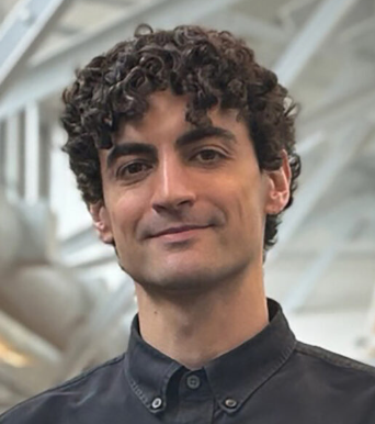 Matthew D. Sacchet合作论文数Harvard Medical School;Massachusetts General Hospital3
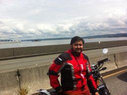 Devapratim Sarma合作论文数3
 Matthew A. Edwardson合作论文数Department of Neurology, University of Washington Seattle3F Aoki合作论文数3
 Sergio Ruiz合作论文数3
 Nikos Logothetis合作论文数Department of Physiology of Cognitive Processes, Max Planck Institute for Biological Cybernetics;International Center for Primate Brain Research, Chinese Academy of Sciences3
 Christoph Braun合作论文数MEG Ctr, Univ Tubingen3Arthur A. Ward合作论文数3
 Gerwin Schalk合作论文数Chen Frontier Lab for Applied Neurotechnology, Tianqiao and Chrissy Chen Institute3Michikazu Matsumura合作论文数3




