Background. We hypothesized that the addition of 4 doses of abatacept to our standard acute graft-versus-host disease (GVHD) prophylaxis would reduce the incidence of day +100 severe acute GVHD in children with transfusion-dependent beta-thalassemia major undergoing a myeloablative allogeneic hematopoietic stem cell transplant (HSCT), without impacting engraftment. Methods. Twenty-four children with beta-thalassemia major received abatacept at a dose of 10 mg/kg intravenously on days –1, +5, +14, and +28 after HSCT in addition to calcineurin inhibitors and methylprednisolone. Outcomes were compared to 8 beta-thalassemia patients who received standard acute GVHD prophylaxis. Results. There was no difference in engraftment between the 2 groups. No patient had grades III–IV acute GVHD by day +100 in the abatacept cohort compared with 50% in the standard acute GVHD prophylaxis group (P = 0.001). Viral reactivation occurred in 5 children in the standard acute GVHD cohort and in 20 children in the abatacept cohort (P = 0.2). Thalassemia-free survival after HSCT was 100% in the abatacept cohort compared to 62.5% in the standard cohort at last follow-up (P = 0.007). Conclusions. Adding abatacept to our routine GVHD prophylaxis reduced the incidence of day +100 severe acute GVHD without impacting engraftment or survival.
We report results of a phase II study of maraviroc to prevent acute graft versus host disease (GVHD) in children undergoing allogeneic hematopoietic stem cell transplant (HSCT). Oral maraviroc was added to standard GVHD prophylaxis of a calcineurin inhibitor with either mycophenolate mofetil, methotrexate or steroids from day −3 until day +30 after HSCT. Maraviroc trough levels were analyzed on day 0, +7, 14, and 21. We assessed functional CCR5 blockade by our previously described pharmacodynamic assay. In total, 17 patients were enrolled prospectively. No patient had liver GVHD by day +100. Four patients developed gastrointestinal (GI) GVHD (Grade II upper GI GVHD n = 2, grade III lower GI GVHD n = 2). No adverse effects of maraviroc were observed. Seven patients discontinued maraviroc at a median of day +14 (range day +1–day +29) due to study rules regarding hepatotoxicity ( n = 5), renal function decline ( n = 1) and withdrawal from study ( n = 1). Maraviroc administration led to CCR5 inhibition but was limited by study rules defining hepatotoxicity, leading to frequent drug discontinuation. We cannot comment on the efficacy of maraviroc with our data but speculate that it could have a role in prevention of acute GI GVHD, with adequate compliance.
BackgroundBlood stream infections (BSI) in hematopoietic stem cell transplant (HSCT) patients with central venous catheters may arise from translocation of bacteria through a non-intact gastrointestinal mucosa and are known as mucosal barrier injury–laboratory confirmed blood stream infections (MBI-LCBI). Central venous catheter maintenance care doesn't prevent MBI-LCB and currently there are no proven strategies to prevent bacteremia secondary to mucosal barrier injury. Gingivitis and periodontitis are features of the mucosal toxicity seen post-HSCT and are associated with BSI frequency. Xylitol is a non-fermentable polysaccharide that reduces dental caries, plaque accumulation, and oral disease progression by inhibiting bacterial growth and community development. We hypothesized that daily dental xylitol, in addition to current oral care practice, is effective at reducing gingivitis, periodontitis and BSI from oral organisms.MethodsWe performed a prospective randomized controlled trial to test our hypothesis. All patients received standard oral care and half were randomized to additionally receive daily xylitol. Oral exams were performed at baseline and at intervals for the first 28 days post-HSCT. Metagenomic shotgun sequencing of gingival samples was performed at these same intervals. For patients who develop BSI, whole genome sequencing of bacterial isolates is being performed to assess for genetic relatedness to corresponding strains present within the patient's oral microbiome preceding the infection.ResultsWe enrolled 35 patients, 17 cases and 18 controls before ending the study early for efficacy. Five patients withdrew from the study (3 cases, 2 controls). Patients who received xylitol had a significantly lower rate of gingivitis, oral plaque and oral ulcers greater than 10 mm. There was a significant decrease in BSI from oral organisms in the first 30 days post HSCT in the xylitol group, 0/14 (0%) vs 5/16 (31%) (p = 0.04). Preliminary microbiome analysis showed decrease relative abundance of Candida albicans in patients receiving xylitol and individual patient analysis shows that xylitol may also impede Streptococcus mitis/oralis dominance in the oral microbiome. Additionally, we found that Staphylococcus aureus, a common cause of Central Line Associated Blood Stream Infection (CLABSI), was prominent in several gingival samples, suggesting BSI with Staphylococcus aureus may indeed be related to mucosal barrier injury.ConclusionsThe addition of xylitol to oral standard care appears to decrease BSI secondary to oral organisms, dental plaque, gingivitis and oral ulcerations in patients undergoing SCT. Blood stream infections (BSI) in hematopoietic stem cell transplant (HSCT) patients with central venous catheters may arise from translocation of bacteria through a non-intact gastrointestinal mucosa and are known as mucosal barrier injury–laboratory confirmed blood stream infections (MBI-LCBI). Central venous catheter maintenance care doesn't prevent MBI-LCB and currently there are no proven strategies to prevent bacteremia secondary to mucosal barrier injury. Gingivitis and periodontitis are features of the mucosal toxicity seen post-HSCT and are associated with BSI frequency. Xylitol is a non-fermentable polysaccharide that reduces dental caries, plaque accumulation, and oral disease progression by inhibiting bacterial growth and community development. We hypothesized that daily dental xylitol, in addition to current oral care practice, is effective at reducing gingivitis, periodontitis and BSI from oral organisms. We performed a prospective randomized controlled trial to test our hypothesis. All patients received standard oral care and half were randomized to additionally receive daily xylitol. Oral exams were performed at baseline and at intervals for the first 28 days post-HSCT. Metagenomic shotgun sequencing of gingival samples was performed at these same intervals. For patients who develop BSI, whole genome sequencing of bacterial isolates is being performed to assess for genetic relatedness to corresponding strains present within the patient's oral microbiome preceding the infection. We enrolled 35 patients, 17 cases and 18 controls before ending the study early for efficacy. Five patients withdrew from the study (3 cases, 2 controls). Patients who received xylitol had a significantly lower rate of gingivitis, oral plaque and oral ulcers greater than 10 mm. There was a significant decrease in BSI from oral organisms in the first 30 days post HSCT in the xylitol group, 0/14 (0%) vs 5/16 (31%) (p = 0.04). Preliminary microbiome analysis showed decrease relative abundance of Candida albicans in patients receiving xylitol and individual patient analysis shows that xylitol may also impede Streptococcus mitis/oralis dominance in the oral microbiome. Additionally, we found that Staphylococcus aureus, a common cause of Central Line Associated Blood Stream Infection (CLABSI), was prominent in several gingival samples, suggesting BSI with Staphylococcus aureus may indeed be related to mucosal barrier injury. The addition of xylitol to oral standard care appears to decrease BSI secondary to oral organisms, dental plaque, gingivitis and oral ulcerations in patients undergoing SCT.
Vulvovaginal graft-versus-host disease (GVHD) is an underdiagnosed and poorly recognized complication of hematopoietic stem cell transplantation (HSCT). Previous studies have reported findings restricted to predominantly adult populations. We report a case series of pediatric and young adult vulvovaginal GVHD, which was identified in 19 patients (median age, 11.8 years; range, 2.4 to 21.9 years) out of a total 302 female patients who underwent transplantation over an 8-year period at a pediatric HSCT center. The majority of patients had concomitant nongenital GVHD; only 1 patient had isolated vulvovaginal GVHD. The median time from bone marrow transplantation to diagnosis of vulvovaginal GVHD was 30 months (range, 2.3 to 97.5 months). A high percentage of the patients in our series were without vulvar or vaginal symptoms (n = 8; 42%), even though 17 patients (89%) presented with grade 3 disease based on current adult grading scales. Vulvar examination findings most frequently included interlabial and clitoral hood adhesions (89%), loss of architecture of the labia minora or clitoral hood (42%), and skin erosions or fissures (37%). Only 5 patients underwent a speculum exam, none of whom had vaginal GVHD. Examination findings of primary ovarian insufficiency (POI) can overlap with those of GVHD, and 6 patients (32%) in our cohort were diagnosed with POI. Only 1 patient was on systemic hormone replacement therapy at the time of vulvovaginal GVHD diagnosis. The majority of patients (n = 16) were treated with topical steroid therapy, with a median time to response of 43 days. Five patients (26%) had a complete response to therapy, and 10 patients (53%) had a partial response. This case series provides valuable insight into pediatric and young adult vulvovaginal GVHD and highlights the need for increased screening for vulvar disease in this population.
BackgroundEarly detection of pulmonary morbidity following haematopoietic stem cell transplantation (HSCT) remains an important challenge for intervention, primarily due to the insensitivity of spirometry to early change, and in paediatrics, patient compliance provides additional challenges. Regional lung ventilation abnormalities in paediatric HSCT patients were quantified using hyperpolarised xenon-129 (129Xe) magnetic resonance imaging (MRI) and compared to spirometry.MethodsMedically stable, paediatric allogeneic HSCT patients (n=23, ages 6–16 years) underwent an outpatient MRI scan where regional ventilation was quantified with a breath-hold of hyperpolarised129Xe gas. Ventilation deficits, regions of the lung that ventilate poorly due to obstruction, were quantified as a ventilation defect percentage (VDP) and compared to forced expiratory volume in 1 s (FEV1), FEV1/forced vital capacity (FVC) ratio, and forced expiratory flow at 25–75% of FVC (FEF25–75%) from spirometry using linear regression.ResultsThe mean±sd129Xe VDP was 10.5±9.4% (range 2.6–41.4%).129Xe VDP correlated with FEV1, FEV1/FVC ratio and FEF25–75%(p≤0.02 for all comparisons). Ventilation deficits were detected in patients with normal spirometry (i.e.FEV1>80%), supporting the sensitivity of129Xe MRI to early obstruction reported in other pulmonary conditions. Seven (30%) patients could not perform spirometry, yet ventilation deficits were observed in five of these patients, detecting abnormalities that otherwise may have gone undetected and untreated until advanced.ConclusionLung ventilation deficits were detected using hyperpolarised129Xe gas MRI in asymptomatic paediatric HSCT patients and in a subgroup who were unable to perform reliable spirometry.129Xe MRI provides a reliable imaging-based assessment of pulmonary involvement in this potentially difficult to diagnose paediatric population.
We sought to define the prevalence of echocardiographic abnormalities in long-term survivors of paediatric hematopoietic stem cell transplantation and determine the utility of screening in asymptomatic patients. We analysed echocardiograms performed on survivors who underwent hematopoietic stem cell transplantation from 1982 to 2006. A total of 389 patients were alive in 2017, with 114 having an echocardiogram obtained >= 5 years post-infusion. A total of 95 patients had echocardiogram performed for routine surveillance. The mean time post-hematopoietic stem cell transplantation was 13 years. Of 95 patients, 77 (82.1%) had ejection fraction measured, and 10/77 (13.0%) had ejection fraction z-scores <= -2.0, which is abnormally low. Those patients with abnormal ejection fraction were significantly more likely to have been exposed to anthracyclines or total body irradiation. Among individuals who received neither anthracyclines nor total body irradiation, only 1/31 (3.2%) was found to have an abnormal ejection fraction of 51.4%, z-score -2.73. In the cohort of 77 patients, the negative predictive value of having a normal ejection fraction given no exposure to total body irradiation or anthracyclines was 96.7% at 95% confidence interval (83.3-99.8%). Systolic dysfunction is relatively common in long-term survivors of paediatric hematopoietic stem cell transplantation who have received anthracyclines or total body irradiation. Survivors who are asymptomatic and did not receive radiation or anthracyclines likely do not require surveillance echocardiograms, unless otherwise indicated.
Pediatric hematology/oncology (PHO) patients receiving therapy or undergoing hematopoietic stem cell transplantation (HSCT) often require a central line and are at risk for bloodstream infections (BSI). There are limited data describing outcomes of BSI in PHO and HSCT patients.
Reduced intensity conditioning (RIC) is increasingly used for hematopoietic stem cell transplantation (HCT). Risk of infertility and primary gonadal insufficiency secondary to standard myeloablative (MAC) HCT is high, but little is known of these complications following use of RIC HCT.
Background: The pathophysiologic progression of bronchiolitis obliterans syndrome (BOS) in the pediatric hematopoietic stem cell transplant (HSCT) population is poorly characterized. We embarked on a prospective BOS screening program to capture early changes in pulmonary function tests (PFTs) after pediatric HSCT. Methods: All pediatric and young adult patients receiving allogeneic HSCT after May 31, 2016 were prospectively screened as outlined by our clinical algorithm (Figure 1). Patients 3 8 years underwent screening via PFTs and six-minute walk testing (6MWT). Patients aged 4 to 7.99 years underwent 6MWT. Results: Sixty of 71 total patients are alive greater than 100 days post-stem cell infusion with 38 of 60 greater than 4 years of age. Fourteen of 16 (87.5%) patients aged 4-7.99 years successfully completed 6MWTs. Fourteen of 22 (63.6%) patients >8 years of age successfully completed screening PFTs at day 100, 3 of which did not have a complete pre-HSCT PFT study. Five of 11 patients with pre- and post-HSCT PFTs were referred for additional work-up to our pulmonary service due to concerning decreased FEV1%-predicted (Figure 2). Only one of the remaining six patients developed chronic skin/vaginal/oral GVHD and subsequently underwent prompt PFTs. The child was found to have findings concerning for BOS requiring intervention that has resulted in improvement to date. Conclusions: Our respiratory screening program is feasible and has resulted in the successful early capture of patients with possible early onset pulmonary pathology including BOS. Patients with abnormal PFTs have received prompt treatment with positive outcomes. Overall, we feel this algorithm allows for improved care of pediatric HSCT patients.
Background: Blood stream infections in patients with central venous catheters may arise from translocation of bacteria through a non-intact mucosa. These infections are known as mucosal barrier injury–laboratory confirmed blood stream infections (MBI-LCBI). Currently there are no proven strategies to prevent bacteremia secondary to mucosal barrier injury after hematopoietic stem cell transplant (HSCT). Xylitol is a non-fermentable sugar alcohol that reduces dental caries, plaque accumulation, and oral disease progression by inhibiting bacterial growth and reducing pathogenic community development. We hypothesized that the addition of Xylitol to standard oral care will decrease bacteremia from oral flora. Methods: We are conducting a prospective randomized study to test our hypothesis. All patients receive standard oral care while half are randomized to also receive daily Xylitol oral wipes. Oral exams are performed at baseline and weekly for the first 28 days post HSCT. Metagenomic shotgun sequencing of gingival samples is performed before and after HSCT. For patients who develop blood stream infection (BSI), whole genome sequencing of pathogenic bacterial isolates is currently being performed in cases and controls, to assess for genetic relatedness to corresponding strains present within the patient's oral microbiome preceding the infection. Results: Preliminary interim analysis of 13 patients demonstrates improved oral health overall (Figure 1). Oral care with Xylitol appeared to prevent Streptococcus mitis/oralis dominance in the oral microbiome (Figure 2). Additionally, the use of Xylitol may prevent BSI from Streptococcus mitis oralis (BSI events n = 0/5) compared to standard oral care (BSI events n = 2/8) within the first 8 days post-transplantation. Interestingly, Streptococcus mitis/oralis comprised 70% of the oral microbiome in one child who subsequently developed a Streptococcus mitis/oralis BSI (Figure 3).Figure 2Relative Streptococcus mitis/oralis abundance in oral microbiome. (A) Standard of care group (SOC) and Xylitol group post-transplant (B) Xylitol group only at baseline and post-transplant and (C) Standard of care group only at baseline and post-transplant.View Large Image Figure ViewerDownload Hi-res image Download (PPT)Figure 3Species abundance represented by bubble chart. (A) Standard of care patient with increase Streptococcus abundance post-transplant (B) Xylitol patient with decrease Streptococcus abundance post-transplant.View Large Image Figure ViewerDownload Hi-res image Download (PPT) The expected study completion date is December 2017. Conclusions: The addition of Xylitol to oral standard care appears to decrease dental plaque, gingivitis and oral ulcerations in patients undergoing HSCT. Xylitol may also impede Streptococcus mitis/oralis dominance in the oral microbiome with potential reduction in blood stream infections.
Background: Early empiric broad-spectrum antibiotic use in hematopoietic stem cell transplant (HSCT) patients with fever to treat a presumed bloodstream infection (BSI) is a common response but may be associated with the loss of gut microbiome diversity, GVHD, poor outcomes and increased mortality. Therefore, we devised a project to explore the true BSI incidence and risks in the peri-transplant time-period. Methods: We performed a 5-year retrospective review of consecutive pediatric HSCT recipients from 3 centers to determine the incidence and risk factors for BSI from the start of HSCT conditioning through stem cell infusion. We reviewed the antibiotic administration in a small cohort (114, 10%) of subjects to determine the percentage of patients receiving empiric broad spectrum antibiotics during this time period. Results: 1135 pediatric and young adult patients underwent HSCT during the study time period. In the small cohort, 83 of 114 (73%) received broad-spectrum antibiotics during chemotherapy for fever or concern for infection. Sixteen (1.4%) developed BSI during conditioning (median of 9 days, IQR: 7-12) prior to stem cell infusion. All patients with BSI underwent allo-HSCT with the majority (69%) receiving transplant for an underlying malignancy or bone marrow failure syndrome. Discussion: BSI are rare in patients receiving conditioning prior to HSCT and prolonged empiric antibiotic use remains commonplace. We recommend conservative and limited antibiotic use in this patient population. Our data suggests treatment may be restricted to certain populations where BSI may be more prevalent. (Table 1)Table 1Demographics (n = 1135)BSI from start of conditioning through day 0 (n = 16)No BSI prior to day 0 (n = 1119)P-valueMale (n = 782)11 (69%)765 (68%)1.00Median Age in Yrs. (IQR)6.0 (2.4-12.5)6.4 (2.8-12.6).98Diagnosis.12Malignancy (n = 660)6 (38%)654 (58%)Immune deficiency (n = 207)3 (19%)204 (18%)Bone Marrow Failure (n = 149)5 (31%)145 (13%)Genetic/Metabolic (n = 50)1 (6%)49 (4%)Benign Hematology (n = 68)1 (6%)67 (7%)Conditioning regimen.33Reduced intensity (n = 193)4 (24%)189 (17%)Myeloablative (n = 942)12 (76%)930 (83%)Stem Cell Source.009Bone Marrow (n = 77)15 (94%)545 (49%)Cord (n = 67)0146 (13%)PBSC (n = 127)1 (6%)428 (38%)Transplant Type.02Autologous (n = 284)0284Allogeneic (n = 851)16 (100%)835 Open table in a new tab
Background: Cardiac injury occurs commonly during hematopoietic stem cell transplant (HSCT) in pediatric patients and may be asymptomatic until very late in long-term survivors. We sought to better define the prevalence of echocardiographic abnormalities in long-term survivors of pediatric HSCT and determine the need for screening in asymptomatic patients. Methods: We analyzed echocardiograms performed on long-term survivors who underwent HSCT at Cincinnati Children's Hospital Medical Center between 1982 and 2006 and had an echocardiogram performed at least five years after cell infusion. Indications for echocardiogram were reviewed, and those patients who had an echocardiogram performed for routine surveillance were included. Echocardiograms were analyzed for left ventricular ejection fraction (EF), end diastolic dimension (LVEDD), septal thickness, and posterior wall thickness. Linear measurements were normalized for age and patient body surface area. Results: A total of 390 patients underwent HSCT between 1982 and 2006 and were alive in 2017, and 117 of these patients had an echo obtained at least five years post-infusion (Table 1). Indications for echocardiograms included: cardiac symptoms 6 (5.1%), congenital cardiac anomalies 9 (7.7%), hypertension 2 (1.7%), known cardiac or pulmonary disease 2 (1.7%), routine post-HSCT surveillance 96 (82.1%), and unknown 2 (1.7%). Among patients who underwent echocardiogram for routine surveillance, the mean EF z-score was −.97 (Figure 1). Mean z-score of LVEDD was −.94, mean septal thickness −1.00, and left ventricular posterior wall thickness −.98. Patients who received anthracycline chemotherapy had a mean z-score EF of −1.31 versus patients who did not received anthracyclines −.34 (P = .004) (Figure 2). Those that received total body irradiation had a mean EF z-score of −1.63 versus though who did not receive TBI −.60 (P = .08). Among individuals who received neither anthracycline chemotherapy nor TBI only 1/31 (3.2%) patient was found to have an abnormal ejection fraction (z-score −2.73, EF 51.4%) (Figure 3).Table 1Patient CharacteristicsLong-Term Echo ObtainedN = 117Long-Term Echo not ObtainedN = 273P-ValueYear of Transplant (median)20031998<.0001Age at Transplant (median)5.475.21.44Years post-HSCT (median, range)11.7 (5.1-30.8)n/an/aFemale43.6%33.8%.07Malignant52.1%44.7%.18Anthracycline Exposure51.3%42.5%.11TBI Prep32.5%35.8%.54Cyclophosphamide Conditioning61.7%71.1%.07 Open table in a new tab Figure 2Routine Surveilance No (Anthra)cycline versus (Anthra)cycline.View Large Image Figure ViewerDownload Hi-res image Download (PPT)Figure 3Routine Surveilance for patients not receiving TBI or Anthracycline (N = 31).View Large Image Figure ViewerDownload Hi-res image Download (PPT) Conclusions: Long term survivors of pediatric HSCT who are asymptomatic and did not receive radiation or anthracyclines likely do not require surveillance echocardiogram, unless otherwise indicated. Patients exposed to these risks require close echocardiographic screening and clinical monitoring for the development of cardiac complications.
Vitamin D deficiency is prevalent among childhood hematopoietic stem cell transplantation (HSCT) recipients and associated with inferior survival at 100 days after transplantation. Achieving and maintaining therapeutic vitamin D levels in HSCT recipients is extremely challenging in the first 3 to 6 months after transplantation due to poor compliance in the setting of mucositis and the concomitant use of critical transplantation drugs that interfere with vitamin D absorption. We sought to evaluate the safety and efficacy of a single, ultra-high-dose of vitamin D given before childhood HSCT to maintain levels in a therapeutic range during the peritransplantation period. Ten HSCT recipients with pretransplantation 25-OH vitamin D (25OHD) level <50 ng/mL and with no history of hypercalcemia, nephrolithiasis, or pathological fractures were enrolled on this pilot study. A single enteral vitamin D dose (maximum 600,000 IU) was administered to each patient based on weight and pretransplantation vitamin D level before the day of HSCT. Vitamin D levels between 30 and 150 ng/mL were considered therapeutic. All patients received close clinical observation and monitoring of 25OHD levels, calcium, phosphate, parathyroid hormone, urine calcium/creatinine ratio, and n-telopeptide for safety and efficacy assessment. The mean age of the study subjects was 5.8 ± 4.9 years, and the mean pretransplantation 25OHD level was 28.9 ± 13.1 ng/mL. All patients tolerated single, ultra-high-oral dose of vitamin D under direct medical supervision. No other oral vitamin D supplements were administered during the observation window of 8 weeks. Three of 10 patients received 400 IU/day of vitamin D in parenteral nutrition only for 5 days during the study window. A mean peak serum vitamin D level of 80.4 ± 28.6 ng/mL was reached at a median of 9 days after the vitamin D dose. All patients achieved a therapeutic vitamin D level of >30 ng/mL. Mean vitamin D levels were sustained at or above 30 ng/mL during the 8-week observation window. There were no electrolyte abnormalities attributed to the ultra-high-dose of vitamin D. Most patients had mildly elevated urine calcium/creatinine ratios during treatment, but none showed clinical or radiologic signs of nephrocalcinosis or nephrolithiasis. Our findings indicate that single ultra-high-oral dose vitamin D treatment given just before HSCT is safe and well tolerated in the immediate peritransplant period in children. Patients in our study were able to achieve and sustain therapeutic vitamin D levels throughout the critical period during which vitamin D insufficiency is associated with decreased overall survival. Larger prospective studies are needed to address the impact of single ultra-high-dose vitamin D treatment on HSCT outcomes.
Background: An accurate and early diagnostic tool identifying the location and severity of suspected obstructive lung pathology following hematopoietic stem cell transplant (HSCT) is desperately needed. Hyperpolarized 129Xe magnetic resonance imaging (MRI) is emerging as a safe, non-ionizing modality that can map and quantify regional airway obstruction in the pediatric population. It has been demonstrated to be more sensitive to early disease than the current clinical gold standard of spirometry. We hypothesized that 129Xe MRI would reveal early lung ventilation deficits in pediatric HSCT patients. Method: Thirteen HSCT patients (4 males/9 females; mean ± SD age at MRI of 10 ± 3 years and time post-HSCT at MRI of 2.8 ± 3.5 years) were recruited for 129Xe MRI. Isotopically-enriched xenon gas (86% 129Xe) was hyperpolarized using a commercial polarizer (Polarean, Durham, NC). 129Xe ventilation MR images were acquired during a breath-hold (≤16 seconds) of up to 1 L of xenon; conventional anatomic MR images also were acquired. 129Xe ventilation defects were assigned to lung lobe and 129Xe ventilation was quantified using a < 60% mean whole-lung 129Xe signal threshold and compared to FEV1%-predicted (forced expiratory capacity in 1 second) from spirometry. Results: Spirometry was completed successfully in 9 of the 12 subjects; FEV1%-predicted was 83% ± 24%. 129Xe MRI was well tolerated by all subjects. Ventilation defects, regions that appeared relatively darker in the 129Xe images, were apparent in 8 of the 12 subjects and varied in regional distribution. The whole-lung 129Xe ventilation defect percentage for the HSCT group was 14% ± 11% (range 3.4% - 41.4%) compared to 6.4% ± 2.8% (range 1.8%-12%; P = .03) for a cohort of age-matched controls. Importantly, 129Xe ventilation deficits were apparent in patients with near-normal FEV1, and there was wide individual variation in ventilation even in cases with identical FEV1s. The figure demonstrates this variation between two HSCT patients at similar time post-transplant with similar FEV1s (total and partial 129Xe ventilation defects are yellow and blue, respectively; purple is hyperventilation). This is consistent with mild lung obstruction observed in other pediatric lung diseases, suggesting that early lung obstruction post-HSCT may also be detected via 129Xe MRI. Conclusions: 129Xe MRI is an emerging modality with strong translational potential for the accurate monitoring of early obstructive pulmonary complications following HSCT. Ventilation deficits were apparent in most patients including those with near-normal FEV1. 129Xe MRI provides regional information that spirometry cannot allowing for a targeted approach to planned procedures like bronchoscopy. Finally, 129Xe MRI's non-ionizing profile allows for repeated imaging making it an attractive and sensitive gauge to assess disease progression or response to intervention.3
Background: We previously reported significant vitamin D (VD) deficiency in pediatric hematopoietic cell transplant (HCT) recipients with 70% of children starting the HCT process with blood 25-OH VD levels <20 ng/mL that resulted in inferior overall survival at 100 days after HCT (Wallace, BBMT 2016). In 2015 we instituted clinical practices for rigorous VD level monitoring and replacement for all HCT recipients to achieve and maintain 25-OH VD level 30-60 ng/ml during first 100 days after HCT. The goal of this quality improvement project was to evaluate if rigorous VD supplementation in the early HCT period has an influence on HCT outcomes as compared to our published historical controls. Method: We reviewed all HCT recipients' transplanted from 2015-2016. All patients received clinical monitoring and supplementation for VD during the first 100 days of transplant (intervention cohort) and compared VD levels and HCT outcomes with patients who received HCT from 2011 (historical controls). Results: We reviewed 131 patients in our intervention cohort and 134 patients in our historical cohort. Patient demographics and disease characteristics were similar in both groups with the exception of more patient receiving myeloablative conditioning regimen in the intervention cohort (P = .02). Patients in both cohorts received similar HCT supportive care. 41.5% of patients in the intervention cohort were VD deficient prior to HCT. 71% of these patients had documented therapeutic VD level at 60 days and 81% at 100 days post HCT (Table 1). In the historical cohort 70% of patients were VD deficient and only 31% had a documented therapeutic VD level at 60 days post HSCT. Overall survival after HCT was significantly better in the intervention cohort at 89% as compared to the historical cohort at 77% at 100 days after HCT (P < .0135, Table 1) with an overall risk of .49 in the intervention cohort after multi- variate analysis (P = .03). The other notable difference was significantly lower incidence of TMA in intervention cohort supplemented with VD, 10% as compared to 19% (P = .03) in the historical group (Table 2).Table 1Overall Survival by CohortTable 2Multivariable Analysis of SurvivalTable 2Multivariable Analysis of Survival Discussion: We improved VD levels in the majority of HCT recipients by rigorous monitoring and blood level-based VD supplementation in the early post-transplant period. We noted significant improvement in overall transplant outcomes and reduction of the HCT-related complication of TMA in our intervention cohort receiving targeted VD supplementation, as compared to our historical control receiving only standard VD doses in parenteral nutrition and enteral feeds. Our experience indicates that a straightforward intervention such as adequate supplementation of the essential vitamin, vitamin D, can significantly impact overall HCT results. Future prospective studies are needed to better understand the impact of VD on HCT outcomes.
Vulvovaginal graft versus host disease is an under-diagnosed and poorly recognized complication of hematopoietic stem cell transplant (HSCT). Previous studies have reported findings restricted to predominantly adult populations. We present the first case series of vulvar GVHD (vGVHD) from a pediatric transplant program.
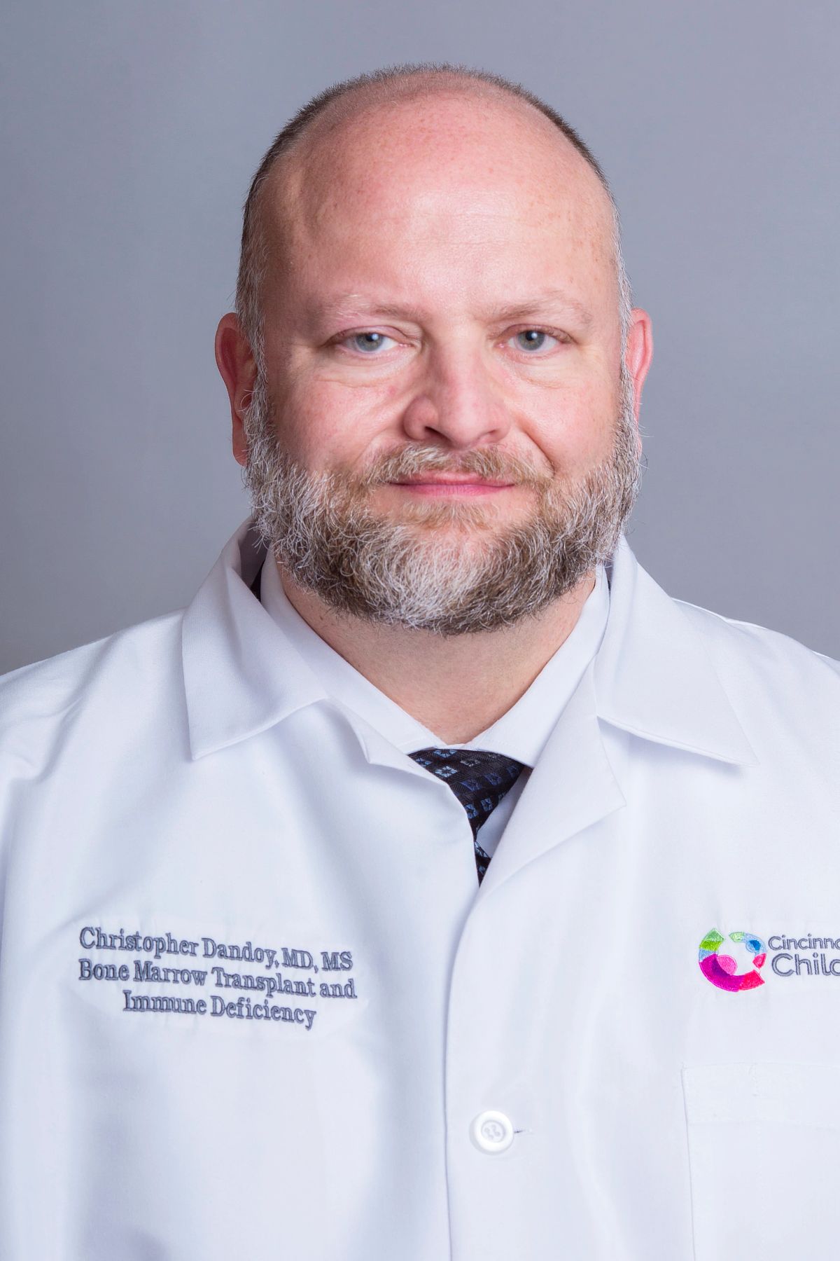 Christopher E. Dandoy合作论文数Cincinnati Children's Hospital Medical Center;University of Cincinnati Cancer Center;University of Cincinnati College of Medicine62
 Stella M. Davies合作论文数59
 Kasiani C. Myers合作论文数Division of Bone Marrow Transplantation and Immune Deficiency, Cincinnati Children's Hospital Medical Center56
 Sonata Jodele合作论文数Division of Bone Marrow Transplantation and Immune Deficiency, Cincinnati Children's Hospital Medical Center49Nelson Adam合作论文数35
 Adam Lane合作论文数Division of Bone Marrow Transplantation and Immune Deficiency, Cincinnati Children's Hospital Medical Center31
 Gregory H Wallace合作论文数27
 Michael S. Grimley合作论文数Cincinnati Children's Hospital Medical Center25
 Pooja Khandelwal合作论文数Division of Bone Marrow Transplantation and Immune Deficiency, Cincinnati Children's Hospital Medical Center20Abigail Pate合作论文数13Ashley Teusink-Cross合作论文数13Rebecca A. Marsh合作论文数13Rotz Seth合作论文数12
 Jacob J.H. Bleesing合作论文数Division of Bone Marrow Transplantation and Immune Deficiency, Cincinnati Children's Hospital Medical Center;Diagnostic Center for Heritable Immunodeficiencies, Cincinnati Children's Hospital Medical Center;Department of Pediatrics, University of Cincinnati11
 Thomas D Ryan合作论文数10
 Ashish Kumar合作论文数9Priscila Badia合作论文数Department of Pediatrics, University of Cincinnati College of Medicine;Division of Bone Marrow Transplantation and Immune Deficiency, Cincinnati Children's Hospital Medical Center;Cincinnati Children's Hospital Medical Center, University of Cincinnati College of Medicine9Chima Ranjit S合作论文数8Howell Jonathan C合作论文数8
 Sharad Chandra合作论文数8Parinda A. Mehta合作论文数8Russel Hirsch合作论文数8Michael B. Jordan合作论文数Division of Bone Marrow Transplantation/Immune Deficiency, Cincinnati Children's Hospital Medical Center8Pauline A. Daniels合作论文数7
 David B. Haslam合作论文数School of Medicine, Washington University7
 John Lynn Jefferies合作论文数6Warren Mikako合作论文数6Flesch Laura合作论文数6Paff Zachary合作论文数6
 Alexandra (Lisa) H. Filipovich合作论文数Department of Pediatrics, UC;Cincinnati Children’s Hospital Medical Center6Sarat Thikkurissy合作论文数5
 Michael D Taylor合作论文数Cincinnati Children's Hospital Medical Center, The Heart Institute5Cynthia B. Taggart合作论文数5
 Jason C. Woods合作论文数5
 Laura L. Walkup合作论文数UC Department of Pediatrics, Cincinnati Children's Hospital Medical Center;UC Department of Biomedical Engineering, Cincinnati Children's Hospital Medical Center5
 Christopher Towe合作论文数5
 Lara A. Danziger-Isakov合作论文数Department of Pediatrics, University of Cincinnati;Cincinnati Children's Hospital Medical Center5Holly R. Hoefgen合作论文数4Cash Michelle合作论文数4Teusink Ashley合作论文数4
 Heidi Lie Andersen合作论文数4
 Tsuyoshi Fukuda合作论文数4
 Susan Rose合作论文数Cincinnati Children's Hospital Medical Center4Janie Benoit合作论文数3Victoria Hickey合作论文数3Kathi Kramer合作论文数3K. Myers合作论文数3Christa Krupski合作论文数3
 Kelly Lake合作论文数3




