Electroencephalography (EEG) recordings often use caps or nets placed on the scalp. However, most are not designed to accommodate curly and coiled hair types, prevalent among individuals of African descent. This limitation results in the collection of suboptimal data that may be excluded from scientific research, raising questions about the generalizability of findings and the applicability of resultant knowledge to these populations. In response, researchers have proposed more inclusive EEG methodology, including using caps with tall pedestals and braiding techniques to improve data quality. Despite this advancement, tall pedestal nets fall short in addressing the need for clear scalp exposure, which is particularly important when capping children with voluminous hair. Similarly, braiding techniques have shown promise in small-scale studies involving adults, but their broader application remains unexplored in younger populations. To bridge these gaps, we introduce a novel approach for advancing representation and validity through the integration of curly hair specialists (CHSs) in pediatric EEG research. We examine the multifaceted role of CHSs and outline the processes involved in conceptualizing, developing, and implementing the CHS Initiative. We also offer a modified design for implementing this model in less resourced contexts and discuss community-based participatory research methods that informed our approach.
The most feared consequence of child abuse is brain injury. Abusive head trauma (AHT) can result in significant brain injuries that can cause death or lifelong handicap of a child, with the full consequence not truly evident until school age or later. AHT is the most common cause of traumatic death for infants with as many as one in four victims of AHT/shaken baby syndrome (SBS) dying (1). Almost all suffer serious health consequences such as sensory impairments as well as cognitive, learning, and behavioral disabilities (2).
The subplate (SP) is a transient fetal brain compartment supporting neuronal migration, axonal ingrowth, and early cortical activity, yet the dynamics of its regional development remain poorly understood in vivo. Using T2-weighted fetal MRI of 68 typically developing fetuses (22 to 32 wk gestational age, GA), we developed a semiautomated pipeline to quantify regional SP morphology (thickness, surface area, and volume). SP characteristics scaled strongly with GA and residual brain volume and showed marked regional differences. After correcting for geometric confounds, regional variation of SP thickness persisted, with highest values in parietal and perisylvian regions, suggesting that SP thickness may serve as a sensitive marker of intrinsic developmental differences. Between the late 2nd and early 3rd trimesters, mean SP thickness increased by 39.2% with large variation across regions (±11.0 SD), whereas surface area growth was more uniform (64.3% ±0.7 SD). Continuous growth trajectories clustered into distinct spatiotemporal profiles: early-developing regions (e.g., pericentral and medial occipital cortices) contrasted with later-developing regions (prefrontal, temporal, and parietal cortices). These patterns partially recapitulate primary-to-association, medial-to-lateral, and posterior-to-anterior maturational hierarchies, pointing to organized developmental programs. SP development also showed region-specific hemispheric asymmetries, including leftward thickness and volume asymmetry in the superior temporal and precentral gyri. Some asymmetries amplified, others attenuated or reversed with age, suggesting both transient states and potential precursors of postnatal lateralization. Together, these findings provide a framework for regional SP quantification and position SP morphology, particularly thickness, as a promising early biomarker that might link fetal SP changes to subsequent cortical development and neurodevelopmental outcomes.
Obtaining consistent quantitative maps of myelin content and relaxation times across different sites and vendors is essential for advancing our understanding of brain development. Herein, we present a harmonized, vendor-agnostic magnetic resonance acquisition method designed for joint T1, T2, and myelin water fraction mapping, along with a method for rapid B1+ and B1- field estimation. We used our dictionary-based fitting and multi-compartment modeling for joint mapping of T1, T2 and myelin water fraction. Self-navigation-based retrospective motion correction was integrated with subspace reconstruction to track and correct rigid head motion during scanning, operating without the need for external hardware. Simulations, phantom and in vivo experiments confirmed the sensitivity and accuracy of the method, particularly for short T2 values corresponding to myelin, and demonstrated consistent performance across multiple scanner types. Coupled with the harmonized calibration scan, the proposed package offers a practical tool for multi-site, multi-vendor neuroimaging studies in both adult and pediatric populations.
Abstract Sickle cell disease (SCD) is characterized by chronic hemolysis, and painful vaso-occlusive episodes (VOE). High levels of fetal hemoglobin (HbF) attenuate the disease phenotype. We used a lentivirus vector (LVV) expressing a short hairpin RNA embedded in a microRNA (shmiR) that targets BCL11A in erythrocytes to induce HbF in a first-in-human study in SCD. The purpose of the study was to assess hematopoietic stem/progenitor cells collection, transduction parameters, safety, HbF induction, and durability. Eleven eligible patients with SCD underwent hematopoietic stem cell (HSC) collection. Plerixafor-mobilized peripheral blood HSCs required for manufacturing were obtained in 1 mobilization cycle for 10 of 11 participants, and 11 of 11 patient products were successfully manufactured with a median time to release of product of 39 days. Ten patients were infused with HSCs transduced with the shmiR vector. Engraftment occurred in all 10 patients. At a median follow-up of 58 months (range, 35-82) after infusion, there were no adverse events attributed to the vector. The transduction efficiency was 93.1%. One patient demonstrated low engraftment of the transduced cells and had suboptimal HbF induction. In the remaining 9 patients, at 2 years after treatment, the peripheral blood demonstrated 71% F cells with 11.9 pg HbF per F cells, and both parameters remained stable in 9 patients with ≥48 months follow-up. All patients who had VOEs before gene therapy demonstrated sustained mitigation of pain events. These data demonstrate excellent manufacturing efficiency, efficacy and safety of targeting BCL11A using a shmiR LVV, and long-term durability of the shmiR vector, leading to a pivotal, multisite, phase 2 trial that is currently underway (NCT05353647). This trial was registered at www.clinicaltrials.gov as NCT03282656.
KCNT1-related epileptic encephalopathy, including epilepsy of infancy with migrating focal seizures, is a severe neurodevelopmental disorder associated with refractory seizures, profound neurologic impairment and premature death. It is caused by de novo genetic variants in KCNT1 that alter the function of Slack, an evolutionarily conserved sodium-gated potassium channel that modulates neuronal firing patterns and excitability. Pathogenic KCNT1 variants lead to overactive Slack channels, boosting total neuronal potassium currents by up to 40%, driving cortical hyperexcitability and causing seizures. Here we investigate antisense oligonucleotide-mediated KCNT1 knockdown as a therapeutic strategy for patients with epilepsy of infancy with migrating focal seizures. Intrathecal delivery of an experimental, non-allele-specific, KCNT1-targeting antisense oligonucleotide by lumbar puncture in two 2-year-old females with KCNT1 p.R474H, a severe, recurrent pathogenic variant, led to a significant reduction in seizure frequency and intensity. However, investigational treatment was also associated with the development of ventricular enlargement or hydrocephalus in both patients, prompting in one case the redirection of goals of care, pointing to a potential monitorable toxicity of some intrathecal antisense oligonucleotides.
OBJECTIVE:Approximately 40% of children undergoing epilepsy surgery have postoperative seizures, underscoring the need for enhanced estimators of the epileptogenic zone (EZ). We hypothesize that visually imperceptible low-entropy activity in the interictal periods, even in the absence of conventional spikes, is a robust signature of the EZ. To test this, we mapped interictal "low-entropy zones" using intracranial electroencephalography (iEEG) in children with drug-resistant epilepsy (DRE) and assessed their value for postsurgical outcome prediction when targeted during surgery, along with their stability over prolonged periods. METHODS:We analyzed iEEG data of 75 DRE children, including brief (5 min) data from patients with known Engel outcome (N = 59; used for outcome prediction) plus prolonged data from a separate recent cohort (N = 16; used for stability assessment). We estimated each contact's entropy across various frequencies (delta to fast-ripple), pinpointed low-entropy zones, and assessed whether their removal predicts outcome (3-fold cross-validation). In addition, the predictive value of entropy during non-epileptiform (spike-free) epochs was also assessed. Furthermore, established interictal estimators (spikes-on-ripple, fast ripples) were tested for outcome prediction. Using the prolonged dataset, we tested whether entropy distribution over brief epochs was similar to prolonged (3 h) data. RESULTS:High overlap between low-entropy zones and resection correlated with low Engel class (p < 0.0001, R = -0.54, N = 59), also during non-epileptiform epochs (R = -0.52). Low-entropy-zone removal predicted outcomes with F1 score of 87% (p < 0.0001, N = 51; Engel I vs III-IV) outperforming spikes-on-ripple (F1 score = 82%, p = 0.002) or fast ripples (F1 score = 80%, p = 0.01). Low-entropy zones retained high predictive value when non-epileptiform epochs were used (F1 score = 89%, N = 44). Entropy distribution over brief epochs was strongly correlated with prolonged data (R > 0.8, p < 0.0001), and its relationship with seizure-onset zone did not differ (brief vs prolonged data: p > 0.6). SIGNIFICANCE:Surgically targeting low-entropy zones accurately predicts the postoperative seizure outcomes of children with DRE. Mapping low-entropy activity using brief iEEG segments shows consistency with using prolonged data and could enhance surgical planning in pediatric DRE.
OBJECTIVE:Lack of participant diversity in pediatric clinical research limits external validity of findings and can exacerbate health inequities. Our objective was to identify evidence-based strategies/interventions that improve enrollment and retention of diversity of pediatric participants in clinical research and provide an evidence-based tool to guide pediatric clinical research protocols. METHODS:This study comprised a scoping review of peer-reviewed literature that reported methods to increase enrollment and retention of pediatric participants in clinical research (2009-2022). Guided by the Preferred Reporting Items for Systematic Reviews and Meta-Analyses (PRISMA) extension for scoping reviews. Data extraction included quantitative and qualitative outcomes. Eligible studies' findings were analyzed and grouped into specific attrition steps that limit pediatric clinical research participants' enrollment and retention. RESULTS:A total of 31 studies met inclusion criteria. Each study's insights were categorized based on attrition steps that limited pediatric participant diversity: study design (n = 1), marketing/outreach strategies (n = 7), participant perceptions (n = 9), consent/enrollment issues (n = 8), incentives/reimbursements mechanisms (n = 4), retention strategies (n = 8), and follow-up/feedback channels (n = 3). Many studies had overlapping attrition categories. Most studies were nonrandomized clinical studies such as retrospective and cohort studies with different strategies/interventions, making comparisons between studies challenging. CONCLUSIONS:The effectiveness in recruiting a diverse cohort of pediatric clinical research participants requires a multifaceted approach addressing issues at every stage of the clinical research process. Enrollment and retention strategies should focus on building trust, providing effective incentives, and incorporating continuous feedback.
Fetal ventriculomegaly (VM) and its severity and associated central nervous system (CNS) abnormalities are important indicators of high risk for impaired neurodevelopmental outcomes. Recently, a novel fetal brain age prediction method using a two-dimensional (2D) single-channel convolutional neural network (CNN) with multiplanar MRI sections showed the potential to detect fetuses with VM. This study examines the diagnostic performance of a deep learning-based fetal brain age prediction model to distinguish fetuses with VM (n = 317) from typically developing fetuses (n = 183), the severity of VM, and the presence of associated CNS abnormalities. The predicted age difference (PAD) was measured by subtracting the predicted brain age from the gestational age in fetuses with VM and typical development. PAD and absolute value of PAD (AAD) were compared between VM and typically developing fetuses. In addition, PAD and AAD were compared between subgroups by VM severity and the presence of associated CNS abnormalities in VM. Fetuses with VM showed significantly larger AAD than typically developing fetuses (P < .001), and fetuses with severe VM showed larger AAD than those with moderate VM (P = .004). Fetuses with VM and associated CNS abnormalities had significantly lower PAD than fetuses with isolated VM (P = .005). These findings suggest that fetal brain age prediction using the 2D single-channel CNN method has the clinical ability to assist in identifying not only the enlargement of the ventricles but also the presence of associated CNS abnormalities. Keywords: MR-Fetal (Fetal MRI), Brain/Brain Stem, Fetus, Supervised Learning, Machine Learning, Convolutional Neural Network (CNN), Deep Learning Algorithms Supplemental material is available for this article. ©RSNA, 2025.
Hypoxic ischemic encephalopathy (HIE) is a brain injury that occurs in 1 ~ 5/1000 term neonates. Accurate identification and segmentation of HIE-related lesions in neonatal brain magnetic resonance images (MRIs) is the first step toward identifying high-risk patients, understanding neurological symptoms, evaluating treatment effects, and predicting outcomes. We release the first public dataset containing neonatal brain diffusion MRI and expert annotation of lesions from 133 patients diagnosed with HIE. HIE-related lesions in brain MRI are often diffuse (i.e., multi-focal), and small (over half the patients in our data having lesions occupying <1% of the brain volume (including ventricles)). Segmentation for HIE MRI data is remarkably different from, and arguably more challenging than, other segmentation tasks such as brain tumors with focal and relatively large lesions. We hope that this dataset can help fuel the development of MRI lesion segmentation methods for HIE and small diffuse lesions in general.
Neonatal encephalopathy (NE) is a significant global health concern. It is a leading cause of long-term neurodevelopmental impairment, with hypoxic-ischaemic perinatal brain injury being the most common underlying contributor. Although therapeutic hypothermia has reduced mortality and improved outcomes for some affected infants, many survivors experience neurodevelopmental disability, including cerebral palsy and/or deficits in cognition, behaviour, and executive functioning. Early and accurate prognostication and identification of injury severity remain a challenge due to evolving clinical signs and multiple etiologies. Magnetic resonance imaging (MRI) is the gold standard for characterizing NE-related brain injury. Diffusion-weighted imaging (DWI) enables early detection of injury, and proton magnetic resonance spectroscopy (1H-MRS), specifically the Lac/NAA peak area ratio from basal ganglia and thalamus, provides robust prognostic indicators of two-year neurodevelopmental outcomes. MRI scoring systems incorporating multiple modalities correlate well with later neurodevelopmental outcomes. Advanced imaging modalities, such as diffusion tensor imaging (DTI), arterial spin labelling (ASL), and blood oxygen level-dependent (BOLD) imaging, offer further insights into microstructural integrity, perfusion, and functional connectivity. By standardizing acquisition protocols and post-processing, MRI biomarkers can serve as reliable, early surrogate endpoints in neuroprotection trials, allowing smaller sample sizes and accelerating clinical translation. MRI and 1H-MRS integration enhances prognostication, guides clinical management, and supports informed decision-making in NE care.
BACKGROUND:In adolescents and adults with complex congenital heart disease (CHD), abnormal cortical folding is a putative predictor of poor neurodevelopmental outcome. However, it is unknown when this relationship first emerges. We test the hypothesis that it begins in utero, when the brain starts to gyrify and folding patterns first become established. METHODS:We carried out a prospective, longitudinal case-control study, acquiring foetal MRIs at two timepoints in utero, (Scan 1 = 20-30 Gestational Weeks (GW) and Scan 2 = 30-39 GW), then followed up participants at two years of age to assess neurodevelopmental outcomes. We used normative modelling to chart growth trajectories of surface features across 60 cortical regions in a control population (n = 157), then quantified the deviance of each foetus with CHD (n = 135) and explored the association with neurodevelopmental outcomes at two years of age. FINDINGS:Differences in cortical development between CHD and Control foetuses only emerged after 30 GW, and lower regional cortical surface area growth was correlated with poorer neurodevelopmental outcomes at two years of age in the CHD group. INTERPRETATION:This work highlights the third trimester specifically as a critical period in brain development for foetuses with CHD, where the reduced surface area expansion in specific cortical regions becomes consequential in later life, and predictive of neurodevelopmental outcome in toddlerhood. FUNDING:This research was supported by the NINDS (R01NS114087, K23NS101120) and NIBIB (R01EB031170) of the NIH, PHN Scholar Award, AAN Clinical Research Training Fellowship, BBRF Young Investigator Awards, and the Farb Family Fund.
Understanding the structural connectivity of the human brain during fetal life is essential for uncovering the early foundations of neural function and vulnerability to developmental disorders. Diffusion-weighted MRI (dMRI) enables noninvasive mapping of white-matter pathways and construction of the brain's structural connectome, but its application to the fetal brain has been constrained by limited data and the technical challenges of fetal dMRI analysis. Here, we present the largest study to date of in utero brain connectivity, analyzing high-quality dMRI data from 198 fetuses between 22 and 37 gestational weeks from the Developing Human Connectome Project. We applied advanced fetal-specific tools for brain segmentation, parcellation, and tractography, and used fiber bundle capacity to weight connections. We reconstructed individual structural connectomes and characterized their developmental trajectories. Graph-theoretical analysis revealed consistent increases in both integration and segregation metrics across gestation, alongside stable small-world properties. Bootstrapping confirmed the robustness of nodal and edge-wise developmental patterns, and a sigmoid growth model identified a narrow time window (approximately 27.5-30.5 weeks) of rapid connectivity strengthening. In addition, we introduced a new method for constructing age-specific connectome templates by aggregating individual subject connectomes. Our analysis shows that this approach outperforms spatial alignment and image-space averaging, yielding templates that preserve individual topology and support accurate age prediction. Together, these findings provide a reasonable normative map of fetal brain structural connectivity and establish a foundation for future studies of atypical development and early indicators of neurological risk.
Hypoxia-ischemia (HI), which disrupts the oxygen supply-demand balance in the brain by impairing blood oxygen supply and the cerebral metabolic rate of oxygen (CMRO2), is a leading cause of neonatal brain injury. However, it is unclear how post-HI hypothermia helps to restore the balance, as cooling reduces CMRO2. Also, how transient HI leads to secondary energy failure (SEF) in neonatal brains remains elusive. Using photoacoustic microscopy, we examined the effects of HI on CMRO2 in awake 10-day-old mice, supplemented by bioenergetic analysis of purified cortical mitochondria. Our results show that while HI suppresses ipsilateral CMRO2, it sparks a prolonged CMRO2-surge post-HI, associated with increased mitochondrial oxygen consumption, superoxide emission, and reduced mitochondrial membrane potential necessary for ATP synthesis—indicating oxidative phosphorylation (OXPHOS) uncoupling. Post-HI hypothermia prevents the CMRO2-surge by constraining oxygen extraction fraction, reduces mitochondrial oxidative stress, and maintains ATP and N-acetylaspartate levels, resulting in attenuated infarction at 24 hr post-HI. Our findings suggest that OXPHOS-uncoupling induced by the post-HI CMRO2-surge underlies SEF and blocking the surge is a key mechanism of hypothermia protection. Also, our study highlights the potential of optical CMRO2 measurements for detecting neonatal HI brain injury and guiding the titration of therapeutic hypothermia at the bedside.
Fetal motion is a critical indicator of neurological development and intrauterine health, yet its quantification remains challenging, particularly at earlier gestational ages (GA). Current methods track fetal motion by predicting the location of annotated landmarks on 3D echo planar imaging (EPI) time-series, primarily in third-trimester fetuses. The predicted landmarks enable simplification of the fetal body for downstream analysis. While these methods perform well within their training age distribution, they consistently fail to generalize to early GAs due to significant anatomical changes in both mother and fetus across gestation, as well as the difficulty of obtaining annotated early GA EPI data. In this work, we develop a cross-population data augmentation framework that enables pose estimation models to robustly generalize to younger GA clinical cohorts using only annotated images from older GA cohorts. Specifically, we introduce a fetal-specific augmentation strategy that simulates the distinct intrauterine environment and fetal positioning of early GAs. Our experiments find that cross-population augmentation yields reduced variability and significant improvements across both older GA and challenging early GA cases. By enabling more reliable pose estimation across gestation, our work potentially facilitates early clinical detection and intervention in challenging 4D fetal imaging settings. Code is available at https://github.com/sebodiaz/cross-population-pose.
OBJECTIVE:Our objective was to investigate prenatal imaging findings, clinical course and outcomes associated with isolated congenital aqueductal stenosis (ICAS). METHOD:A retrospective study was conducted in the period of 2010-2023, including patients with ICAS confirmed postnatally who were imaged prenatally with ≥ 1 year of follow-up. Patients with additional anomalies (structural or genetic) were excluded. Neurodevelopmental outcomes were verified by pediatricians, and imaging underwent standardized measurement by a neuroradiologist. RESULTS:Twenty-one patients were prenatally diagnosed with ICAS, at a median gestational age (GA) of 19.7 weeks. Overall, 13/14 patients exhibited a fronto-occipital horn ratio (FOHR) > 0.5, indicating clinically significant ventriculomegaly in initial MRI at 18-32 weeks GA. There was an increase in the median size of the third ventricular coronal width from 7 mm in prenatal imaging to 12 mm in postnatal imaging (p = 0.01). Twenty patients (95.2%) required shunting or endoscopic third ventriculostomy and bilateral choroid plexus cauterization (ETV/CPC), with 10 undergoing multiple CSF diversion procedures during follow-up. Among the study group, nine patients experienced epilepsy, 6/8 aged < 5 years exhibited global developmental delay, and 6/12 aged ≥ 5 years required special education services. CONCLUSION:Our findings indicate a progressive increase in prenatal ventricular sizes, with most children requiring hydrocephalus treatment and experiencing neurodevelopmental impairment.
BACKGROUND:3-T MRI can improve image quality of fetal imaging compared to 1.5-T MRI. However, concerns exist regarding increased local tissue heating at 3-T. PURPOSE:To assess fetal MRI radiofrequency (RF) safety at 3-T by comparing simulated tissue heating to 1.5-T (using constant RF exposure) and by simulating tissue heating at 3-T using RF exposures from clinical fetal examinations. STUDY TYPE:Retrospective. POPULATION:Seven voxelized anatomical pregnant body models (gestational age [GA] 30 ± 3 weeks [mean ± standard deviation], maternal body mass index [BMI] 27.8 ± 8.5 kg/m2) were used. Maternal whole-body average specific absorption rate (wbSAR) logs were collected from 85 clinical examinations at 3-T (GA 25 ± 6 weeks, BMI 30.3 ± 6.8 kg/m2). FIELD STRENGTH/SEQUENCE:3-T, 1.5-T, HASTE, VIBE, TRUFISP, EPI, DTI. ASSESSMENT:Simulated maternal and fetal peak and average SAR, temperature, and peak thermal dose were compared at 3-T and 1.5-T for 60 min 2 W/kg wbSAR using 7 body models and a 16-rung band-pass RF coil. Temperature and thermal dose were simulated in one body model using clinical wbSAR exposures at 3-T. STATISTICAL TESTS:Factorial analysis of variance was performed using 28 maternal and fetal temperature measurements from 7 body models to detect a difference between 3-T and 1.5-T. p < 0.05 was considered statistically significant. RESULTS:For constant RF exposure, we found no difference between 3-T and 1.5-T in peak maternal (1.5-T:40.38 ± 0.21°C; 3-T:40.40 ± 0.20°C; p = 0.85), peak fetal (1.5-T:39.21 ± 0.17°C; 3-T:39.09 ± 0.16°C; p = 0.19), and average maternal (1.5-T:37.32 ± 0.05°C; 3-T:37.33 ± 0.04°C; p = 0.68) temperature. We observed significantly higher average fetal temperatures at 1.5-T (1.5-T:37.75 ± 0.06°C; 3-T:37.70 ± 0.05°C). For 3-T clinical RF exposures, simulated peak temperatures exceeded the recommended limits. However, the thermal dose was below the recommended limit. DATA CONCLUSION:For the same RF coil geometry, local heating was similar at 3-T and 1.5-T for constant RF exposure. Although realistic 3-T RF exposures could cause peak temperatures above the recommended limits, thermal dose was below the recommended limit. EVIDENCE LEVEL:1. TECHNICAL EFFICACY:Stage 1.
Hypoxic ischemic encephalopathy (HIE) is a brain dysfunction occurring in approximately 1-5/1000 term-born neonates. Accurate segmentation of HIE lesions in brain MRI is crucial for prognosis and diagnosis but presents a unique challenge due to the diffuse and small nature of these abnormalities, which resulted in a substantial gap between the performance of machine learning-based segmentation methods and clinical expert annotations for HIE. To address this challenge, we introduce ParadiseNet, an algorithm specifically designed for HIE lesion segmentation. ParadiseNet incorporates global-local learning, progressive uncertainty learning, and self-evolution learning modules, all inspired by clinical interpretation of neonatal brain MRIs. These modules target issues such as unbalanced data distribution, boundary uncertainty, and imprecise lesion detection, respectively. Extensive experiments demonstrate that ParadiseNet significantly enhances small lesion detection (< 1%) accuracy in HIE, achieving an over 4% improvement in Dice, 6% improvement in NSD compared to U-Net and other general medical image segmentation algorithms.
Objectives:To develop a data harmonization framework for neonatal hypoxic-ischemic encephalopathy (HIE) studies and demonstrate its suitability for prognostic biomarker development. Materials and Methods:Variables were first categorized by chronological stages and then by medical topics. We created a dictionary to harmonize variable names and value coding. We began by merging comprehensive data from 2 landmark nationwide therapeutic hypothermia for HIE trials (2008-2016, 21 sites) in the Eunice Kennedy Shriver National Institute of Child Health and Human Development (NICHD) Neonatal Research Network (NRN). The 2 datasets differ in available variables, variable naming and coding, necessitating harmonization. To demonstrate the utility of this data harmonization framework, we computed the distributions of variables and ranked them by the strength of associations with 18- to 22-month outcomes. Associations were measured using Pearson's correlation analysis. Outcomes were defined as (a) a 5-class variable: survivors with normal, mild, moderate, severe disability, or death and (b) the Bayley-III Scales. Results:We created a dictionary of 1181 variables on 532 patients across 5 chronologic categories and 60 medical subcategories. The distribution of major predictive and outcome variables, and the variables strongly associated with neurodevelopmental outcomes at 18-22 months were presented. The modified Sarnat scores at the Post-intervention and NICU-discharge stage, and the NRN pattern of MRI injury score showed strong associations with outcome variables. Conclusion:We designed a data harmonization framework specifically for HIE. Our initial effort in merging 2 iconic clinical trials shows strong predictor-outcome associations, allowing subsequent development of advanced prognostic biomarkers of neonatal HIE.
 Borjan Gagoski合作论文数Boston Children's Hospital71
 Yangming Ou合作论文数Image, Informatics & Intelligence Research Lab, Boston Children's Hospital, Harvard Medical School;Department of Radiology, Boston Children's Hospital, Harvard Medical School47
 Esra A. Turk合作论文数46Kiho Im合作论文数45
 Polina Golland合作论文数Department of Electrical Engineering and Computer Science, Massachusetts Institute of Technology;Computer Science & Artificial Intelligence Laboratory, Massachusetts Institute of Technology;Institute for Medical Engineering & Science, Massachusetts Institute of Technology43
 Elfar Adalsteinsson合作论文数Research Laboratory of Electronics, Massachusetts Institute of Technology;Institute for Medical Engineering & Science, Massachusetts Institute of Technology42
 Rudolph Pienaar合作论文数36
 Christos Papadelis合作论文数Lab for Human Brain Dynamics;Brain Science Institue;Lab for Human Brain Dynamics, Brain Science Institue34
 Tamilia Eleonora合作论文数Center for Integrated Research, Università Campus Bio-Medico di Roma29Yun Hyuk Jin合作论文数Department of Biomedical Engineering, Hanyang University29
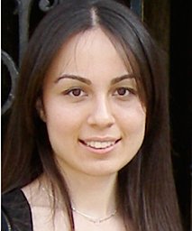 Ahtam Banu合作论文数Division of Newborn Medicine, Fetal Neonatal Neuroimaging and Developmental Science Center29
 Maria Angela Franceschini合作论文数Massachusetts General Hospital;Athinoula A. Martinos Center for Biomedical Imaging, Mass General Research Institute;Harvard Medical School29
 Lawrence Wald合作论文数Athinoula A. Martinos Center for Biomedical Imaging, Massachusetts General Hospital, Harvard Medical School26
 Phillip L. Pearl合作论文数School of Medicine and Health Sciences, George Washington University26
 Caitlin Rollins合作论文数26
 Joseph Madsen合作论文数Department of Neurosurgery, Boston Children's Hospital;Harvard Medical School25
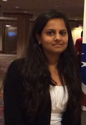 Rutvi Vyas合作论文数Div Newborn Med, Boston Childrens Hosp22
 Lilla Zöllei合作论文数Athinoula A. Martinos Center for Biomedical Imaging, Mass General Research Institute, Massachusetts General Hospital;Harvard Medical School21
 Simon K. Warfield合作论文数Harvard Medical School;Computational Radiology Laboratory, Department of Radiology, Boston Children’s Hospital, Harvard CTSC Translational Technologies Imaging Consortium21Pei-Yi Lin合作论文数Boston Children's Hospital20
 Jane Newburger合作论文数Department of Cardiology, Boston Children's Hospital;Harvard Medical School20
 Henry Feldman合作论文数Institutional Centers for Clinical and Translational Research, Boston Children's Hospital;Biostatistics and Research Design Center, Boston Children's Hospital;Harvard Medical School18Mathieu Dehaes合作论文数Département de Génie Électrique and Institut de génie biomédical, École Polytechnique de Montréal18
 Georgios Ntolkeras合作论文数Div Newborn Med, Boston Childrens Hosp17
 Nadege Roche-Labarbe合作论文数17Janet S. Soul合作论文数17
 Lana Vasung合作论文数Department of Pediatrics, University of Geneva16Sarah U. Morton合作论文数16
 Stout Jeffrey N合作论文数Radiology, Athinoula A. Martinos Center for Biomedical Imaging;Massachusetts Institute of Technology, Harvard-MIT Health Sciences and Technology;Massachusetts Institute of Technology, Harvard-MIT Health Sciences and Technology15
 Emi Takahashi合作论文数14Kalpathy S. Krishnamoorthy合作论文数14
 Steven M Stufflebeam合作论文数MGH-MIT-HMS Athinoula A. Martinos Center for Biomedical Imaging, Massachusetts General Hospital14
 Nadine Gaab合作论文数Department of Neurology, Music and Neuroimaging Laboratory, Beth Israel Deaconess Medical Center and Harvard Medical School14
 Junshen Xu合作论文数13
 Bruce Fischl合作论文数Department of Radiology, Harvard Medical School;Martinos Center, Massachusetts General Hospital;Department of Radiology, Massachusetts General Hospital13William H. Barth合作论文数13Jason Sutin合作论文数Department of Biomedical Engineering;Boston UniVersity;Department of Biomedical Engineering, Boston UniVersity13
 Silvina Ferradal合作论文数Universidad Nacional de Rosario13
 Tomo Tarui合作论文数12Ortinau Cynthia合作论文数12
 Angela Fenoglio合作论文数12
 Jim Barkovich合作论文数Department of Radiology and Biomedical Imaging, University of California, San Francisco12
 Kawin Setsompop合作论文数Department of Radiology, School of Medicine, Stanford University;Stanford Cancer Institute, Stanford University;Harvard-MIT Division of Health Sciences and Technology;Department of Electrical Engineering, School of Engineering, Stanford University;Athinoula A. Martinos Center for Biomedical Imaging12
 Ali Gholipour合作论文数Boston Children's Hospital, Harvard Medical School12
 Judy Estroff合作论文数Department of Radiology, Children's Hospital Boston12
 Danielle D. Sliva合作论文数Fetal-Neonatal Neuroimaging and Developmental Science Center, Boston Children's Hospital Boston;Laboratories of Cognitive Neuroscience, Boston Children's Hospital;Boston Children's Hospital Boston, Boston Children's Hospital11
 Christopher A Walsh合作论文数The Paul G. Allen Frontiers Group Discovery Center for Human Brain Evolution;Howard Hughes Medical Institute;Division of Genetics & Genomics, Boston Children's Hospital;Harvard Medical School11Drucilla J. Roberts合作论文数11
 Yogesh Rathi合作论文数Harvard Medical School11




