PURPOSE:The IEC 60601-2-33 standard provides consensus-based safety provisions for MRI equipment. Protection of patients against cardiac stimulation (CS) is based on limiting the maximum E-field induced by MRI gradient coils. In practice, this is achieved by imposing a conservative dB/dt threshold on any gradient waveform. The dB/dt-over-E-field conversion ratio currently used in IEC 60601-2-33 was derived in a homogeneous ellipsoid exposed to a uniform B-field and is 10 (T/s)*(V/m)-1. This limit is becoming increasingly restrictive in high performance clinical systems. We therefore evaluate dB/dt-over-E-field ratios in realistic body models and coils using state-of-the-art electromagnetic simulations. METHODS:We performed two independent simulation studies in a total of 75 realistic body models and 13 commercial gradient systems and derived dB/dt-over-E-field ratios in the heart. We thresholded the E-field maps to mitigate the impact of staircasing artifacts in boundary voxels between the myocardium and the lungs. RESULTS:Thresholding the E-field maps at the 99th percentile E-field value (E99) eliminates staircasing artifacts in both simulation studies. Study #1 predicts a larger range of dB/dt-over-E99 ratios (13-53 (T/s)*(V/m)-1) than study #2 (12-35 (T/s)*(V/m)-1). Despite differences in EM solvers, body models, coils, mesh resolution, and post-processing, both studies find similar worst-case ratios of dB/dt-over-E99 of 12-13 (T/s)*(V/m)-1. CONCLUSION:Our simulations of dB/dt-over-E-field ratios for cardiac safety in MRI cover a large range of realistic clinical scenarios. An increase of the allowable dB/dt beyond the current CS limit in IEC 60601-2-33 may be feasible.
Cerebrospinal fluid (CSF) flow is a key component of the brain's waste clearance system. However, our understanding of CSF flow in the human brain, particularly within the brain-wide subarachnoid space (SAS), is limited due to a lack of non-invasive tools for measuring slow flow. Here, we propose a CSF flowmetry technique using phase-contrast MRI combined with a slow-flow-sensitized acquisition. It achieves high sensitivity in measuring slow CSF flow (e.g., 100 μm/s), and enables quantitative measurement of the velocity and direction with whole-brain coverage, spanning from ventricles to SAS. Our proof-of-concept results demonstrate repeatable flow measurements and show that cardiac pulsation induces coherent CSF flow changes within the SAS. Our data also suggest that cardiac pulsation has a stronger driving effect on brain-wide CSF flow compared to respiration. This technique provides a valuable tool for investigating CSF dynamics and pathways to advance a holistic understanding of brain-wide CSF flow.
The hypothalamus plays an important role in the regulation of the bodys metabolic state and behaviors related to survival. Despite its importance however, many questions exist regarding the intrinsic and extrinsic connections of the hypothalamus in humans, especially its relationship with the cortex. As a heterogeneous structure, it is possible that the hypothalamus is composed of different subregions, which have their own distinct relationships with the cortex. Previous work on functional connectivity in the human hypothalamus have either treated it as a unitary structure or relied on methodological approaches that are limited in modeling its intrinsic functional architecture. Here, we used resting state data from ultrahigh field 7 Tesla fMRI and a data driven analytical approach to identify functional subregions of the human hypothalamus. Our approach identified four functional hypothalamic subregions based on intrinsic functional connectivity, which in turn showed distinct patterns of functional connectivity with cortex. Overall, all hypothalamic subregions showed stronger connectivity with a cortical network, Cortical Network 1 composed primarily of frontal, midline, and limbic cortical areas and weaker connectivity with a second cortical network composed largely of posterior sensorimotor regions, Cortical Network 2. Of the hypothalamic subregions, the anterior hypothalamus showed the strongest connection to Cortical Network 1, while a more ventral subregion containing the anterior hypothalamus extending to the tuberal region showed the weakest connectivity. The findings support the use of ultrahigh field, high resolution imaging in providing a more incisive investigation of the human hypothalamus that respects its complex internal structure and extrinsic functional architecture.
Magnetic fields switching at kilohertz frequencies induce electric fields in the body, which can cause peripheral nerve stimulation (PNS). Although magnetostimulation has been extensively studied below 10 kHz, the behavior of PNS at higher frequencies remains poorly understood. This study aims to investigate PNS thresholds at frequencies up to 88.1 kHz and to explore deviations from the widely accepted hyperbolic strength-duration curve (SDC).PNS thresholds were measured in the head of 8 human volunteers using a solenoidal coil at 16 distinct frequencies, ranging from 200 Hz to 88.1 kHz. A hyperbolic SDC was used as a reference to compare the frequency-dependent behavior of PNS thresholds.Contrary to the predictions of the hyperbolic SDC, PNS thresholds did not decrease monotonically with frequency. Instead, thresholds reached a minimum near 25 kHz, after which they increased by an average of 39% from 25 kHz to 88.1 kHz across subjects. This pattern indicates a significant deviation from previously observed behavior at lower frequencies.Our results suggest that PNS thresholds exhibit a non-monotonic frequency dependence at higher frequencies, diverging from the traditional hyperbolic SDC. These findings offer critical data for refining neurodynamic models and provide insights for setting PNS safety limits in applications like MRI gradient coils and magnetic particle imaging (MPI). Further investigation is needed to understand the biological mechanisms driving these deviations beyond 25 kHz.Clinical impact—These findings call for further basic research into biological mechanisms underlying high frequency PNS threshold trends, and supports refinement of safety guidelines for MRI and MPI systems for clinical implementation.
The brain continuously anticipates the energetic needs of the body and prepares to meet those needs before they arise, called allostasis. In support of allostasis, the brain continually models the sensory state of the body, called interoception. We replicated and extended a large-scale system supporting allostasis and interoception in the human brain using ultra-high precision 7 Tesla functional magnetic resonance imaging (fMRI) (N = 90), improving the precision of subgenual and pregenual anterior cingulate topography combined with extensive brainstem nuclei mapping. We observed over 90% of the anatomical connections published in tract-tracing studies in non-human animals. The system also included regions of dense intrinsic connectivity broadly throughout the system, some of which were identified previously as part of the backbone of neural communication across the brain. These results strengthen previous evidence for a whole-brain system supporting the modeling and regulation of the internal milieu of the body.
BACKGROUND:3-T MRI can improve image quality of fetal imaging compared to 1.5-T MRI. However, concerns exist regarding increased local tissue heating at 3-T. PURPOSE:To assess fetal MRI radiofrequency (RF) safety at 3-T by comparing simulated tissue heating to 1.5-T (using constant RF exposure) and by simulating tissue heating at 3-T using RF exposures from clinical fetal examinations. STUDY TYPE:Retrospective. POPULATION:Seven voxelized anatomical pregnant body models (gestational age [GA] 30 ± 3 weeks [mean ± standard deviation], maternal body mass index [BMI] 27.8 ± 8.5 kg/m2) were used. Maternal whole-body average specific absorption rate (wbSAR) logs were collected from 85 clinical examinations at 3-T (GA 25 ± 6 weeks, BMI 30.3 ± 6.8 kg/m2). FIELD STRENGTH/SEQUENCE:3-T, 1.5-T, HASTE, VIBE, TRUFISP, EPI, DTI. ASSESSMENT:Simulated maternal and fetal peak and average SAR, temperature, and peak thermal dose were compared at 3-T and 1.5-T for 60 min 2 W/kg wbSAR using 7 body models and a 16-rung band-pass RF coil. Temperature and thermal dose were simulated in one body model using clinical wbSAR exposures at 3-T. STATISTICAL TESTS:Factorial analysis of variance was performed using 28 maternal and fetal temperature measurements from 7 body models to detect a difference between 3-T and 1.5-T. p < 0.05 was considered statistically significant. RESULTS:For constant RF exposure, we found no difference between 3-T and 1.5-T in peak maternal (1.5-T:40.38 ± 0.21°C; 3-T:40.40 ± 0.20°C; p = 0.85), peak fetal (1.5-T:39.21 ± 0.17°C; 3-T:39.09 ± 0.16°C; p = 0.19), and average maternal (1.5-T:37.32 ± 0.05°C; 3-T:37.33 ± 0.04°C; p = 0.68) temperature. We observed significantly higher average fetal temperatures at 1.5-T (1.5-T:37.75 ± 0.06°C; 3-T:37.70 ± 0.05°C). For 3-T clinical RF exposures, simulated peak temperatures exceeded the recommended limits. However, the thermal dose was below the recommended limit. DATA CONCLUSION:For the same RF coil geometry, local heating was similar at 3-T and 1.5-T for constant RF exposure. Although realistic 3-T RF exposures could cause peak temperatures above the recommended limits, thermal dose was below the recommended limit. EVIDENCE LEVEL:1. TECHNICAL EFFICACY:Stage 1.
Objective. Magnetic particle imaging (MPI) was introduced in 2005 as a promising, tracer-based medical imaging modality with the potential for high sensitivity and spatial resolution. Since then, numerous preclinical devices have been built but only a few human-scale devices, none of which targeted functional neuroimaging. In this work, we probe the challenges of scaling the technology to meet the needs of human functional neuroimaging with sufficient sensitivity for detecting the hemodynamic changes following brain activation with a spatio-temporal resolution comparable to current functional magnetic resonance imaging approaches. Approach. We built a human brain-scale MPI system using a mechanically-rotated, permanent-magnet-based field-free line (FFL) ( 1.1Tm-1) with a water-cooled, 26 kHz drive coil producing a field of up to 7 mT peak , and receive coil that can fit over a human head. Images are acquired continuously at a temporal resolution of 5 s/image, controlled by in-house LabView-based acquisition software with online reconstruction. We used a dilution series to quantify the detection limit, a series of parallel-line phantoms to assess the spatial resolution, and a large 'G' shaped phantom to demonstrate the human-scale field of view (FOV). Main results. The imager has a sensitivity of about 1 mu g Fe over a 2D imaging FOV of 181 mm diameter(132 pixels) in a 5 s image. Depending on the image reconstruction used, the spatial resolution defined by 50% contrast between adjacent lines was 5-7 mm. Significance. This proof-of-concept system demonstrates a pathway for human MPI functional neuroimaging with the potential for an order of magnitude increase of sensitivity compared to the other human hemodynamic imaging methods. It demonstrates the successful transition of the FFL based MPI architecture from the rodent to human scale and identifies areas which could benefit from further work.
PURPOSE:We characterize electromagnetic (EM) radiation patterns and levels in conventional MRI systems as a function of field strength and load symmetry, providing a framework for mitigation strategies allowing operation without a shielded room. METHODS:We simulated the far-field radiation pattern and fields at a 10 m radius (|E|10m and |B|10m) for a solenoidal superconducting MRI with a body birdcage coil operated between 0.25T and 6.5T. Five load configurations probed the impact of load-symmetry, ranging from a sphere to a body load (least-symmetric). We also assessed simple layered EM absorbers at the bore-ends. RESULTS:All configurations exceeded regulatory limits for realistic transmit levels. At 1.5T, a 300 Vrms RF-pulse is 2700-fold the |E|10m limit. Field strength and load symmetry strongly modulate radiation patterns and levels. The radiated power increased by more than four orders of magnitude from 0.25T to 6.5T. Spherical load radiation transitioned from a peak gain at the bore-ends (0.25-0.5T) to a donut-shaped pattern, suggesting current loops around the bore (1 T-1.5T), back to bore-axis-directed gain, suggesting propagating waves along the bore (2T-6.5T). Transition patterns were seen between these regimes; uniform radiation at 0.75T and a combined donut/bore-directed pattern at 1.75T. Load asymmetry increased both strength and pattern asymmetry, with the body load having the highest and least symmetric radiation with the legs facilitating wave propagation at high-fields. A simple optimized layered absorber at scanner's service-end reduced 3T peak radiation by 11 dB. CONCLUSION:Radiation from unshielded scanners far exceeds regulatory limits, particularly at high-field. Mitigation strategies must address load-symmetry, field strength, and wave effects.
Defining the connectome, the complete matrix of structural connections between the nervous system nodes, is a challenge for human systems neuroscience due to the range of scales that must be bridged. Here we report the design of the Connectome 2.0 human magnetic resonance imaging (MRI) scanner to perform connectomics at the mesoscopic and microscopic scales with strong gradients for in vivo human imaging. We construct a 3-layer head-only gradient coil optimized to minimize peripheral nerve stimulation while achieving a gradient strength of 500 mT m−1 and a slew rate of 600 T m−1 s−1, corresponding to a 5-fold greater gradient performance than state-of-the-art research gradient systems, including the original Connectome (Connectome 1.0) scanner. We find that gains in sensitivity of up to two times were achieved by integrating a 72-channel in vivo head coil and a 64-channel ex vivo whole-brain radiofrequency coil with built-in field monitoring for data fidelity. We demonstrate mapping of fine white matter pathways and inferences of cellular and axonal size and morphology approaching the single-micron level, with at least a 30
PURPOSE:High-resolution ex vivo diffusion-weighted imaging (dMRI) with high b $$ b $$ -values presents significant challenges, including low signal-to-noise ratio (SNR), magnetic field perturbations, and temperature-related measurement shifts. This work introduces a hardware-based solution to address these limitations in human ex vivo brain imaging. METHODS:A customized anatomically conformal 64-channel receive array coil with a dedicated Tx birdcage coil was developed for 3T diffusion-weighted imaging of whole human ex vivo brain specimens. Field monitoring capabilities were integrated to correct spatiotemporal field perturbations caused by gradient-induced eddy currents. Temperature stability throughout extended acquisition periods was achieved through an integrated stabilization system. Coil performance was validated through comprehensive measurement of SNR, g-factor maps, field camera free induction decays (FIDs), temperature, mean diffusivity, and fractional anisotropy across multiple diffusion-weighted scans. RESULTS:The system demonstrated 73% higher SNR compared with a 72-channel in vivo head coil. Integration of the field camera maintained its FID quality without SNR penalties or significant receive coil coupling effects. Temperature stabilization improved the reliability of quantitative diffusion-weighted measurements by eliminating measurement drift during a 13-hour acquisition, where mean diffusivity and mean kurtosis would have increased by 22% and decreased by 19%, respectively. CONCLUSION:We describe an integrated hardware approach for addressing higher order field perturbations, thermal instability, and SNR challenges in human ex vivo whole brain dMRI under high-diffusion sensitizing gradient conditions. This approach combines an anatomically optimized multichannel receive array, concurrent field monitoring, and active temperature stabilization. Enhanced image quality and improved reliability of quantitative MR imaging were demonstrated with this comprehensive hardware solution.
Combining brain imaging methods with non-invasive brain stimulation such as transcranial magnetic stimulation (TMS) is a rapidly expanding field with the potential to drastically improve the understanding of brain function. However, currently there is no generally applicable hardware solution optimized for these types of acquisitions. To make concurrent TMS/fMRI experiments at 3 T feasible without sacrificing imaging quality, we have designed, constructed, and tested the first of its kind "RF Cap": a 26-channel flexible RF coil cap. The RF Cap achieves full brain coverage with high sensitivity while allowing the administration of TMS at most targets over the scalp with an easy setup and possibility of using a neuronavigation system. The RF Cap consists of a FLEXIBLE and a RIGID part. The FLEXIBLE part is a neoprene cap with 26 flexible RF coaxial cable loops sewn onto it and distributed following a soccer ball layout. The RF elements were interfaced using flexible PCBs and incorporating a BALUN to minimize common modes on the short cables connecting the elements to their preamplifiers placed on the RIGID part. This solution provides a user-friendly approach for concurrent TMS/fMRI acquisitions while ensuring optimal patient comfort. We show that the RF Cap offers at least 4 times more SNR than a birdcage coil at the center of the brain and 10-16 times more SNR on the cortex. The effects of the TMS on the SNR of the RF Cap are between 10% and 25% loss over the region where the TMS coil is placed. The RF Cap has the potential to transform concurrent TMS/fMRI into a practical and useful neuroscientific tool as well as to pave the way for future clinical applications.
PURPOSE:To develop and test two high-density MRI coil arrays with integrated field monitoring systems for enhanced diffusion imaging with strong diffusion-sensitizing gradients. METHODS:Two multichannel head coils were constructed for first- and second-generation 3T Connectome MRI scanners, incorporating 64 and 72 receive channels, respectively. The array coils were evaluated using RF bench-level metrics, including quality factor, tuning, matching, and coupling measurements. Imaging performance was comprehensively assessed through metrics such as SNR, B 1 + $$ {B}_1^{+} $$ efficiency, and inter-channel noise correlations, and compared with and without field camera integration. Parallel imaging capability was evaluated using geometry (g)-factors. The field camera performance was characterized by quantifying phase errors and field probe FID lifetimes. In vivo DWI acquisitions with high b $$ b $$ -values were performed to evaluate the system's ability to correct higher-order field perturbations. RESULTS:The developed arrays demonstrated up to 1.4-fold higher SNR and superior g-factor performance when compared to a commercially available 32-channel head coil. Integration of the field camera was achieved without compromising the performance of either system. In vivo imaging with concurrent field monitoring enabled accurate spatiotemporal field corrections, significantly reducing geometric distortions, blurring, and ghosting in high b $$ b $$ -value DWI. CONCLUSION:The integration of high-density MRI arrays with field monitoring systems facilitated the capture and correction of spatiotemporal field perturbations during strong gradient activity, substantially enhancing image quality and diffusion parameter mapping quality. These advancements provide a robust platform for exploring the structural intricacies of the human connectome.
We report experimental PNS threshold measurements of an asymmetric PNS optimized whole-body gradient coil and compare it to a standard symmetric coil designed without PNS optimization. Stimulation thresholds were measured in 10 healthy adult subjects for five clinically relevant scan positions. The optimized design raised thresholds by up to 47% in four out of the five studied scan positions (head, cardiac, pelvic, and knee imaging positions). These results support the potential value of PNS-optimized asymmetric whole-body gradients for maximizing image encoding performance
Advances in the spatiotemporal resolution and field-of-view of neuroimaging tools are driving mesoscale studies for translational neuroscience. On October 10, 2023, the Center for Mesoscale Mapping (CMM) at the Massachusetts General Hospital (MGH) Athinoula A. Martinos Center for Biomedical Imaging and the Massachusetts Institute of Technology (MIT) Health Sciences Technology based Neuroimaging Training Program (NTP) hosted a symposium exploring the state-of-the-art in this rapidly growing area of research. "Mesoscale Brain Mapping: Bridging Scales and Modalities in Neuroimaging" brought together researchers who use a broad range of imaging techniques to study brain structure and function at the convergence of the microscopic and macroscopic scales. The day-long event centered on areas in which the CMM has established expertise, including the development of emerging technologies and their application to clinical translational needs and basic neuroscience questions. The in-person symposium welcomed more than 150 attendees, including 57 faculty members, 61 postdoctoral fellows, 35 students, and four industry professionals, who represented institutions at the local, regional, and international levels. The symposium also served the training goals of both the CMM and the NTP. The event content, organization, and format were planned collaboratively by the faculty and trainees. Many CMM faculty presented or participated in a panel discussion, thus contributing to the dissemination of both the technologies they have developed under the auspices of the CMM and the findings they have obtained using those technologies. NTP trainees who benefited from the symposium included those who helped to organize the symposium and/or presented posters and gave "flash" oral presentations. In addition to gaining experience from presenting their work, they had opportunities throughout the day to engage in one-on-one discussions with visiting scientists and other faculty, potentially opening the door to future collaborations. The symposium presentations provided a deep exploration of the many technological advances enabling progress in structural and functional mesoscale brain imaging. Finally, students worked closely with the presenting faculty to develop this report summarizing the content of the symposium and putting it in the broader context of the current state of the field to share with the scientific community. We note that the references cited here include conference abstracts corresponding to the symposium poster presentations.
Eliminating the traditional RF-shielded cabin (Faraday cage) used in almost all clinical magnetic resonance imaging (MRI) suites would lower installation costs and facilitate placement in diverse settings. However, this necessitates both a reduction in the electromagnetic (EM) radiation emitted by the RF body transmit system and an attenuation of EM interference (EMI) detected by the RF receive system. While recent advancements have successfully addressed the receive problem through external EMI detection coils and post-processing algorithms to remove interference from the MR images, the transmit problem is less studied and is the primary focus of this work. To tackle this challenge, we introduce a novel approach incorporating a second RF body birdcage coil and an EM absorber system, effectively reducing RF radiation from a 3T MRI system operating without a Faraday cage. We model the radiation from two configurations and compare it to a conventional body coil. The approaches use either a second mirrored-birdcage coil or a second overlapped-birdcage coil to reduce far-field radiation while minimally affecting spin-excitation. Our results demonstrate that both dual-birdcage strategies achieve a 3.1-fold reduction in radiation. Successfully mitigating EM radiation from MRI systems operated without shielded rooms will advance cost-effective MRI installations in diverse clinical/research environments. Our findings encourage further exploration of transmit coil design technology to control and minimize MRI-induced EM radiation. This research contributes to the ongoing efforts in optimizing MRI technology for broader applications and accessibility.
PURPOSE:Echo planar time-resolved imaging (EPTI) is a new imaging approach that addresses the limitations of EPI by providing high-resolution, distortion- and T2/ T 2 * $$ {\mathrm{T}}_2^{\ast } $$ blurring-free imaging for functional MRI (fMRI). However, as in all multishot sequences, intershot phase variations induced by physiological processes can introduce temporal instabilities to the reconstructed time-series data. This study aims to reduce these instabilities in multishot EPTI. THEORY AND METHODS:In conventional multishot EPTI, the time intervals between the shots comprising each slice can introduce intershot phase variations. Here, the fast low-angle excitation echo-planar technique (FLEET), in which all shots of each slice are acquired consecutively with minimal time delays, was combined with a variable flip angle (VFA) technique to improve intershot consistency and maximize signal. A recursive Shinnar-Le Roux RF pulse design algorithm was used to generate pulses for different shots to produce consistent slice profiles and signal intensities across shots. Blipped controlled aliasing in parallel imaging simultaneous multislice was also combined with the proposed VFA-FLEET EPTI to improve temporal resolution and increase spatial coverage. RESULTS:The temporal stability of VFA-FLEET EPTI was compared with conventional EPTI at 7 T. The results demonstrated that VFA-FLEET can provide spatial-specific increase of temporal stability. We performed high-resolution task-fMRI experiments at 7 T using VFA-FLEET EPTI, and reliable BOLD responses to a visual stimulus were detected. CONCLUSION:The intershot phase variations induced by physiological processes in multishot EPTI can manifest as specific spatial patterns of physiological noise enhancement and lead to reduced temporal stability. The VFA-FLEET technique can substantially reduce these physiology-induced instabilities in multishot EPTI acquisitions. The proposed method provides sufficient stability and sensitivity for high-resolution fMRI studies.
PurposePeripheral nerve stimulation (PNS) limits the usability of state-of-the-art whole-body and head-only MRI gradient coils. We used detailed electromagnetic and neurodynamic modeling to set an explicit PNS constraint during the design of a whole-body gradient coil and constructed it to compare the predicted and experimentally measured PNS thresholds to those of a matched design without PNS constraints.MethodsWe designed, constructed, and tested two actively shielded whole-body Y-axis gradient coil winding patterns: YG1 is a conventional symmetric design without PNS-optimization, whereas YG2's design used an additional constraint on the allowable PNS threshold in the head-imaging landmark, yielding an asymmetric winding pattern. We measured PNS thresholds in 18 healthy subjects at five landmark positions (head, cardiac, abdominal, pelvic, and knee).ResultsThe PNS-optimized design YG2 achieved 46% higher average experimental thresholds for a head-imaging landmark than YG1 while incurring a 15% inductance penalty. For cardiac, pelvic, and knee imaging landmarks, the PNS thresholds increased between +22% and +35%. For abdominal imaging, PNS thresholds did not change significantly between YG1 and YG2 (-3.6%). The agreement between predicted and experimental PNS thresholds was within 11.4% normalized root mean square error for both coils and all landmarks. The PNS model also produced plausible predictions of the stimulation sites when compared to the sites of perception reported by the subjects.ConclusionThe PNS-optimization improved the PNS thresholds for the target scan landmark as well as most other studied landmarks, potentially yielding a significant improvement in image encoding performance that can be safely used in humans.
 Kawin Setsompop合作论文数Department of Radiology, School of Medicine, Stanford University;Stanford Cancer Institute, Stanford University;Harvard-MIT Division of Health Sciences and Technology;Department of Electrical Engineering, School of Engineering, Stanford University;Athinoula A. Martinos Center for Biomedical Imaging118
 Elfar Adalsteinsson合作论文数Research Laboratory of Electronics, Massachusetts Institute of Technology;Institute for Medical Engineering & Science, Massachusetts Institute of Technology113
 Jonathan Polimeni合作论文数Harvard Medical School;Athinoula A. Martinos Center for Biomedical Imaging, Massachusetts General Hospital;Division of Health Sciences and Technology, Harvard-MIT96
 Boris Keil合作论文数Klinik für Strahlendiagnostik, Philipps-Universität Marburg87
 Bastien Guerin合作论文数Harvard Medical School;Massachusetts General Hospital84
 Thomas Witzel合作论文数School of Medicine, University of California-San Diego71
 Stephen F. Cauley合作论文数School of Electrical Engineering, Purdue University60
 Susie Huang合作论文数Division of Neuroradiology, Massachusetts General Hospital;Harvard Medical School;Athinoula A. Martinos Center59
 Stockmann Jason P合作论文数Massachusetts General Hospital Martinos Center for Imaging58
 Berkin Bilgic合作论文数Athinoula A. Martinos Center for Biomedical Imaging, Massachusetts General Hospital;Harvard Medical School56
 Bruce Rosen合作论文数Harvard Medical School;Athinoula A. Martinos Center for Biomedical Imaging, Massachusetts General Hospital;Division of Health Sciences and Technology, Harvard-MIT54
 Christina Triantafyllou合作论文数43
 Azma Mareyam合作论文数42Wiggins G C合作论文数41
 Bruce Fischl合作论文数Department of Radiology, Harvard Medical School;Martinos Center, Massachusetts General Hospital;Department of Radiology, Massachusetts General Hospital37
 Mathias Davids合作论文数Commonwealth Fusion Systems;Hochebene UG, MediMeisterschaften GmbH36Marta Bianciardi合作论文数NINDS, National Institutes of Health34
 Borjan Gagoski合作论文数Boston Children's Hospital32
 Fuyixue Wang合作论文数Massachusetts Gen Hosp, Athinoula A Martinos Ctr Biomed Imaging, Dept Radiol, Charlestown, MA 02129 USA30
 Aapo Nummenmaa合作论文数29
 Jennifer A. Mcnab合作论文数Radiology Department, Stanford University28
 Patricia Ellen Grant合作论文数Harvard Medical School;Maternal Fetal Care Center, Boston Children’s Hospital;Brain Injury Center, Boston Children’s Hospital26
 Julien Cohen-Adad合作论文数Département de génie électrique, Polytechnique;Montréal26
 Valerie Klein合作论文数Department of Radiology, Massachusetts General Hospital;Harvard Medical School24
 Vivek K Goyal合作论文数Department of Electrical and Computer Engineering, College of Engineering, Boston University;Center for Information and Systems Engineering, Boston University23
 Qiuyun Fan (范秋筠)合作论文数Medical School of Tianjin University22
 Clarissa Cooley合作论文数Athinoula A. Martinos Center for Biomedical Imaging, Mass General Research Institute, Massachusetts General Hospital;Department of Radiology, Harvard Medical School19Vijayanand Alagappan合作论文数19
 Wim Vanduffel合作论文数Lab Neuro & Psychophysiol, Katholieke Univ Leuven18Andreas Potthast合作论文数18
 Qiyuan Tian (田启源)合作论文数School of Biomedical Engineering, Tsinghua University17Augustinack Jean C合作论文数17Himanshu Bhat合作论文数Northwestern University17
 Andre Van Der Kouwe合作论文数Laboratory for Computational Neuroimaging, Athinoula A. Martinos Center for Biomedical Imaging, Massachusetts General Hospital, Harvard Medical School;Department of Human Biology, University of Cape Town16
 Giorgio Bonmassar合作论文数Harvard Medical School;Analog Brain Imaging Lab, Athinoula A. Martinos Center for Biomedical Imaging, Massachusetts General Hospital16J. R. Polimeni合作论文数15
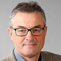 Lothar R. Schad合作论文数Medizinische Fakultät Mannheim, Universität Heidelberg15
 Eichner Cornelius合作论文数15Adam C. Zelinski合作论文数15Esra A. Turk合作论文数15Monika Sliwiak合作论文数Dept Radiol, AA Martinos Ctr Biomed Imaging14
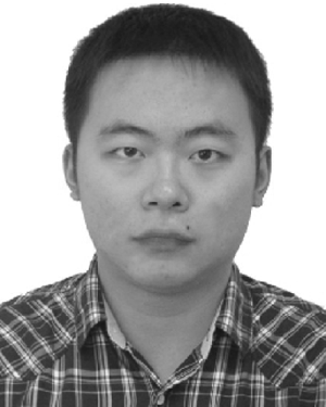 Congyu Liao合作论文数Zhejiang Univ, Coll Biomed Engn & Instrumental Sci, Ctr Brain Imaging Sci & Technol, Key Lab Biomed Engn,Minist Educ, Hangzhou, Zhejiang, Peoples R China14
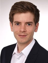 Daniel Polak合作论文数A. A. Martinos Center for Biomedical Imaging, Massachusetts General Hospital14
 Laleh Rad合作论文数Biomedical Engineering, McCormick School of Engineering and Applied Science, Northwestern University14
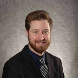 Clifford Bryan合作论文数14Franz Schmitt合作论文数14Wei-Ching Lo合作论文数13J. Polimeni合作论文数13
 Ehsan Kazemivalipour合作论文数Dept Radiol, Northwestern Univ13




