Oligodendrocytes, the myelinating cells of the central nervous system (CNS), are generated from oligodendrocyte precursor cells (OPCs) that express neurotransmitter receptors. However, the mechanisms that affect OPC activity in vivo and the physiological roles of neurotransmitter signaling in OPCs are unclear. In this study, we generated a transgenic mouse line that expresses membrane-anchored GCaMP6s in OPCs and used longitudinal two-photon microscopy to monitor OPC calcium (Ca 2+ ) dynamics in the cerebral cortex. OPCs exhibit focal and transient Ca 2+ increases within their processes that are enhanced during locomotion-induced increases in arousal. The Ca 2+ transients occur independently of excitatory neuron activity, rapidly decline when OPCs differentiate and are inhibited by anesthesia, sedative agents or noradrenergic receptor antagonists. Conditional knockout of α1A adrenergic receptors in OPCs suppresses spontaneous and locomotion-induced Ca 2+ increases and reduces OPC proliferation. Our results demonstrate that OPCs are directly modulated by norepinephrine in vivo to enhance Ca 2+ dynamics and promote population homeostasis.
Astrocytes are a direct target of neuromodulators and can influence neuronal activity on broad spatial and temporal scales through their close proximity to synapses. However, our knowledge about how astrocytes are functionally recruited during different animal behaviors and their diverse effects on the CNS remains limited. To enable measurement of astrocyte activity patterns in vivo during normative behaviors, we developed a high-resolution, long working distance, multi-core fiber optic imaging platform that allows visualization of cortical astrocyte calcium transients through a cranial window in freely moving mice. Using this platform, we defined the spatiotemporal dynamics of astrocytes during diverse behaviors, ranging from circadian fluctuations to novelty exploration, showing that astrocyte activity patterns are more variable and less synchronous than apparent in head-immobilized imaging conditions. Although the activity of astrocytes in visual cortex was highly synchronized during quiescence to arousal transitions, individual astrocytes often exhibited distinct thresholds and activity patterns during explorative behaviors, in accordance with their molecular diversity, allowing temporal sequencing across the astrocyte network. Imaging astrocyte activity during self-initiated behaviors revealed that noradrenergic and cholinergic systems act synergistically to recruit astrocytes during state transitions associated with arousal and attention, which was profoundly modulated by internal state. The distinct activity patterns exhibited by astrocytes in the cerebral cortex may provide a means to vary their neuromodulatory influence in response to different behaviors and internal states.
Oligodendrocytes are generated from a widely distributed population of progenitors that express neurotransmitter receptors, but the mechanisms that alter activity of these oligodendrocyte precursor cells (OPCs) in vivo have not been determined. We generated a novel line of transgenic mice to express membrane-anchored GCaMP6s in OPCs and used longitudinal two-photon microscopy to monitor their Ca 2+ changes in the cerebral cortex of awake mice. OPCs exhibited high rates of spontaneous activity, consisting of focal, transient Ca 2+ increases within their highly ramified processes. Unexpectedly, these events occurred independent of excitatory neuron activity, but were inhibited by anesthesia, sedative agents, and antagonists of noradrenergic signaling. These norepinephrine enhanced Ca 2+ dynamics rapidly declined as with differentiation. Selective knockout of α 1A adrenergic receptors in OPCs suppressed both spontaneous and locomotion-induced Ca 2+ increases, indicating that OPCs are directly modulated by norepinephrine in vivo , providing a means to alter their dynamics and lineage progression during distinct brain states.
Mood-altering compounds hold promise for the treatment of many psychiatric disorders, such as depression, but connecting their molecular, circuit, and behavioral effects has been challenging. Here we find that, analogous to effects in rodent learned helplessness models, ketamine pre-exposure persistently suppresses futility-induced passivity in larval zebrafish. While antidepressants are thought to primarily act on neurons, brain-wide imaging in behaving zebrafish showed that ketamine elevates intracellular calcium in astroglia for many minutes, followed by persistent calcium downregulation post-washout. Calcium elevation depends on astroglial α1-adrenergic receptors and is required for suppression of passivity. Chemo-/optogenetic perturbations of noradrenergic neurons and astroglia demonstrate that the aftereffects of glial calcium elevation are sufficient to suppress passivity by inhibiting neuronal-astroglial integration of behavioral futility. Imaging in mouse cortex reveals that ketamine elevates astroglial calcium through conserved pathways, suggesting that ketamine exerts its behavioral effects by persistently modulating evolutionarily ancient neuromodulatory systems spanning neurons and astroglia.### Competing Interest StatementDEO is a co-founder of Delix Therapeutics, Inc., serves as the Chief Innovation Officer and Head of the Scientific Advisory Board, and has sponsored research agreements with Delix Therapeutics. Delix Therapeutics has licensed technology from the University of California, Davis. AEC is a co-founder of Q-State Biosciences. All other authors declare that they have no competing interests.
Parkinson’s disease (PD) is mediated, in part, by intraneuronal accumulation of α-synuclein aggregates andsubsequent death of dopamine (DA) neurons in the substantia nigra pars compacta (SNpc). Microglial hyperactivation of the NOD-like receptor protein 3 (NLRP3) inflammasome has been well-documented in various neurodegenerative diseases, including PD. We show here that loss of parkin activity in mouse and human DA neurons results in spontaneous neuronal NLRP3 inflammasome assembly, leading to DA neuron death. Parkin normally inhibits inflammasome priming by ubiquitinating and targeting NLRP3 for proteasomal degradation. Loss of parkin activity also contributes to the assembly of an active NLRP3 inflammasome complex via mitochondrial-derived reactive oxygen species (mitoROS) generation through the accumulation of another parkin ubiquitination substrate, ZNF746/PARIS. Inhibition of neuronal NLRP3 inflammasome assembly prevents degeneration of DA neurons in familial and sporadic PD models. Strategies aimed at limiting neuronal NLRP3 inflammasome activation hold promise as a disease-modifying therapy for PD.
Monitoring brain activity associated with natural physiological behavior is at the core of many neuroscience studies, yet many labs utilize imaging modalities that require anesthesia or immobilization of animals that cause unnatural stress and perturbed behavior. Instead, we separated the physiological system from the optical one using a fiber-optic tether, which allows the use of larger, optimized optical components while allowing mice to move freely thereby minimizing interference to their behavior. In vivo astrocyte activity, both spontaneous and stimulus-evoked was observed. The developed dual-color fluorescence system extended functionality to study multiple cell types concurrently.
Chronic traumatic encephalopathy (CTE) is a neurodegenerative disease associated with repeated head traumas. Using immunohistochemistry for glial fibrillary acidic protein as a marker, plus automated quantitative analysis, we examined the characteristics and extent of astrogliosis present in stage III and IV CTE, along with Alzheimer’s disease (AD), and frontotemporal dementia (FTD) cases. Astrogliosis in CTE patients was more diffuse compared to that of AD and FTD patients, which was concentrated in the sulcal depths. Of 14 patients with CTE, 10 exhibited signs of a degenerating astrocyte pathology, characterized by beaded, broken astrocytic processes. This astrocytic degeneration was typically found to be diffuse throughout the white matter, although two cases demonstrated astrocytic degeneration in the gray matter. The degeneration was also observed in 2 of 3 AD and 2 of 3 FTD brains, with overall similar characteristics across diseases. There was minimal to no astrocytic degeneration in six age-matched controls with no neurodegenerative disease. We found that the extent of the white matter astrocytic degeneration was strongly correlated with the level of overall astrogliosis in both the white and gray matter. However, astrocytic degeneration was not correlated with the overall extent of tau pathology. Specifically, there was no correlation between levels of p-tau in the sulcal depths and astrocytic degeneration in the white matter adjacent to the sulcal depths. Thus, astrocytic degeneration and overall astrogliosis appear to represent distinct pathological features of CTE. Further investigation into these astroglial pathologies could provide new insights into underlying disease mechanisms and represent a potential target for in vivo assessment of CTE as well as other neurodegenerative disorders.
 Dwight E. Bergles合作论文数Department of Otolaryngology-Head and Neck Surgery, School of Medicine, Johns Hopkins University;Solomon H. Snyder Department of Neuroscience, School of Medicine, Johns Hopkins University5
 Priyanka Hanumaihgari合作论文数2Rebecca W. Pak合作论文数Dept Biomed Engn, Johns Hopkins Univ2
 Jin U. Kang合作论文数Department of Electrical and Computer Engineering, Whiting School of Engineering, Johns Hopkins University;Department of Dermatology, School of Medicine, Johns Hopkins University;Kavli Neuroscience Discovery Institute, Johns Hopkins University;Laboratory for Computational Sensing and Robotics, Johns Hopkins University;Intelligent Optical Imaging and Vision Laboratory, Whiting School of Engineering, Johns Hopkins University2
 Amit Agarwal合作论文数Sanjay Gandhi Post Graduate Institute of Medical Sciences2Tsai-Yi Lu合作论文数2Shiran Su合作论文数1Stewart Neifert合作论文数1
 Aanishaa Jhaldiyal合作论文数Department of Physiology, Johns Hopkins University School of Medicine1
 Marc Duque合作论文数Mol Neurobiol Lab, Salk Inst Biol Studies1Shahinoor Begum合作论文数1
 Gian Carlo Molina-Castro合作论文数1Yung-Tian A Gau合作论文数Johns Hopkins University1
 Jared T Hinkle合作论文数Johns Hopkins Univ, Sch Med, Med Scientist Training Program, Baltimore, MD 21205 USA1
 Manoj Kumar合作论文数Institute for Cell Engineering, Johns Hopkins University School of Medicine1Enquan Xu合作论文数1
 Valina Dawson合作论文数Institute for Cell Engineering, School of Medicine, The Johns Hopkins University;Department of Neurology, School of Medicine, The Johns Hopkins University1
 Xiaobo Mao合作论文数Department of Otolaryngology-Head and Neck Surgery, School of Medicine, Johns Hopkins University1
 Sung Ung Kang合作论文数Department of Otolaryngology-Head and Neck Surgery, School of Medicine, Johns Hopkins University1
 Tae-In Kam合作论文数Departments of Neurology and Neurosurgery, School of Medicine, Biomedical Engineering1Brahmachari Saurav合作论文数Department of Neurological Sciences, Rush University Medical Center1
 Ahrens Misha B合作论文数Howard Hughes Med Inst, Janelia Res Campus1
 Thor D. Stein合作论文数Alzheimer's Disease Research Center, Boston University;Chobanian & Avedisian School of Medicine, Boston University1
 Jaepyeong Cha合作论文数1
 Chen Alex合作论文数1
 Laurena Holleran合作论文数1
 Chou Shih-Ching合作论文数Nuclear Science &Technology Development Center, National Tsing Hua University;Nuclear Science & Technology Development Center, National Tsing Hua University1
 Victor Alvarez合作论文数Laboratory Operations, Chronic Traumatic Encephalopathy Center, Boston University;Chobanian & Avedisian School of Medicine, Boston University1
 Nikhil Panicker合作论文数Iowa Center for Advanced Neurotoxicology, Iowa State University1Hu Wang合作论文数1
 David M. Olson合作论文数Department of Obstetrics and Gynaecology, Faculty of Medicine & Dentistry, University of Alberta1
 Peter Calabresi合作论文数Department of Neurological Sciences, Larner College of Medicine, University of Vermont1
 Juan C. Troncoso合作论文数School of Medicine, Johns Hopkins University;Brain Resource Center, Johns Hopkins University1Akkentli Fatih合作论文数1Narayan Sujatha合作论文数1Mihika Gangolli合作论文数1
 Adam Cohen合作论文数Departments of Chemistry and Chemical Biology, and Physics, Harvard University1
 Mark C. Fishman合作论文数Fishman Lab, Department of Stem Cell & Regenerative Biology, Harvard University1
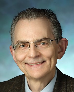 Ted M. Dawson合作论文数Institute for Cell Engineering, School of Medicine, Johns Hopkins University1
 Riki Kawaguchi合作论文数David Geffen School of Medicine, University of California Los Angeles1
 Florian Engert合作论文数Department of Molecular and Cellular Biology, Harvard University1
 Ann C. McKee合作论文数Chobanian & Avedisian School of Medicine, Boston University1
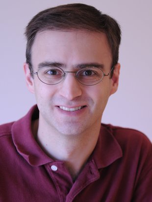 Prober David Aaron合作论文数Division of Biology and Biological Engineering, California Institute of Technology1Olga Pletnikova合作论文数Johns Hopkins University1
 Loren Looger合作论文数Department of Neurosciences, School of Medicine, University of California, San Diego1
 Gesine Saher合作论文数Max Planck Institute for Multidisciplinary Sciences1
 Senthilkumar Karuppagounder合作论文数Neuroregenerat Program, Johns Hopkins Univ1
 Robert E. Schmidt合作论文数Department of Pathology & Immunology, School of Medicine, Washington University;Division of Neuropathology, Washington University;Electron Microscopy Facility, Washington University1
 David Brody合作论文数Department of Neurology, School of Medicine, Uniformed Services University of the Health Sciences1




