Sleep is an essential and evolutionarily conserved behavior. While mammals and several other species have been shown to exhibit well-defined sleep sub-states, it remains unclear to what extent such differentiation exists across the animal kingdom. Here we show, using long-term behavioral data combined with Hidden Markov Modeling, that larval zebrafish display two behaviorally defined sleep-related sub-states: a low-activity state consistent with light sleep and a quiescent state consistent with deep sleep. These are likely analogous to light and deep NREM sleep in mammals. Although both states occur primarily at night, arousal responses differ by sleep sub-state, and sleep deprivation induces deep sleep-dominated rebound. Moreover, the proportions of deep and light sleep are selectively altered by genetic and pharmacological manipulations of melatonin, serotonin, and norepinephrine signaling, offering new insights into how these neuromodulators shape sleep architecture. These results support zebrafish as a tractable model for dissecting the regulation and function of sleep sub-states. More broadly, they demonstrate that structured, multi-state sleep is a conserved feature of vertebrate behavior.
Neurofibromatosis type 1 (NF1) is a common neurodevelopmental disorder that is associated with several developmental and behavioral phenotypes, including reduced and fragmented sleep at night. Here we describe the role of NF1 in sleep in zebrafish, a diurnal vertebrate whose mechanisms of sleep control are broadly conserved with those of mammals. The zebrafish genome encodes for two nf1 paralogs, nf1a and nf1b, that function in a partially redundant manner. We found that zebrafish with reduced nf1 function exhibit decreased and delayed sleep at night, similar to humans with the NF1 disorder, in a dose-dependent manner. Zebrafish nf1 mutants also exhibit shorter sleep bouts, decreased arousal threshold, and decreased sleep depth, consistent with disrupted sleep, and show increased sleep in the morning. Finally, we found that zebrafish nf1 mutants have intact behavioral circadian rhythms, and the nf1 mutant sleep phenotype does not require entrained circadian rhythms. These results establish a vertebrate nf1 model that recapitulates key aspects of human NF1-associated sleep disturbances, and provides a basis to explore mechanisms through which NF1 regulates sleep. This study also suggests that disrupted sleep in the human NF1 disorder is directly due to reduced NF1 function, rather than a secondary symptom of the disorder.
Classical studies identified a critical role for the hypothalamus in regulating sleep and wake states, but few such hypothalamic neuronal populations have been identified. Here, we describe a sleep-promoting population of hypothalamic neurons that expresses the neuropeptides QRFP and parathyroid hormone 4 (Pth4) in zebrafish. Optogenetic stimulation of these neurons results in a large increase in sleep that requires pth4 but not qrfp. Noradrenergic locus coeruleus (LC) neurons and serotonergic (5HT) raphe nuclei (RNs) in the hindbrain express distinct pth receptors, and genetic epistasis and cell ablation experiments revealed that Pth4 neuron-induced sleep is suppressed in mutants that lack noradrenaline in the LC or lack the 5HT RNs. Pth4 neuron-induced sleep is also suppressed in serine/threonine kinase 32a (stk32a) mutants, possibly via stk32a-expressing neurons in the prethalamus that express pth receptors. These results identify QRFP/Pth4 neurons as a novel hypothalamic sleep-promoting population and support a model in which distinct sleep- and wake-promoting hypothalamic populations act via monoaminergic neurons in the hindbrain to control vigilance state.
Both neurons and glia communicate through diffusible neuromodulators; however, how neuron-glial interactions in such neuromodulatory networks influence circuit computation and behavior is unclear. During futility-induced behavioral transitions in the larval zebrafish, the neuromodulator norepinephrine (NE) drives fast excitation and delayed inhibition of behavior and circuit activity. We found that astroglial purinergic signaling implements the inhibitory arm of this motif. In larval zebrafish, NE triggers astroglial release of adenosine triphosphate (ATP), extracellular conversion of ATP into adenosine, and behavioral suppression through activation of hindbrain neuronal adenosine receptors. Our results suggest a computational and behavioral role for an evolutionarily conserved astroglial purinergic signaling axis in NE-mediated behavioral and brain state transitions and position astroglia as important effectors in neuromodulatory signaling.
Transient exposure to ketamine can trigger lasting changes in behavior and mood. We found that brief ketamine exposure causes long-term suppression of futility-induced passivity in larval zebrafish, reversing the “giving-up” response that normally occurs when swimming fails to cause forward movement. Whole-brain imaging revealed that ketamine hyperactivates the norepinephrine-astroglia circuit responsible for passivity. After ketamine washout, this circuit exhibits hyposensitivity to futility, leading to long-term increased perseverance. Pharmacological, chemogenetic, and optogenetic manipulations show that norepinephrine and astrocytes are necessary and sufficient for ketamine’s long-term perseverance-enhancing aftereffects. In vivo calcium imaging revealed that astrocytes in adult mouse cortex are similarly activated during futility in the tail suspension test and that acute ketamine exposure also induces astrocyte hyperactivation. The cross-species conservation of ketamine’s modulation of noradrenergic-astroglial circuits and evidence that plasticity in this pathway can alter the behavioral response to futility hold promise for identifying new strategies to treat affective disorders.
Congenital disorders of glycosylation (CDG) comprise a class of inborn errors of metabolism resulting from pathogenic variants in genes coding for enzymes involved in the asparagine-linked glycosylation of proteins. Unexpectedly to date, no CDG has been described for ALG10 , encoding the alpha-1,2-glucosyltransferase catalyzing the final step of lipid-linked oligosaccharide biosynthesis. Genome-wide association studies (GWAS) of human traits in the UK Biobank revealed significant SNP associations with short sleep duration, reduced napping frequency, later sleep timing and evening diurnal preference as well as cardiac traits at a genomic locus containing a pair of paralogous enzymes ALG10 and ALG10B . Modeling Alg10 loss in Drosophila, we identify an essential role for the N -glycosylation pathway in maintaining appropriate neuronal firing activity, healthy sleep, preventing seizures, and cardiovascular homeostasis. We further confirm the broader relevance of neurological findings associated with Alg10 from humans and flies using zebrafish and nematodes and demonstrate conserved biochemical roles for N -glycosylation in Arabidopsis . We report a human subject homozygous for variants in both ALG10 and ALG10B arising from a consanguineous marriage, with epilepsy, brain atrophy, and sleep abnormalities as predicted by the fly phenotype. Quantitative glycoproteomic analysis in our Drosophila model identifies potential key molecular targets for neurological symptoms of CDGs.
Both neurons and glia communicate via diffusible neuromodulatory substances, but the substrates of computation in such neuromodulatory networks are unclear. During behavioral transitions in the larval zebrafish, the neuromodulator norepinephrine drives fast excitation and delayed inhibition of behavior and circuit activity. We find that the inhibitory arm of this feedforward motif is implemented by astroglial purinergic signaling. Neuromodulator imaging, behavioral pharmacology, and perturbations of neurons and astroglia reveal that norepinephrine triggers astroglial release of adenosine triphosphate, extracellular conversion into adenosine, and behavioral suppression through activation of hindbrain neuronal adenosine receptors. This work, along with a companion piece by Lefton and colleagues demonstrating an analogous pathway mediating the effect of norepinephrine on synaptic connectivity in mice, identifies a computational and behavioral role for an evolutionarily conserved astroglial purinergic signaling axis in norepinephrine-mediated behavioral and brain state transitions.
Animals need to rapidly learn to recognize and avoid predators. This ability may be especially important for young animals due to their increased vulnerability. It is unknown whether, and how, nascent vertebrates are capable of such rapid learning. Here, we used a robotic predator-prey interaction assay to show that 1 week after fertilization—a developmental stage where they have approximately 1% the number of neurons of adults—zebrafish larvae rapidly and robustly learn to recognize a stationary object as a threat after the object pursues the fish for ∼1 min. Larvae continue to avoid the threatening object after it stops moving and can learn to distinguish threatening from non-threatening objects of a different color. Whole-brain functional imaging revealed the multi-timescale activity of noradrenergic neurons and forebrain circuits that encoded the threat. Chemogenetic ablation of those populations prevented the learning. Thus, a noradrenergic and forebrain multiregional network underlies the ability of young vertebrates to rapidly learn to recognize potential predators within their first week of life.
With the oncoming age of big data, biologists are encountering more use cases for cloud-based computing to streamline data processing and storage. Unfortunately, cloud platforms are difficult to learn, and there are few resources for biologists to demystify them. We have developed a guide for experimental biologists to set up cloud processing on Amazon Web Services to cheaply outsource data processing and storage. Here we provide a guide for setting up a computing environment in the cloud and showcase examples of using Python and Julia programming languages. We present example calcium imaging data in the zebrafish brain and corresponding analysis using suite2p software. Tools for budget and user management are further discussed in the attached protocol. Using this guide, researchers with limited coding experience can get started with cloud-based computing or move existing coding infrastructure into the cloud environment.
Sleep is a nearly universal feature of animal behaviour, yet many of the molecular, genetic, and neuronal substrates that orchestrate sleep/wake transitions lie undiscovered. Employing a viral insertion sleep screen in larval zebrafish, we identified a novel gene, dreammist (dmist), whose loss results in behavioural hyperactivity and reduced sleep at night. The neuronally expressed dmist gene is conserved across vertebrates and encodes a small single-pass transmembrane protein that is structurally similar to the Na+,K+-ATPase regulator, FXYD1/Phospholemman. Disruption of either fxyd1 or atp1a3a, a Na+,K+-ATPase alpha-3 subunit associated with several heritable movement disorders in humans, led to decreased night-time sleep. Since atpa1a3a and dmist mutants have elevated intracellular Na+ levels and non-additive effects on sleep amount at night, we propose that Dmist-dependent enhancement of Na+ pump function modulates neuronal excitability to maintain normal sleep behaviour.
Over the past decade, zebrafish have emerged as a powerful model for the study of vertebrate sleep and wake behaviors. Experimental evidence has demonstrated behavioral, anatomical, genetic, and pharmacological conservation of sleep between zebrafish and mammals, suggesting that discoveries in zebrafish can inform our understanding of mammalian sleep. Here, we describe a protocol for performing sleep behavioral experiments in larval zebrafish, using a high-throughput video tracking system. We explain how to set up a sleep behavioral experiment and provide guidelines on how to analyze the data. Using this protocol, a typical experiment can be completed in less than five days, and this method provides a scalable platform to perform genetic and pharmacological screens in a simple and cost-effective vertebrate model. By combining high-throughput behavioral assays with several advantageous features of zebrafish, this model system provides new opportunities to make discoveries that clarify the genetic and neurological mechanisms that regulate sleep.
Mood-altering compounds hold promise for the treatment of many psychiatric disorders, such as depression, but connecting their molecular, circuit, and behavioral effects has been challenging. Here we find that, analogous to effects in rodent learned helplessness models, ketamine pre-exposure persistently suppresses futility-induced passivity in larval zebrafish. While antidepressants are thought to primarily act on neurons, brain-wide imaging in behaving zebrafish showed that ketamine elevates intracellular calcium in astroglia for many minutes, followed by persistent calcium downregulation post-washout. Calcium elevation depends on astroglial α1-adrenergic receptors and is required for suppression of passivity. Chemo-/optogenetic perturbations of noradrenergic neurons and astroglia demonstrate that the aftereffects of glial calcium elevation are sufficient to suppress passivity by inhibiting neuronal-astroglial integration of behavioral futility. Imaging in mouse cortex reveals that ketamine elevates astroglial calcium through conserved pathways, suggesting that ketamine exerts its behavioral effects by persistently modulating evolutionarily ancient neuromodulatory systems spanning neurons and astroglia.### Competing Interest StatementDEO is a co-founder of Delix Therapeutics, Inc., serves as the Chief Innovation Officer and Head of the Scientific Advisory Board, and has sponsored research agreements with Delix Therapeutics. Delix Therapeutics has licensed technology from the University of California, Davis. AEC is a co-founder of Q-State Biosciences. All other authors declare that they have no competing interests.
Sleep is a fundamental behavioral state that is observed throughout the animal kingdom, yet our understanding of this state remains limited. Zebrafish are a useful vertebrate animal model to dissect genetic and neuronal mechanisms underlying the regulation of sleep and wake states. The diurnal circadian pattern of sleep–wake cycles in zebrafish is similar to that of humans, and many sleep-regulating neuropeptides and hormones that are found in humans, such as melatonin and hypocretin, play similar roles in zebrafish. Due to their small size, rapid development, and optical transparency, larval zebrafish are well suited for high-throughput screens to identify genes, neurons, and drugs that regulate sleep. Numerous techniques and genetic tools have been established that enable powerful approaches to explore mechanisms that underlie zebrafish sleep. In this chapter, we will discuss current approaches to studying sleep in larval zebrafish, including detailed experimental procedures, and describe the advantages and limitations of each approach.
Salient sensory stimuli are perceived by the brain, which guides both the timing and outcome of behaviors in a context-dependent manner. Light is such a stimulus, which is used in treating mood disorders often associated with a dysregulated hypothalamic-pituitary-adrenal stress axis. Relationships between the emotional valence of light and the hypothalamus, and how they interact to exert brain-wide impacts remain unclear. Employing larval zebrafish with analogous hypothalamic systems to mammals, we show in free-swimming animals that hypothalamic corticotropin releasing factor (CRFHy) neurons promote dark avoidance, and such role is not shared by other hypothalamic peptidergic neurons. Single-neuron projection analyses uncover processes extended by individual CRFHy neurons to multiple targets including sensorimotor and decision-making areas. In vivo calcium imaging uncovers a complex and heterogeneous response of individual CRFHy neurons to the light or dark stimulus, with a reduced overall sum of CRF neuronal activity in the presence of light. Brain-wide calcium imaging under alternating light/dark stimuli further identifies distinct and distributed photic response neuronal types. CRFHy neuronal ablation increases an overall representation of light in the brain and broadly enhances the functional connectivity associated with an exploratory brain state. These findings delineate brain-wide photic perception, uncover a previously unknown role of CRFHy neurons in regulating the perception and emotional valence of light, and suggest that light therapy may alleviate mood disorders through reducing an overall sum of CRF neuronal activity.
Sleep disorders and chronic sleep disturbances are common and are associated with cardio-metabolic diseases and neuropsychiatric disorders. Several genetic pathways and neuronal mechanisms that regulate sleep have been described in animal models, but the genes underlying human sleep variation and sleep disorders are largely unknown. Identifying these genes is essential in order to develop effective therapies for sleep disorders and their associated comorbidities. To address this unmet health problem, genome-wide association studies (GWAS) have identified numerous genetic variants associated with human sleep traits and sleep disorders. However, in most cases, it is unclear which gene is responsible for a sleep phenotype that is associated with a genetic variant. As a result, it is necessary to experimentally validate candidate genes identified by GWAS using an animal model. Rodents are ill-suited for this endeavor due to their poor amenability to high-throughput sleep assays and the high costs associated with generating, maintaining, and testing large numbers of mutant lines. Zebrafish (Danio rerio), an alternative vertebrate model for studying sleep, allows for the rapid and cost-effective generation of mutant lines using the CRISPR/Cas9 system. Numerous zebrafish mutant lines can then be tested in parallel using high-throughput behavioral assays to identify genes whose loss affects sleep. This process identifies a gene associated with each GWAS hit that is likely responsible for the human sleep phenotype. This strategy is a powerful complement to GWAS approaches and holds great promise to identify the genetic basis for common human sleep disorders.
BACKGROUND AND AIMS:The liver is a highly regenerative organ, but its regenerative capacity is compromised in severe liver injury settings. In chronic liver diseases, the number of liver progenitor cells (LPCs) correlates proportionally to disease severity, implying that their inefficient differentiation into hepatocytes exacerbates the disease. Moreover, LPCs secrete proinflammatory cytokines; thus, their prolonged presence worsens inflammation and induces fibrosis. Promoting LPC-to-hepatocyte differentiation in patients with advanced liver disease, for whom liver transplantation is currently the only therapeutic option, may be a feasible clinical approach because such promotion generates more functional hepatocytes and concomitantly reduces inflammation and fibrosis. APPROACH AND RESULTS:Here, using zebrafish models of LPC-mediated liver regeneration, we present a proof of principle of such therapeutics by demonstrating a role for the epidermal growth factor receptor (EGFR) signaling pathway in differentiation of LPCs into hepatocytes. We found that suppression of EGFR signaling promoted LPC-to-hepatocyte differentiation through the mitogen-activated ERK kinase (MEK)-extracellular signal-regulated kinase (ERK)-sex-determining region Y-box 9 (SOX9) cascade. Pharmacological inhibition of EGFR or MEK/ERK promoted LPC-to-hepatocyte differentiation as well as genetic suppression of the EGFR-ERK-SOX9 axis. Moreover, Sox9b overexpression in LPCs blocked their differentiation into hepatocytes. In the zebrafish liver injury model, both hepatocytes and biliary epithelial cells contributed to LPCs. EGFR inhibition promoted the differentiation of LPCs regardless of their origin. Notably, short-term treatment with EGFR inhibitors resulted in better liver recovery over the long term. CONCLUSIONS:The EGFR-ERK-SOX9 axis suppresses LPC-to-hepatocyte differentiation during LPC-mediated liver regeneration. We suggest EGFR inhibitors as a proregenerative therapeutic drug for patients with advanced liver disease.
Experiments with biological samples require precise control of environmental conditions. In our work we use zebrafish ( Danio rerio ) to understand the neurobiology of sleep, which requires precise control of temperature and lighting. Like many labs, lighting and temperature in the animal facility are centrally controlled in the building. During behavioral experiments and microscopy sessions, we use custom-built heating systems and perform occasional manual checks of conditions. However, without a system to precisely record conditions, gradual changes in temperature can go unnoticed for a long time, and temporary failures may be missed entirely. Here we present the design and characterization of affordable open-source tools to record temperature and light conditions during animal experiments using an Arduino microcontroller or a Raspberry Pi compact computer. The waterproof temperature sensor has high stability over 50 days of recording and is precise to 0.1°C. The Arduino device can be used through a common serial port interface for which we present code in Python and MATLAB. The Raspberry Pi version can be accessed through a web interface, for which we provide an installation guide. We use the device to record and review temperature and lighting conditions in two zebrafish animal facilities. We use our platform to add a water heating system to maintain temperature at 28°C during in vivo light-sheet imaging of larval zebrafish. We show that a change in temperature from 28°C to 32°C affects resting heart rate of the animal, highlighting the importance of maintaining and recording conditions. The protocols presented here do not require advanced engineering, fabrication, or software skills, and provide an approach to accurately record and report experimental conditions.
Prokineticin receptors (PROKR1 and PROKR2) are G protein-coupled receptors which control human central and peripheral reproductive processes. Importantly, allelic variants of PROKR2 in humans are associated with altered migration of GnRH neurons, resulting in congenital hypogonadotropic hypogonadism (CHH), a heterogeneous disease characterized by delayed/absent puberty and/or infertility. Although this association is established in humans, murine models failed to fully recapitulate the reproductive and olfactory phenotypes observed in patients harboring PROKR2 mutations. Here, taking advantage of zebrafish model we investigated the role of prokr1b (ortholog of human PROKR2) during early stages of GnRH neuronal migration. Real-Time PCR and whole mount in situ hybridization assays indicate that prokr1b spatial-temporal expression is consistent with gnrh3. Moreover, knockdown and knockout of prokr1b altered the correct development of GnRH3 fibers, a phenotype that is rescued by injection of prokr1b mRNA. These results suggest that prokr1b regulates the development of the GnRH3 system in zebrafish. Analysis of gonads development and mating experiments indicate that prokr1b is not required for fertility in zebrafish, although its loss determine changes also at the testis level. Altogether, our results support the thesis of a divergent evolution in the control of vertebrate reproduction and provide a useful in vivo model for deciphering the mechanisms underlying the effect of PROKR2 allelic variants on CHH.
Although several sleep-regulating neuronal populations have been identified, little is known about how they interact with each other to control sleep/wake states. We previously identified neuropeptide VF (NPVF) and the hypothalamic neurons that produce it as a sleep-promoting system (Lee et al., 2017). Here we show using zebrafish that npvf-expressing neurons control sleep via the serotonergic raphe nuclei (RN), a hindbrain structure that is critical for sleep in both diurnal zebrafish and nocturnal mice. Using genetic labeling and calcium imaging, we show that npvf-expressing neurons innervate and can activate serotonergic RN neurons. We also demonstrate that chemogenetic or optogenetic stimulation of npvf-expressing neurons induces sleep in a manner that requires NPVF and serotonin in the RN. Finally, we provide genetic evidence that NPVF acts upstream of serotonin in the RN to maintain normal sleep levels. These findings reveal a novel hypothalamic-hindbrain neuronal circuit for sleep/wake control.
Sleep is a cross-species phenomenon whose evolutionary and biological function remain poorly understood. Clinical and animal studies suggest that sleep disturbance is significantly associated with disruptions in protein homeostasis-or proteostasis-in the brain, but the mechanism of this link has not been explored. In the cell, the protein kinase R (PKR)-like endoplasmic reticulum kinase (PERK) pathway modulates proteostasis by transiently inhibiting protein synthesis in response to proteostatic stress. In this study, we examined the role of the PERK pathway in sleep regulation and provide the first evidence that PERK signaling is required to regulate normal sleep in both vertebrates and invertebrates. We show that pharmacological inhibition of PERK reduces sleep in both Drosophila and zebrafish, indicating an evolutionarily conserved requirement for PERK in sleep. Genetic knockdown of PERK activity also reduces sleep in Drosophila, whereas PERK overexpression induces sleep. Finally, we demonstrate that changes in PERK signaling directly impact wake-promoting neuropeptide expression, revealing a mechanism through which proteostatic pathways can affect sleep and wake behavior. Taken together, these results demonstrate that protein synthesis pathways like PERK could represent a general mechanism of sleep and wake regulation and provide greater insight into the relationship between sleep and proteostasis.
 Grigorios Oikonomou合作论文数16
 Alexander F. Schier合作论文数The Center for Molecular Life Sciences, University of Basel14
 Chanpreet Singh合作论文数12
 Daniel A Lee合作论文数CALTECH, Tianqiao & Chrissy Chen Inst Neurosci12
 Jason Rihel合作论文数Department of Cell and Developmental Biology, University College London10Chiu Cindy N合作论文数8
 Florian Engert合作论文数Department of Molecular and Cellular Biology, Harvard University6
 Andrey Andreev合作论文数CALTECH, Tianqiao & Chrissy Chen Inst Neurosci5
 Ahrens Misha B合作论文数Howard Hughes Med Inst, Janelia Res Campus5
 Paula Suarez-Bregua合作论文数5
 José Miguel Cerdá-Reverter合作论文数Department of Fish Physiology and Biotechnology, Instituto de Acuicultura de Torre de la Sal5
 Cristian Cañestro合作论文数Institute of Neuroscience, University of Oregon5Josep Rotllant合作论文数5Eric A. Mosser合作论文数4
 Marc Duque合作论文数Mol Neurobiol Lab, Salk Inst Biol Studies4Justin Liu合作论文数4
 Chen Alex合作论文数4
 Tran Steven合作论文数Department of Cell and Systems Biology, University of Toronto Mississauga4Narayan Sujatha合作论文数4Chen Shijia合作论文数4
 Pedro M. Guerreiro合作论文数Centre of Marine Sciences (CCMAR),, University of Algarve4
 Audrey Chen合作论文数3Brett J. Niles合作论文数3Uyen Pham合作论文数Dept Biochem, Duke Univ3Jae Engle合作论文数Dept Psychol, Univ Calif San Diego3Steven Zimmerman合作论文数3
 Young Hong合作论文数Cooper University Hospital, Rowan University3Torres-Núñez Eva合作论文数3
 Avni Gandhi合作论文数3
 Shaojun Du合作论文数Department of Biochemistry and Molecular Biology, School of Medicine, University of Maryland3
 Marianne Elizabeth Bronner合作论文数Division of Biology and Biological Engineering, California Institute of Technology3
 Paul W. Sternberg合作论文数Sternberg Lab, Division of Biology and Biological Engineering, California Institute of Technology3
 Bruce A. Edgar合作论文数Department of Oncological Sciences, School of Medicine, University of Utah;Huntsman Cancer Institute, University of Utah3Francesca Luzzani合作论文数2
 Ivan Bassi合作论文数2Ludovica Cotellessa合作论文数2Rong-wei Zhang合作论文数2Christin J. Montz合作论文数2Andrew J Hill合作论文数2Shahinoor Begum合作论文数2Xuelong Mi合作论文数2Yali Zhao合作论文数2
 Nancy Wayne合作论文数University of California, Los Angeles2
 Dwight E. Bergles合作论文数Department of Otolaryngology-Head and Neck Surgery, School of Medicine, Johns Hopkins University;Solomon H. Snyder Department of Neuroscience, School of Medicine, Johns Hopkins University2Choy Margaret合作论文数2Marelli Federica合作论文数2
 Marc Vidal合作论文数Dana-Farber Cancer Institute;Harvard Medical School;Division of Medical Sciences, Harvard Kenneth C. Griffin Graduate School of Arts and Sciences, Harvard University2
 Jacqueline Lane合作论文数2
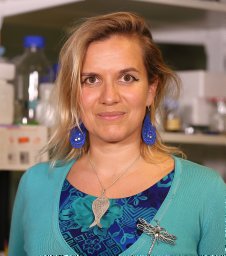 Wyart Claire合作论文数Institut du Cerveau et de la Moelle Épinière, Hôpital de la Pitié-Salpêtrière Paris;UMR 1127 Paris, Institut National de la Santé et de la Recherche Médicale;UMR 7225 Paris, Centre National de la Recherche Scientifique;UPMC Univ.;Institut du Cerveau et de la Moelle Épinière, UPMC Univ.2




