The amino acid, polyamine, and organocation (APC) superfamily is the second largest superfamily of membrane proteins forming secondary transporters that move a range of organic molecules across the cell membrane. Each transporter in the APC superfamily is specific for a unique subset of substrates, even if they possess a similar structural fold. The mechanism of substrate selectivity remains, by and large, elusive. Here, we report two crystal structures of an APC member from Methanococcus maripaludis, the alanine or glycine: cation symporter (AgcS), with L- or D-alanine bound. Structural analysis combined with site-directed mutagenesis and functional studies inform on substrate binding, specificity, and modulation of the AgcS family and reveal key structural features that allow this transporter to accommodate glycine and alanine while excluding all other amino acids. Mutation of key residues in the substrate binding site expand the selectivity to include valine and leucine. These studies provide initial insights into substrate selectivity in AgcS symporters.
The flavin mononucleotide (FMN) riboswitch is an emerging target for the development of novel RNA-targeting antibiotics. We previously discovered an FMN derivative, 5FDQD, that protects mice against diarrhea-causing Clostridium difficile bacteria. Here, we present the structure-based drug design strategy that led to the discovery of this fluoro-phenyl derivative with antibacterial properties. This approach involved the following stages: (1) structural analysis of all available free and bound FMN riboswitch structures; (2) design, synthesis, and purification of derivatives; (3) in vitro testing for productive binding using two chemical probing methods; (4) in vitro transcription termination assays; and (5) resolution of the crystal structures of the FMN riboswitch in complex with the most mature candidates. In the process, we delineated principles for productive binding to this riboswitch, thereby demonstrating the effectiveness of a coordinated structure-guided approach to designing drugs against RNA.
The flavin mononucleotide (FMN) riboswitch is an emerging target for the development of novel RNA-targeting antibiotics. We previously discovered an FMN derivative, 5FDQD, that protects mice against diarrhea-causing Clostridium difficile bacteria. Here, we present the structure-based drug design strategy that led to the discovery of this fluoro-phenyl derivative with antibacterial properties. This approach involved the following stages: (1) structural analysis of all available free and bound FMN riboswitch structures; (2) design, synthesis, and purification of derivatives; (3) in vitro testing for productive binding using two chemical probing methods; (4) in vitro transcription termination assays; and (5) resolution of the crystal structures of the FMN riboswitch in complex with the most mature candidates. In the process, we delineated principles for productive binding to this riboswitch, thereby demonstrating the effectiveness of a coordinated structure-guided approach to designing drugs against RNA.
ABSTRACT The flavin mononucleotide (FMN) riboswitch is an emerging target for the development of novel RNA-targeting antibiotics. We previously discovered an FMN derivative —5FDQD— that protects mice against diarrhea-causing Clostridium difficile bacteria. Here, we present the structure-based drug design strategy that led to the discovery of this fluoro-phenyl derivative with antibacterial properties. This approach involved the following stages: (1) structural analysis of all available free and bound FMN riboswitch structures; (2) design, synthesis and purification of derivatives; (3) in vitro testing for productive binding using two chemical probing methods; (4) in vitro transcription termination assays; (5) resolution of the crystal structures of the FMN riboswitch in complex with the most mature candidates. In the process, we delineated principles for productive binding to this riboswitch, thereby demonstrating the effectiveness of a coordinated structure-guided approach to designing drugs against RNA. GRAPHICAL ABSTRACT Exploring the chemical structure landscape of FMN riboswitch binders.
The cryoEM method micro-electron diffraction (MicroED) utilizes 3D crystals only a few hundred nanometers thick for atomic structure determination, thus shrinking the crystal size limits set by synchrotron micro-focus beamlines. MicroED is ideal for 3D nanocrystals, such as those formed from amyloid protein segments. Amyloid proteins form fibers that are the hallmarks of a class of more than 25 amyloid diseases, ranging from Parkinson's disease to Type II Diabetes (T2D). Although full-length amyloid proteins have so far been resistant to crystallization, select protein segments that form the spines of amyloid fibrils do form such tiny 3D crystals. Gaining knowledge of the atomic structures of these protein segments is critical to our understanding of amyloid fibril structures that contribute to disease onset and progression. Here, we use MicroED to determine the structures of two 11-residue segments from Islet Amyloid Polypeptide (IAPP) at 1.4 Å and 2.0 Å resolution, respectively. Amyloid fibrils of IAPP are found in approximately 90% of T2D patients and the formation of these fibrils is tightly linked to pancreatic β-cell death and insulin dependence. The two segments overlap in sequence by 5 residues. Their structures add to the range of polymorphism observed previously in crystal structures of shorter IAPP segments. One segment, 15-25WT, forms an unusual arrangement of single, out-of-register β-sheets. The second segment, 19-29S20G, contains the familial S20G mutation that leads to early-onset T2D. It forms pairs of β-sheets mated by a dry interface that share structural features with and are similarly cytotoxic to full-length IAPP fibrils. Thus, this structure may serve as a model for the toxic spine of IAPP aggregates. These findings, facilitated by MicroED, give us insight into disease-relevant structures that can be utilized for structure-based design of therapeutics.
hIAPP fibrils are associated with Type-II Diabetes, but the link of hIAPP structure to islet cell death remains elusive. Here we observe that hIAPP fibrils are cytotoxic to cultured pancreatic β-cells, leading us to determine the structure and cytotoxicity of protein segments composing the amyloid spine of hIAPP. Using the cryoEM method MicroED, we discover that one segment, 19–29 S20G, forms pairs of β-sheets mated by a dry interface that share structural features with and are similarly cytotoxic to full-length hIAPP fibrils. In contrast, a second segment, 15–25 WT, forms non-toxic labile β-sheets. These segments possess different structures and cytotoxic effects, however, both can seed full-length hIAPP, and cause hIAPP to take on the cytotoxic and structural features of that segment. These results suggest that protein segment structures represent polymorphs of their parent protein and that segment 19–29 S20G may serve as a model for the toxic spine of hIAPP.
Spider venom toxins, such as Protoxin-II (ProTx-II), have recently received much attention as selective Nav1.7 channel blockers, with potential to be developed as leads for the treatment of chronic nocioceptive pain. ProTx-II is a 30-amino acid peptide with three disulfide bonds that has been reported to adopt a well-defined inhibitory cystine knot (ICK) scaffold structure. Potential drawbacks with such peptides include poor pharmacodynamics and potential scrambling of the disulfide bonds in vivo. In order to address these issues, in the present study we report the solid-phase synthesis of lanthionine-bridged analogues of ProTx-II, in which one of the three disulfide bridges is replaced with a thioether linkage, and evaluate the biological properties of these analogues. We have also investigated the folding and disulfide bridging patterns arising from different methods of oxidation of the linear peptide precursor. Finally, we report the X-ray crystal structure of ProTx-II to atomic resolution; to our knowledge this is the first crystal structure of an ICK spider venom peptide not bound to a substrate.
Traditionally, crystallographic analysis of macromolecules has depended on large, well-ordered crystals, which often require significant effort to obtain. Even sizable crystals sometimes suffer from pathologies that render them inappropriate for high-resolution structure determination. Here we show that fragmentation of large, imperfect crystals into microcrystals or nanocrystals can provide a simple path for high-resolution structure determination by the cryoEM method MicroED and potentially by serial femtosecond crystallography.
 Robert Batey合作论文数The Batey Laboratory, Department of Biochemistry, College of Arts and Sciences, University of Colorado33
 Tamir Gonen合作论文数Department of Biological Chemistry, University of California, Los Angeles28
 Shi Dan合作论文数25
 Hattne Johan合作论文数Lawrence Berkeley Natl Lab, Univ Calif Berkeley16
 Duilio Cascio合作论文数Department of Biological Chemistry, University of California, Los Angeles14J. Hattne合作论文数10M.J. de la Cruz合作论文数8M. Sawaya合作论文数8
 Paul Seidler合作论文数7
 Jose A. Rodriguez合作论文数Biochemistry Division, Department of Chemistry and Biochemistry, College of Physical Sciences, University of California, Los Angeles;Institute for Genomics and Proteomics, Department of Energy Office of Science, University of California, Los Angeles;California NanoSystems Institute, University of California, Los Angeles;Molecular Biology Institute, University of California, Los Angeles;NSF-STROBE Science and Technology Center7
 Nannenga Brent L合作论文数Chem Engn, Arizona State Univ7
 David Eisenberg合作论文数Department of Chemistry and Biochemistry, College of Physical Sciences, University of California, Los Angeles7M. Jason De La Cruz合作论文数7D. Eisenberg合作论文数6Michael Sawaya合作论文数Molecular Biology Institute, University of California, Los Angeles;Institute for Genomics and Proteomics, US Department of Office Energy of Science6WICKENS P合作论文数5J.J. Trausch合作论文数5Philip D. G. Coish合作论文数5Silvia Sánchez-Martínez合作论文数4
 David Lilley合作论文数Division of Molecular Cell and Developmental Biology, School of Life Sciences, University of Dundee4D.A. Robinson合作论文数3C.M. Hammond合作论文数3Q. Vicens合作论文数3P. Daldrop合作论文数3SCHOSTAREZ H合作论文数3E. Mondragon合作论文数3R. Brenk合作论文数3B.L. Nannenga合作论文数3
 Andrea L. Halweg-Edwards合作论文数3
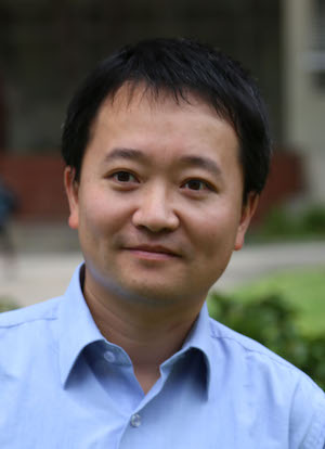 Lin Jiang合作论文数Molecular Biology Institute, University of California, Los Angeles3Blount K F合作论文数3S.D. Gilbert合作论文数2Robert C Gadwood合作论文数2
 Estefania Mondragon Tenorio合作论文数2James E Johnson合作论文数2Stephen McCarthy合作论文数2
 Marc Messerschmidt合作论文数SLAC Natl Accelerator Lab, Linac Coherent Light Source, Menlo Pk, CA 94025 USA2
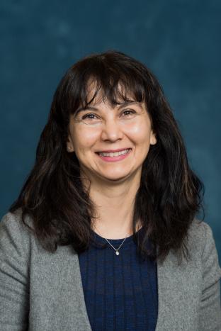 Magdalena Ivanova合作论文数Department of Neurology, University of Michigan Medical School2Lisa M. Johnson合作论文数2Elizabeth L. Guenther合作论文数2
 Aaron S. Brewster合作论文数Physical Biosciences Division, Lawrence Berkeley National Laboratory;Physical Biosciences Division, Lawrence Berkeley National Laboratory2Trausch Jeremiah J合作论文数2Oskarsson Marie E合作论文数2
 Konstantinos Thalassinos合作论文数Structural and Molecular Biology, Division of Biosciences, Faculty of Life Sciences, University College London2
 Smriti Sangwan合作论文数UCLA DOE Inst, Univ Calif Los Angeles2Suto Robert K合作论文数Xtal BioStructures Inc.2Krotee P合作论文数2Maya Topf合作论文数Birkbeck College, University of London2Guillermo Calero合作论文数2




