Detailed methods on cell culture, cell viability assays, RNAseq, western blot, and Elastic net regression analysis.
Supplementary Figure S5 shows the strategy of senescent cell depletion in the KPPC-IA (INK-ATTAC) mouse model and its effects on different cell type numbers.
Motivation Multiomics data analysis is essential for scientific discovery in precision medicine. However, translating analysis results of omics data analysis into novel scientific hypotheses remains a significant challenge. Human experts must manually review analysis results and generate new hypotheses based on extensive and interconnected biomedical prior knowledge, which is subjective and not scalable. While large language models can accelerate the discovery, their reasoning improves when grounded in structured, auditable, and comprehensive biomedical prior knowledge. However, biomedical knowledge is scattered across heterogeneous databases that use diverse and inconsistent nomenclature systems, making it difficult to integrate resources into a unified format for scalable analysis. This fragmentation limits the ability of artificial intelligence systems to fully leverage biomedical data for scientific discovery.Results We developed BioMedGraphica, a novel all-in-one platform that harmonizes fragmented biomedical resources by integrating 11 entity types and 30 relation types from 43 databases into a unified textual prior knowledge graph containing 2 306 921 entities and 27 232 091 relations. In addition, we present a novel textual-numeric graph (TNG) data structure concept, where textual information captures prior biological knowledge (e.g. transcription start sites, functions, mechanisms), numeric values represent quantitative biomedical features, and the integrated relations can help uncover mechanisms. By bridging prior knowledge with user-specific data, TNG is a novel and ideal data structure for developing novel graph analysis models.Availability and implementation The code is available at: https://github.com/FuhaiLiAiLab/BioMedGraphica and BioMedGraphica knowledge graph database can be downloaded from huggingface dataset: https://huggingface.co/datasets/FuhaiLiAiLab/BioMedGraphica
Effective antitumor immunity ultimately depends on the priming and activation of tumor-specific cytotoxic CD8+ T cells; however, the role of intratumoral cell-cell immune interactions remains incompletely understood. Recent work has revealed that the temporospatial co-localization of dendritic cells (DC), T helper (Th) cells, and cytotoxic T lymphocytes (CTL) within the tumor immune microenvironment following immune checkpoint blockade correlates with clinical response. In this study, we report the integration of more than 1 million spatially resolved single-cell profiles across six spatial proteomic and transcriptomic assays, which demonstrated that DC:Th:CTL three-cell-type clusters were common even in immunotherapy-naïve and highly desmoplastic tumors, such as fibrolamellar carcinoma and pancreatic ductal adenocarcinoma. We found that these immune triads were enriched for functionally important type 1 conventional DC, mature DCs enriched in immunoregulatory molecules, CXCL13+ Th, and GZMK+ effector CTL phenotypes. Subsequent multiplex immunofluorescence imaging of more than 450 primary pancreatic ductal adenocarcinoma tumors showed that the density of antigen-presenting cell:Th:CTL three-cell-type clusters was correlated with intratumoral T-cell clonal expansion and improved overall survival. These findings suggest that DC:Th:CTL triads are conserved across solid tumors and highlight the importance of intratumoral spatial niches in mediating endogenous antitumor immunity.
BACKGROUND & AIMS:Targeting the transforming growth factor-β (TGF-β) pathway to reverse the immunologically "cold" tumor microenvironment of pancreatic ductal adenocarcinoma (PDAC) remains clinically unsuccessful, warranting novel therapeutic strategies. METHODS:We performed multiplex immunohistochemistry on human PDAC samples to correlate cell-type-specific TGF-β pathway activation and CD8+ T-cell abundance and developed a tumor and T-cell coculture to interrogate the TGF-β pathways that promote T-cell-mediated cytotoxicity. We employed newly generated genetically engineered mouse models and a specific pathway inhibitor and confirmed our findings using single-cell RNA sequencing, flow cytometry, and multiplex immunohistochemistry. We performed proteomics and various in vitro and in vivo assays to establish the mechanisms. RESULTS:We found TGF-β-activated kinase 1 (TAK1, Map3k7) to be aberrantly activated in PDAC cells and correlates with T-cell dysfunction. Pharmacological inhibition with Takinib, or genetic deletion of tumor Map3k7 in an autochthonous p48-Cre/Trp53f/f/LSL-KrasG12D genetically engineered mouse model, enhances CD4+ and CD8+ effector T-cell infiltration and renders immune checkpoint blockade effective. Mechanistically, TAK1 inhibition induces DNA damage and cytoplasmic DNA leakage, which activates the cyclic GMP-AMP synthase-Stimulator of Interferon Genes DNA sensing pathway, triggering inflammatory responses that promote adaptive immune cell infiltration. At the molecular level, TAK1 phosphorylates Ephrin Receptor A2 at Serine 897, which in turn phosphorylates RAD51 at Tyrosine 315, a key DNA repair protein involved in homologous recombination. CONCLUSIONS:We uncover TAK1 as a critical mediator in maintaining genomic integrity and highlight its potential as a therapeutic target to induce an inflamed tumor microenvironment that sensitizes PDAC to immune checkpoint blockade.
Abstract Background: Calreticulin (CALR) is an endoplasmic reticulum chaperone protein that can translocate to the plasma membrane during cellular stress. Despite its known roles in cancer, CALR levels in the blood have not been extensively studied in cancer patients. We hypothesized that serum CALR is elevated in cancer patients and may serve as a noninvasive biomarker for early detection. Methods: Serum samples were collected from patients diagnosed with pancreatic ductal adenocarcinoma (PDAC), breast cancer (BC), colorectal cancer (CRC), and healthy donors. CALR concentration was quantified using a RayBiotech Human CALR Sandwich ELISA. Statistical analyses were performed using Mann-Whitney U tests for group comparisons and ROC analysis for evaluating diagnostic performance on GraphPad Prism. Results: 80 PDAC patients, 25 BC patients, 25 CRC patients, and 60 healthy donors were included in the study. Median (IQR) CALR level increased from 36.22 pg/mL (5.09-264.6) in healthy individuals to 363.1 pg/mL (226.5-729.7) in PDAC patients, 182.5 pg/mL (108.1-344.0) in CRC patients, and 584.9 pg/mL (381.2-823.4) in BC patients. Pairwise testing confirmed significantly higher CLAALR levels in PDAC vs. healthy (p <0.0001), BC vs. healthy (p < 0.0001), and CRC vs. healthy (p = 0.0021).ROC analysis demonstrated strong diagnostic potential of serum CALR for distinguishing PDAC from healthy individuals (AUC = 0.799). The optimal cutoff, determined using Youden’s Index, was 147 pg/mL with a sensitivity of 89% and specificity of 71.7%. Differentiating healthy donors from BC was also significant (AUC = 0.844) with sensitivity of 88% and specificity of 73.33% at the optimal cutoff value of 308.7 pg/mL. CRC was also distinguishable from healthy donors with an optimal cutoff of 52.81 pg/mL, yielding a sensitivity of 96% and specificity of 58.3% (AUC = 0.72). Conclusion: Serum CALR is significantly elevated in patients with PDAC, BC, and CRC compared to healthy individuals and demonstrates strong diagnostic accuracy. These findings identify CALR as a promising noninvasive biomarker for cancer detection and justifies future validation studies assessing combinatorial biomarker panels. Citation Format: Jasmine Watts, Lucinda Ann Hall, Lorenzo Thompson, Jacqueline L. Mudd, Steven Forsythe, Yuvasri Golivi, Roheena Panni, William E. Gillanders, Li Ding, Ryan C. Fields, Benjamin Larimer, Rachael Guenter, John B. Rose. Serum calreticulin as a promising biomarker for cancer screening [abstract]. In: Proceedings of the American Association for Cancer Research Annual Meeting 2026; Part 1 (Regular Abstracts); 2026 Apr 17-22; San Diego, CA. Philadelphia (PA): AACR; Cancer Res 2026;86(7 Suppl):Abstract nr 2543.
With the rapid growth of large-scale single-cell omic datasets, omic foundation models (FMs) have emerged as powerful tools for advancing research in life sciences and precision medicine. However, most existing omic FMs rely primarily on numerical transcriptomic data by sorting genes as sequences, while lacking explicit integration of biomedical prior knowledge and signaling interactions that are critical for scientific discovery. Here, we introduce the Text-Omic Signaling Graph (TOSG), a novel data structure that unifies human-interpretable biomedical textual knowledge, quantitative omic data, and signaling network information. Using this framework, we construct OmniCellTOSG, a large-scale resource comprising approximately half million meta-cell TOSGs derived from around 80 million single-cell and single-nucleus RNA-seq profiles across organs and diseases. We further develop CellTOSG-FM, a multimodal graph language FM, to jointly analyze textual, omic and signaling network context. Across diverse downstream tasks, CellTOSG-FM outperforms existing omic FMs, and provides interpretable insights into disease-associated targets and signaling pathways.
Supplementary Table S1 shows the list of antibodies, reagents, mouse models, and software that are being utilized in this study.
Breast and prostate cancers are both hormone-driven adenocarcinomas that undergo analogous invasion programs. Using lightsheet microscopy on intact tumors, we identified transitional junctions between precancerous and invasive regions. We then developed a multimodal serial-section workflow integrating volumetric reconstruction with spatial transcriptomics. Analysis of 319 spatial assays from 51 cases revealed gene expression features and novel structural insights defining the shift from precancer to invasive disease. In breast cancer, loss of MGP and PLAT was associated with invasive transition and promoted tumorigenesis in functional assays. In prostate cancer, GDF15, ALDH1A3, ANPEP, and FASN were upregulated along invasive progression, and their knockdown in PC-3 cells suppressed proliferation and migration. Enrichment of tumor-associated macrophages (SPP1+ and MS4A6A+) along non-triple-negative breast cancer breast cancer transitions highlights immune involvement as a potential driver of invasiveness. SIGNIFICANCE:Our method of defining precise spatial locations of invasive transition allows for the direct interrogation of transition drivers, presenting new therapeutic targets for the two most prevalent cancers and providing a framework for studying spatially defined mechanisms of tumor progression. See related commentary by Jing and Li, p. 1720.
Supplementary Figure S4 shows the enrichment of FAK signaling in senCAFs and the effect of FAK inhibition on senCAF number and phenotype, both in vivo and in vitro.
Supplementary Figure S3 shows transcriptional signatures concerning SASPs and ECM/Matrisome in different subsets of CAFs in human PDAC using scRNA-seq.
Cellular senescence, a stress-induced program causing stable cell-cycle arrest, is a hallmark of liver aging, fibrosis, and cancer. However, the cell-type-specific mechanisms, spatial organization, and cancer-associated alterations in the liver remain unclear. We profiled 43 normal human livers spanning ages and fibrosis stages using a single-cell multiome, Xenium spatial transcriptomics, and CODEX, complemented by fibrotic mouse models and 24 colorectal cancer liver metastases. We found CDKN1A+ senescent hepatocytes, fibroblasts, cholangiocytes, and endothelial cells associated with age, liver disease, or cancer. Senescence differed between aged and fibrotic livers, with similar patterns in mice. Spatially, CDKN1A+ hepatocytes localized periportally, while SERPINE1+ aging-associated hepatocytes formed spatial clusters, potentially mediated by Claudins and THBS1. Fibrotic regions contained CXCL12+ senescent fibroblasts interacting with CXCR4+ immune cells. Chemotherapy intensified senescence in hepatocytes by 5-fold relative to aging and led to unique CDKN2A+ populations. Across conditions, senescent cells shared AP-1 activation, pro-inflammatory cytokines, and apoptosis resistance, suggesting therapeutic opportunities.
INTRODUCTION:PNETs are rare pancreatic malignancies originating from islet cells and exhibit a strong co-occurrence with Diabetes Mellitus (DM), associated with worse survival outcomes. However, studies have yet to delineate the impact of insulin dependent (IDDM) and non-insulin dependent (NIDDM) on poor oncological outcomes. METHODS:Utilizing the U.S. Neuroendocrine Tumor Study Group database (1999-2016), we performed a retrospective cohort study of adult patients who underwent primary surgical resection of PNETs. Patients were categorized based on preoperative diagnosis into non-DM, NIDDM, and IDDM cohorts. We used the Kaplan-Meier method and log-rank test to study cancer-specific survival (CSS). Cox proportional Hazards models were used to assess the impact of IDDM on CSS. RESULTS:Of the 1122 patients included in the analysis, 870 (77%) were non-DM, 168 (15%) were NIDDM, and 84 (8%) were IDDM. The groups were similar in tumor stage and grade. However, they differed in sex, BMI, age, ASA class, tumor location, preoperative HbA1c, and serum glucose (p-value < 0.05). Patients with IDDM had significantly decreased 5-year CSS compared to patients without IDDM (CSS: IDDM 85%, NIDDM 94%, non-DM 93%, NIDDM + non-DM 93%; p < 0.01). On multivariate analysis, IDDM was independently associated with worse CSS (HR 2.27, 95% Confidence Interval 1.15-4.45, p = 0.02). CONCLUSION:Insulin dependence is associated with worse cancer-specific survival in PNET patients following surgical resection compared to PNET patients with NIDDM or without DM.
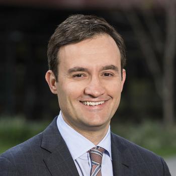 George A. Poultsides合作论文数Division of General Surgery, School of Medicine, Stanford University243
 Shishir Maithel合作论文数Division of Surgical Oncology, Department of Surgery, Emory University School of Medicine;Winship Cancer Institute, Emory University202
 Sharon M Weber合作论文数Division of Surgical Oncology, Department of Surgery, School of Medicine and Public Health, University of Wisconsin-Madison;Carbone Cancer Center, University of Wisconsin-Madison155
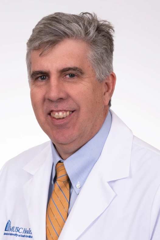 William Gabriel Hawkins合作论文数Department of Surgery, Medical University of South Carolina;Hollings Cancer Center, Medical University of South Carolina154
 Timothy M Pawlik合作论文数Department of Surgery, College of Medicine, The Ohio State University Wexner Medical Center147
 Bradley A Krasnick合作论文数Washington Univ, Sch Med, Dept Surg, St Louis, MO 63110 USA114
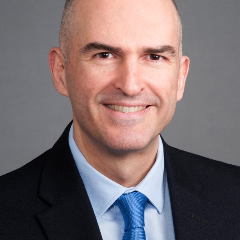 Konstantinos Votanopoulos合作论文数School of Medicine, Wake Forest University;Wake Forest Organoid Research Center, Wake Forest Institute for Regenerative Medicine;Atrium Health Wake Forest Baptist112Kamran Idrees合作论文数Memorial Sloan-Kettering Cancer Center107
 Tran Thuy合作论文数School of Medicine, Stanford University102
 Schmidt Carl合作论文数Wexner Med Ctr, Ohio State Univ100
 Cecilia G. Ethun合作论文数81
 Steven Strasberg合作论文数Department of Surgery, School of Medicine, Washington University in St Louis81
 Li Ding合作论文数Ding Lab, Division of Oncology, John T. Milliken Department of Medicine, School of Medicine, Washington University in St. Louis80
 Ioannis Hatzaras合作论文数James Cancer Hospital, The Ohio State University Medical Center77
 Clifford Cho合作论文数Division of Hepatopancreatobiliary and Advanced Gastrointestinal Surgery, Medical School, University of Michigan66
 Eliza Beal合作论文数Karmanos Cancer Center;Michael & Marian Ilitch Department of Surgery, School of Medicine, Wayne State University66
 David G. Denardo合作论文数Division of Oncology, John T. Milliken Department of Medicine, School of Medicine, Washington University in St. Louis;Siteman Cancer Center66Kenneth Cardona合作论文数66
 Charles R. Scoggins合作论文数Surgical Oncology Division, Department of Surgery, School of Medicine, University of Louisville65
 Perry Shen合作论文数School of Medicine, Wake Forest University60Sanford Dominic E合作论文数59
 Linda X Jin合作论文数Department of Surgery, Washington University in St. Louis58
 Harveshp D Mogal合作论文数57
 Ahmed Salem合作论文数Division of Cancer Sciences, School of Medical Sciences, Faculty of Biology, Medicine and Health, The University of Manchester56
 Robert C.G. Martin合作论文数Division of Surgical Oncology, Department of Surgery, School of Medicine, University of Louisville;Upper GI and HPB Multi-Disciplinary Clinic54Alexandra G. Lopez-Aguiar合作论文数Winship Canc Inst, Emory Univ Hosp51Shenoy Rivfka合作论文数51
 Malcolm (Hart) Squires合作论文数Division of Surgical Oncology, Levine Cancer Institute, Atrium Health50
 Edward A. Levine合作论文数Department of Surgical Sciences, School of Medicine, Wake Forest University;Atrium Health Wake Forest Baptist Comprehensive Cancer Center50Chelsea A. Isom合作论文数49
 Flavio G. Rocha合作论文数Surgical Oncology Clinic, Knight Cancer Institute, Oregon Health and Science University;Division of Surgical Oncology, School of Medicine, Oregon Health and Science University49
 S. Peter Goedegebuure合作论文数Division of General Surgery, Department of Surgery, Washington University in St. Louis46Daniel E. Abbott合作论文数43
 Hammill Chet W合作论文数Barnes Jewish Hosp, Washington Univ42William C. Chapman合作论文数41
 Kevin K. Roggin合作论文数Hollings Cancer Center, Medical University of South Carolina;Department of Surgery, College of Medicine, Medical University of South Carolina39
 Winslow Emily R合作论文数39
 Mary Dillhoff合作论文数Department of Surgery, The Ohio State University39Jacqueline Mudd合作论文数Washington Univ, Sch Med, St Louis, MO USA38
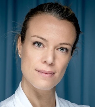 Gaya Spolverato合作论文数Azienda Ospedaliera Università di Padova;Memorial Sloan Kettering Cancer Center38Worhunsky David合作论文数38
 Mark Bloomston合作论文数38
 David A. Kooby合作论文数Winship Cancer Institute, Emory University;Emory Saint Joseph's Hospital;Division of Surgical Oncology, Department of Surgery, School of Medicine, Emory University;Emory Johns Creek Hospital38Paula Marincola Smith合作论文数36
 Lauren Postlewait合作论文数36Grignol Valerie P合作论文数Department of Surgery, Medical College of Wisconsin34
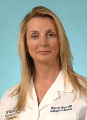 Maria B. Majella Doyle合作论文数Department of Surgery, Washington University in St. Louis;Barnes-Jewish Hospital;St. Louis Children’s Hospital34Howard J Harrison合作论文数33David C. Linehan合作论文数33




