PBMCs recovered from ex vivo ELISpot were stimulated with 9-10mer Mam-A pulsed APCs overnight and stained for ICS and specific peptide-HLA tetramers
Tumor-associated antigen (TAA) vaccines are being explored as a strategy to induce antitumor immune responses. Mammaglobin-A (Mam-A) is a TAA expressed in >50% of patients with breast cancer. Previously, we have shown that Mam-A DNA vaccines induce antitumor immune responses in patients with stable metastatic disease. To further evaluate the potential of the Mam-A vaccine, we initiated a phase Ib clinical trial in patients with estrogen receptor-positive breast cancer prior to surgery. Eight patients were assigned to arm 1 (neoadjuvant endocrine therapy alone) and 17 to arm 2 (neoadjuvant endocrine therapy plus Mam-A vaccination); the final analysis included 8 patients from arm 1 and 13 from arm 2. Ex vivo enzyme-linked immunospot (ELISpot) analysis of peripheral blood mononuclear cells demonstrated that Mam-A vaccination induced Mam-A-specific T cells in 8 of 13 patients. Intracellular cytokine staining and Mam-A-specific tetramer staining revealed that vaccine-induced Mam-A-specific T cells included both CD4(+) and CD8(+) polyfunctional T cells. Finally, high-throughput imaging mass cytometry identified 24 cellular metaclusters with features of tumor, immune, stromal, and endothelial cells and revealed an increased CD8(+) T-cell prevalence in the tumor after Mam-A vaccination. In particular, vaccination was associated with the infiltration of PD-1(+)CD8(+) T cells. In addition, postvaccination tumor samples exhibited close spatial interactions between cytotoxic CD8(+) T cells (CTL) and Mam-A(+) tumor cells and between CTL and antigen-experienced CD4(+) T cells. Together, these results suggest that Mam-A DNA vaccination elicits both systemic and intratumoral antitumor immune responses.
Abstract Background: Calreticulin (CALR) is an endoplasmic reticulum chaperone protein that can translocate to the plasma membrane during cellular stress. Despite its known roles in cancer, CALR levels in the blood have not been extensively studied in cancer patients. We hypothesized that serum CALR is elevated in cancer patients and may serve as a noninvasive biomarker for early detection. Methods: Serum samples were collected from patients diagnosed with pancreatic ductal adenocarcinoma (PDAC), breast cancer (BC), colorectal cancer (CRC), and healthy donors. CALR concentration was quantified using a RayBiotech Human CALR Sandwich ELISA. Statistical analyses were performed using Mann-Whitney U tests for group comparisons and ROC analysis for evaluating diagnostic performance on GraphPad Prism. Results: 80 PDAC patients, 25 BC patients, 25 CRC patients, and 60 healthy donors were included in the study. Median (IQR) CALR level increased from 36.22 pg/mL (5.09-264.6) in healthy individuals to 363.1 pg/mL (226.5-729.7) in PDAC patients, 182.5 pg/mL (108.1-344.0) in CRC patients, and 584.9 pg/mL (381.2-823.4) in BC patients. Pairwise testing confirmed significantly higher CLAALR levels in PDAC vs. healthy (p <0.0001), BC vs. healthy (p < 0.0001), and CRC vs. healthy (p = 0.0021).ROC analysis demonstrated strong diagnostic potential of serum CALR for distinguishing PDAC from healthy individuals (AUC = 0.799). The optimal cutoff, determined using Youden’s Index, was 147 pg/mL with a sensitivity of 89% and specificity of 71.7%. Differentiating healthy donors from BC was also significant (AUC = 0.844) with sensitivity of 88% and specificity of 73.33% at the optimal cutoff value of 308.7 pg/mL. CRC was also distinguishable from healthy donors with an optimal cutoff of 52.81 pg/mL, yielding a sensitivity of 96% and specificity of 58.3% (AUC = 0.72). Conclusion: Serum CALR is significantly elevated in patients with PDAC, BC, and CRC compared to healthy individuals and demonstrates strong diagnostic accuracy. These findings identify CALR as a promising noninvasive biomarker for cancer detection and justifies future validation studies assessing combinatorial biomarker panels. Citation Format: Jasmine Watts, Lucinda Ann Hall, Lorenzo Thompson, Jacqueline L. Mudd, Steven Forsythe, Yuvasri Golivi, Roheena Panni, William E. Gillanders, Li Ding, Ryan C. Fields, Benjamin Larimer, Rachael Guenter, John B. Rose. Serum calreticulin as a promising biomarker for cancer screening [abstract]. In: Proceedings of the American Association for Cancer Research Annual Meeting 2026; Part 1 (Regular Abstracts); 2026 Apr 17-22; San Diego, CA. Philadelphia (PA): AACR; Cancer Res 2026;86(7 Suppl):Abstract nr 2543.
Amino acid sequence and HLA specificity of Mam-A peptides used for ICS and tetramer analysis
Breast and prostate cancers are both hormone-driven adenocarcinomas that undergo analogous invasion programs. Using lightsheet microscopy on intact tumors, we identified transitional junctions between precancerous and invasive regions. We then developed a multimodal serial-section workflow integrating volumetric reconstruction with spatial transcriptomics. Analysis of 319 spatial assays from 51 cases revealed gene expression features and novel structural insights defining the shift from precancer to invasive disease. In breast cancer, loss of MGP and PLAT was associated with invasive transition and promoted tumorigenesis in functional assays. In prostate cancer, GDF15, ALDH1A3, ANPEP, and FASN were upregulated along invasive progression, and their knockdown in PC-3 cells suppressed proliferation and migration. Enrichment of tumor-associated macrophages (SPP1+ and MS4A6A+) along non-triple-negative breast cancer breast cancer transitions highlights immune involvement as a potential driver of invasiveness. SIGNIFICANCE:Our method of defining precise spatial locations of invasive transition allows for the direct interrogation of transition drivers, presenting new therapeutic targets for the two most prevalent cancers and providing a framework for studying spatially defined mechanisms of tumor progression. See related commentary by Jing and Li, p. 1720.
Pancreatic ductal adenocarcinoma (PDAC) is unresponsive to standard immunotherapies despite harboring cancer neoantigens capable of eliciting T cell responses. We completed two phase 1 clinical trials (NCT03956056 and NCT03122106) evaluating safety and immunogenicity of synthetic long peptide (SLP) and DNA personalized cancer vaccines (PCVs). PCVs were administered after resection and adjuvant chemotherapy. Tumor/normal whole-exome sequencing, RNA sequencing, and pVACtools were used to identify and prioritize candidate PCV neoantigens. PCVs were well tolerated without any grade ≥3 adverse events. Neoantigen-specific responses were demonstrated by interferon-γ enzyme-linked immunospot and intracellular cytokine staining. Expanded T cell receptor clonotypes were sequenced and transduced into autologous peripheral blood mononuclear cells to confirm neoantigen specificity. When compared with a contemporaneous institutional propensity-matched cohort, PCV patients demonstrated a trend toward prolonged median overall survival (4.4 versus 3.5 years, log-rank P = 0.23). Overall, PDAC PCVs are safe and feasible and elicit polyclonal T cell responses, linking prioritized cancer neoantigens to functional antitumor immunity.
Representative examples of IF images depicting specific staining of different antibodies in different human tissues
Vaccines composed of mRNA and lipid nanoparticles (LNPs) activate B cells and T cells by inducing in vivo production of specific protein antigens. While B cells can be activated directly by antigens, T cell activation requires antigen processing and presentation by MHC molecules on specialized antigen-presenting cells (APCs). In response to viral infections, tumours, and protein- and cDNA-based vaccines, antigen presentation to CD8+ T cells is particularly dependent on type 1 conventional dendritic (cDC1) cells, which are specialized for efficient cross-presentation of exogenous antigens1-4. However, whether similar mechanisms have a role in mRNA-LNP vaccination is unclear. Here we report that mRNA-LNP vaccines do not require cDC1 cells or the WDFY4-dependent cross-presentation pathway for CD8+ T cell priming but instead engage both cDC1 and cDC2 cells redundantly. While CD8+ T cells primed exclusively by either cDC1 or cDC2 cells showed phenotypic differences, both could mediate anti-tumour responses and memory formation. Importantly, acquisition by cDCs of peptide-MHC-I complexes from non-haematopoietic cells, called cross-dressing, provides a substantial component of CD8+ T cell priming, in a manner dependent on type I interferon. mRNA-LNP induction of cross-dressing might explain their ability to activate CD8+ T cells against antigens not encoded by the vaccine.
Advancements in immunogenomics and immuno-oncology have enabled the development of personalized cancer vaccines (PCVs) that target cancer cell-specific somatic variants. A subset of these variants produce neoantigens that, when presented on tumor cells by MHC molecules, have the potential to elicit a robust and specific immune response. To date, there are over one hundred interventional studies listed on clinicaltrials.gov that explore the use of PCVs. We have supported a number of these trials through the creation of bioinformatic pipelines, tools, and procedures for the identification of patient-specific neoantigen candidates. While many of these steps have been automated, the final selection of neoantigen candidates often relies on expert manual review, creating a bottleneck that limits scalability and full automation of PCV workflows. Addressing this challenge, we introduce NEAT (Neoantigen Evaluation & Automated Triage), a machine learning-based approach that enables automated neoantigen candidate prioritization and supports the transition toward more scalable and reproducible PCV design. We implemented a prediction model trained and tested on existing vaccine design results from 33 patients and 1,943 peptides, across 3 clinical trials, including 439 peptides prioritized for PCV inclusion. This model uses features such as tumor variant allele frequency, RNA expression, driver gene status, binding/presentation scores, and transcript support level to automatically predict whether a peptide will be accepted, rejected, or require further human review before inclusion in a vaccine. The model achieved a sensitivity of 0.847 and specificity of 0.924, with an area under the curve of 0.955. The model predictions have been incorporated in pVACtools v7.0.0. By integrating this model into the vaccine development pipeline, we foresee a significant reduction in the time required to transition from patient sample collection to vaccine manufacturing, thereby enhancing the efficiency and scalability of PCV production.
Gating strategy for the various cell types and functional status based on the presence of specific markers captured by the antibody panel
Mam-A vaccination increases the leukocyte infiltration in vaccinated BC tissues compared to non-vaccinated BC tissues
 S. Peter Goedegebuure合作论文数Division of General Surgery, Department of Surgery, Washington University in St. Louis87
 Rebecca L. Aft合作论文数63Li Lijin合作论文数44Julie A. Margenthaler合作论文数44
 Timothy Fleming合作论文数Norton Thoracic Institute44Zhang Xiuli合作论文数43
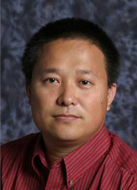 Feng Gao合作论文数Mary Culver Department of Surgery, Washington University School of Medicine in St. Louis43
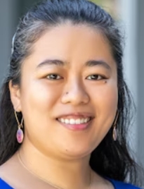 Cynthia Ma合作论文数36Timothy J Eberlein合作论文数34Rama Suresh合作论文数29
 Ian S Hagemann合作论文数Departments of Medicine and of Molecular Biology & Pharmacology, 660 S. Euclid Ave., Campus Box 8127, St. Louis, Missouri 63110, USA29David J. Cole合作论文数28
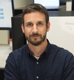 Malachi Griffith合作论文数Genome Sciences Centre, BC Cancer Agency28Nancy B. Myers合作论文数26
 John M Herndon合作论文数25Amy E. Cyr合作论文数23
 Jasreet Hundal合作论文数Natera22
 William Gabriel Hawkins合作论文数Department of Surgery, Medical University of South Carolina;Hollings Cancer Center, Medical University of South Carolina22
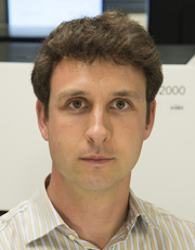 Obi L. Griffith合作论文数Divisions of Hematology & Oncology, Department of Medicine, Washington University School of Medicine in St. Louis22Foluso Ademuyiwa合作论文数19Ademuyiwa Foluso O合作论文数19
 Ryan C. Fields合作论文数Division of General Surgery, Department of Surgery, School of Medicine, Washington University;Barnes-Jewish Hospital;Siteman Cancer Center, Barnes-Jewish West County Hospital19
 Matthew Ellis合作论文数Department of Psychiatry, Washington University School of Medicine In St. Louis19Michael J. Naughton合作论文数19Donna Jeffe合作论文数School of Medicine, Washington University18
 Mario Schootman合作论文数1;Department of Medicine and Pediatrics;Washington University;School of Medicine17
 Elaine R. Mardis合作论文数Departments of Pediatrics, College of Medicine, The Ohio State University;Institute for Genomic Medicine, Nationwide Children's Hospital15Sankpal Narendra V合作论文数14
 Savas Soysal合作论文数Chirurgie im Zentrum13
 Li Ding合作论文数Ding Lab, Division of Oncology, John T. Milliken Department of Medicine, School of Medicine, Washington University in St. Louis13Michael Mitas合作论文数Department of Surgery, Medical University of South Carolina13Sherri R Davies合作论文数Washington Univ, Sch Med, Dept Med, St Louis, MO 63110 USA12Salem Mohamed Labib合作论文数12
 Darren R Cullinan合作论文数Department of Surgery, University of Tennessee Health Science Center12
 David G. Denardo合作论文数Division of Oncology, John T. Milliken Department of Medicine, School of Medicine, Washington University in St. Louis;Siteman Cancer Center12
 Ted Howard Hansen合作论文数Department of Pathology and Immunology, Washington University in St. Louis12Jeremy Hoog合作论文数12
 Kenneth Murphy合作论文数Division of Immunobiology, Department of Pathology & Immunology, School of Medicine, Washington University in St. Louis;Howard Hughes Medical Institute11Kaidi Mikhitarian合作论文数11
 Reyka G Jayasinghe合作论文数Department of Medicine, Washington University School of Medicine10Zachary L Skidmore合作论文数Washington Univ, Sch Med, McDonnell Genome Inst, St Louis, MO 63108 USA10
 Maria Pérez合作论文数Department of Medicine, Washington University School of Medicine10
 Imran Zoberi合作论文数10Gavin P. Dunn合作论文数10
 Robert Schreiber合作论文数Department of Pathology & Immunology, School of Medicine, Washington University;Center for Human Immunology and Immunotherapy Programs, Washington University;Siteman Comprehensive Cancer Center, Washington University10
 Kelsy Cotto合作论文数Department of Medicine, Washington University School of Medicine9
 Siqi Chen (陈斯琦)合作论文数Washington Univ St Louis9Tanner M. Johanns合作论文数9
 Souzan Sanati合作论文数8




