Awareness of action (AoA), or conscious awareness of an action just performed, is an important part of daily experience with major practical and ethical relevance, yet the neural mechanisms of AoA remain largely unknown. The main barrier to studying AoA is a lack of experimental paradigms to directly compare neural activity in aware versus unaware actions. Borrowing from the field of perceptual awareness, where exciting progress has been made by contrastive analysis of aware versus unaware stimuli, we developed a game where participants repeatedly perform nearly identical moves while engaged in a distractor task, and the participants then report awareness or unawareness of the moves they just performed. We found that on short timescales, aware actions had larger neurophysiological signals both preceding and following movement. The differences included both volitional and perceptual event-related potentials (premovement positivity, N140, and P300), as well as frontal midline theta, event-related alpha/beta desynchronization, and postmove blink rates. On longer time scales, we identified a novel positive event-related potential only preceding unaware moves and found behavioral and pupillometric evidence for decreased attention and arousal over minutes concurrent with AoA loss. Our findings reveal three neural mechanisms that may synergistically contribute to AoA: (i) long-term increases in arousal/attentional state at time of action; (ii) increased action-related motor volitional signals; and (iii) increased action-related sensory perceptual signals. Deeper understanding of AoA may ultimately elucidate the causes of variable AoA in daily life and lead to better treatments for impaired AoA in neuropsychiatric disorders.
Despite many years of research, the quest to identify neural correlates of perceptual consciousness (NCC) remains unresolved. One major obstacle lies in methodological limitations: most studies rely on non-invasive neural measures with limited spatial or temporal resolution making it difficult to disentangle proper NCCs from concurrent cognitive processes. Additionally, the relatively low sensitivity of non-invasive neural measures limits the interpretation of null findings in studies targeting proper NCCs. In this review, we discuss how human intracranial recordings can advance the search for NCCs, by offering high spatiotemporal resolution, improved signal sensitivity, and broad cortical and subcortical coverage. We review studies that have examined NCCs at the level of single neurons and populations of neurons, and evaluate their implications on the debates between cognitive and sensory theories of consciousness. Finally, we highlight the limits of current intracranial human recordings and propose future directions based on emerging technologies and novel experimental paradigms.
We present a functional magnetic resonance (fMRI) dataset collected as part of an adversarial collaboration aimed at arbitrating between the Global Neuronal Workspace theory (GNWT) and the Integrated Information Theory (IIT) of consciousness. Participants (N = 118) were presented with suprathreshold visual stimuli belonging to four different categories (faces, objects, letters, false fonts) with three orientations (front, left, right view), and three durations (0.5, 1.0, 1.5 seconds). Participants were asked to identify infrequent targets that changed in each block, thereby rendering two categories task-relevant and two task-irrelevant. The simplicity of the experimental design and of the task given to the participants ensures that these data are broadly reusable. Besides testing predictions from other theories of consciousness, these data can be used to examine various aspects of visual processing. The anonymized data were converted to Brain Imaging Data Structure (BIDS), and can be easily accessed through a web platform or an API. The dataset contains quality reports, demographics, behavioral performance, and eye-tracking data. We also provide code for preprocessing and analyzing the data.
Neurovascular coupling links calcium (Ca2+)-dependent neuronal activity to cerebral blood volume changes, whereas neurometabolic coupling describes alterations of neuronal activity and glucose uptake. While mesoscale optical imaging of neurovascular coupling is prevalent, neurometabolic coupling has been explored much less. We describe a multiplexed optical system with a closed cranial window setup for longitudinal studies in Thy1-jRGECO1a mice where neuronal activity is measured with Ca2+-dependent red fluorescence, glucose uptake with bolus injections of 2NBDG with green fluorescence, and cerebral blood volume (CBV) with near-infrared spectroscopy (NIRS). Genetically encoded calcium indicators (GECIs) provide strong fluorescent signals for assessing Ca2+-dependent neuronal activity. Thy1-jRGECO1a, a novel GECI with red fluorescence emission that penetrates deeper into tissue, allows for simultaneous imaging of metabolic activity using a green-fluorescent glucose analog, 2-(N-(7-nitrobenz-2-oxa-1,3-diazol-4-yl)amino)-2-deoxyglucose (2NBDG), which is taken up like glucose and then phosphorylated. Dual-fluorescent (red, green) and NIRS recordings confirm strong neurovascular coupling during hindpaw stimuli (Ca2+-CBV; P = 0.0033, r(2) = 0.91), whereas neurometabolic coupling (Ca2+-2NBDG; P < 0.001) was three times stronger during stimulation (r(2) = 0.75; slope = 0.6) compared to rest (r(2) = 0.49; slope = 0.23). In summary, multiplexed optical imaging can be used to reveal mechanisms of neurovascular and neurometabolic (un)couplings during ischemia, traumatic brain injury, aging, and Alzheimer's disease.
Thalamic stimulation has emerged as a promising neuromodulation target for treating disorders of consciousness. Impaired consciousness, a debilitating outcome in temporal lobe epilepsy (TLE) remains a central problem for patients whose seizures cannot be treated pharmacologically and cannot be stopped with conventional surgery or responsive hippocampal stimulation. Although prior studies suggest an essential role of the thalamic intralaminar central lateral (CL) nucleus in arousal and sleep, evidence for a direct effect of thalamic intralaminar stimulation on human arousal has been limited. To address these gaps, the START (stimulation of the thalamus for arousal restoral in TLE) clinical trial investigated the efficacy of bilateral CL thalamic stimulation to restore consciousness during human sleep and TLE seizures. Five patients with medically refractory mesial temporal lobe epilepsy were implanted with an investigational neurostimulator, the Medtronic Summit RC+STM. Optimal CL stimulation parameters were obtained through individualized titration in slow wave sleep, evaluated through analysis of patient movement from video recordings and electrophysiology from simultaneously recorded scalp and hippocampal electroencephalography (EEG). We found that bilateral CL stimulation led to robust arousal from sleep characterized by increased body movements and decreased low frequency power (2-15 Hz) in both cortical and hippocampal EEG during 5 minute epochs of stimulation compared to baseline slow wave sleep. We evaluated impaired consciousness during seizures using verbal and non-verbal behavioral tests administered automatically by smartwatch, and found significant behavioral impairment in three of five patients during seizures with hippocampal stimulation. Administering CL stimulation in patients with impaired consciousness during TLE seizures showed significant improvement in behavioral outcomes in two of three patients, with one patient reaching their baseline performance comparable to non-seizure times. Overall, we found that stimulation of the thalamic CL in TLE patients increased arousal during both sleep and seizures. These findings demonstrate the potential of CL as a therapeutic target for mitigating impaired consciousness in TLE and can serve as a foundation for additional studies to test generalizability of CL stimulation effects on other disorders of consciousness. ### Competing Interest Statement The authors have declared no competing interest. ### Clinical Trial NCT04897776 ### Funding Statement This work was supported by NIH/NINDS UG3/UH3 NS112826. ### Author Declarations I confirm all relevant ethical guidelines have been followed, and any necessary IRB and/or ethics committee approvals have been obtained. Yes The details of the IRB/oversight body that provided approval or exemption for the research described are given below: Ethics committee/IRB of Yale University School of Medicine, Mayo Clinic, and Dartmouth-Hitchcock medical center gave ethical approval for this work. I confirm that all necessary patient/participant consent has been obtained and the appropriate institutional forms have been archived, and that any patient/participant/sample identifiers included were not known to anyone (e.g., hospital staff, patients or participants themselves) outside the research group so cannot be used to identify individuals. Yes I understand that all clinical trials and any other prospective interventional studies must be registered with an ICMJE-approved registry, such as ClinicalTrials.gov. I confirm that any such study reported in the manuscript has been registered and the trial registration ID is provided (note: if posting a prospective study registered retrospectively, please provide a statement in the trial ID field explaining why the study was not registered in advance). Yes I have followed all appropriate research reporting guidelines, such as any relevant EQUATOR Network research reporting checklist(s) and other pertinent material, if applicable. Yes All data generated in the present study will be made available from the corresponding author upon reasonable request.
Impaired consciousness is a debilitating and unpredictable outcome of mesial temporal lobe seizures whose mechanisms to date remain unclear. Moreover, questions about the relationship between impaired consciousness and lateralization, hemispheric spread and electrophysiological characteristics of seizures are yet to be answered. To address these gaps, we conducted in-depth investigation of behavioral and intracranial EEG data from 186 mesial temporal lobe seizures of 51 patients with intractable mesial temporal lobe epilepsy. We found that bilateral mesial temporal spread of seizures is not a necessary condition for impaired consciousness, although seizures with bilateral mesial temporal involvement were significantly more likely to have impaired consciousness than unilateral seizures. Contrary to prior belief, we found no relationship between the onset side (left vs. right temporal lobe or language dominant vs. non-dominant lobe) of seizures and the probability of impaired consciousness. Lastly, we established that widespread increases in slow-wave activity (delta band) in extratemporal cortical areas, as well as increases in fast activity (beta band) in the temporal lobes were both robust markers of seizures with impaired consciousness and could predict ictal impairment with up to 86% accuracy. Our findings shed new light on networks that underlie impaired consciousness in temporal lobe epilepsy and may help guide deep brain stimulation of such systems (e.g. via thalamic nuclei) as a potential intervention to improve consciousness during seizures. ### Competing Interest Statement The authors have declared no competing interest. National Institutes of Health, https://ror.org/01cwqze88, 5UH3NS112826-05
BACKGROUND AND OBJECTIVES:Impaired consciousness in epilepsy negatively affects quality of life. Previous work has focused on temporal lobe seizures, where cortical slow waves are associated with depressed subcortical arousal and impaired consciousness. However, it is unknown whether frontal lobe seizures also show cortical slow waves or a different activity pattern with impaired consciousness. METHODS:Intracranial EEG (icEEG) recordings from patients at 3 centers were retrospectively assessed to identify seizures originating in the frontal lobe. Seizures were classified as focal preserved consciousness (FPC), focal impaired consciousness (FIC), or focal to bilateral tonic-clonic (FBTC) based on video review. Changes in icEEG power from preictal baseline were calculated in different cortical regions and across frequency ranges in these 3 seizure categories. RESULTS:Sixty-five seizures in 30 patients (mean age 27.7 years, 43% female) were analyzed. Frontal lobe FPC seizures showed approximately 40% icEEG power increases in the frontal lobe of onset across frequency ranges, with smaller changes in other regions. Frontal lobe FIC seizures showed approximately 50% power increases, not significantly different from FPC seizures in the lobe of onset (p = 0.519, 95% CI -25.8 to 50.4), but with significantly greater power increase in other widespread cortical regions (p < 0.001, 95% CI 14.1-45.3). It is important to note that the widespread icEEG power increases in FIC seizures occurred not just in the slow-wave frequency range, but broadly across other frequencies including fast activity. However, the widespread power increases in FIC seizures differed from those of FBTC seizures where icEEG power increases were much greater at approximately 600%, significantly greater than in FIC seizures in both the frontal lobe of onset and other cortical regions (p < 0.001, 95% CI 330.1-781.9 and 375.3-818.2, respectively). DISCUSSION:The widespread power increases across frequencies in frontal lobe FIC seizures contrast with those in focal temporal lobe epilepsy, where impaired consciousness is associated with cortical slow waves. These findings suggest that different focal seizure types produce impaired consciousness by affecting widespread cortical regions but through different physiologic mechanisms. Insights gained by studying the physiology of impaired consciousness may be the first step toward developing novel treatments to prevent this significant negative consequence of epilepsy and improve quality of life.
OBJECTIVE:Impaired consciousness during seizures significantly impacts the quality of life for individuals with epilepsy, and recent research has introduced the network inhibition hypothesis, suggesting that impaired consciousness results from the active inhibition of subcortical arousal mechanisms. However, direct evidence in awake animals has been lacking. Our study aimed to address this gap by recording the activity of individual neurons in crucial brainstem and basal forebrain nuclei in a novel behaving mouse model. METHODS:We conducted recordings in head-fixed mice running on a freely moving wheel with implanted electrodes in the orbitofrontal cortex and hippocampi. Focal limbic seizures were induced by applying current pulses, and simultaneously we obtained juxtacellular single unit activity recordings from arousal nuclei in the brainstem and basal forebrain. Double immunofluorescence was performed postrecording to confirm cell locations and cholinergic identities. RESULTS:Our findings revealed that focal seizure activity suppressed behavior based on decreased running wheel speed, and the orbitofrontal cortex exhibited slow waves resembling encephalopathy or deep sleep. Single unit recordings showed diverse firing patterns during seizures, with some neurons reducing firing, others increasing, and some remaining relatively stable. Importantly, cholinergic neurons in the brainstem pedunculopontine and laterodorsal tegmental nuclei exhibited significant reductions in firing during focal limbic seizures. SIGNIFICANCE:Our findings provide direct evidence that focal limbic seizures are associated with decreased cholinergic neuronal firing in brainstem arousal nuclei, linking subcortical suppression to cortical impairment. Further exploration of these pathways promises a deeper understanding of ictal unconsciousness and potential novel treatments for people with epilepsy.
Although recent work has made headway in understanding the neural temporospatial dynamics of conscious perception, much of that work has focused on visual paradigms. To determine whether there are shared mechanisms for perceptual consciousness across sensory modalities, here we test within the auditory domain. Participants completed an auditory threshold task while undergoing intracranial electroencephalography. Recordings from >2,800 grey matter electrodes were analyzed for broadband gamma power (a range which reflects local neural activity). For perceived trials, we find nearly simultaneous activity in early auditory regions, the right caudal middle frontal gyrus, and the non-auditory thalamus; followed by a wave of activity that sweeps through auditory association regions into parietal and frontal cortices. For not perceived trials, significant activity is restricted to early auditory regions. These findings show the cortical and subcortical networks involved in auditory perception are similar to those observed with vision, suggesting shared mechanisms for conscious perception.
Studies of human perception have shown early cortical signals for primary information encoding, and later signals for higher order processing. An important late signal is the cortical event-related desynchronization (ERD) in the alpha (8-12 Hz) and beta (12-30 Hz) frequency band, which has been linked to human perceptual awareness. Detailed mechanistic investigation of the ERD would be greatly facilitated by availability of a suitable animal model. We conducted local field potential recordings in the mouse frontal association cortex (FrA), thalamic intralaminar centrolateral nucleus (CL), primary auditory cortex (A1), and primary visual cortex (V1) during two auditory tasks. Fully audible brief 50 ms stimuli with both tasks produced early broadband gamma (30-100 Hz) frequency activity at 0-250ms, followed by a late cortical alpha/beta ERD 250-750 ms after stimulus onset. The ERD was statistically significant in FrA and A1, but not in V1. Interestingly, a significant ERD was also observed in thalamic CL. The magnitude of the ERD at full stimulus intensity, and the slope of the relationship between stimulus intensity versus ERD magnitude, were both largest in FrA, and smaller in CL and A1. Conversely, for early broadband gamma activity the magnitude at full intensity and slopes were largest in A1, smaller in CL and smaller still in FrA. These findings strongly support mice as a promising platform for further investigation of the ERD to better understand the origin and function of this robust yet understudied electrophysiological phenomenon.
Temporal lobe epilepsy (TLE) is the most common form of epilepsy and is characterized by focal seizures originating from limbic structures, including the hippocampus. Patients with TLE often experience impaired consciousness. A recent awake mouse model study demonstrated decreased cortical cholinergic innervation during focal seizures with impaired consciousness, based on cortical slow wave activity and decreased behavioral responsiveness. But the underlying mechanisms for reduced cortical cholinergic activity are not fully understood. This study employs the same awake mouse model combined with electrophysiology recordings in key network nodes, cell-specific calcium imaging in the lateral septum, and neurotransmitter sensing in one of the major subcortical cholinergic systems, the nucleus basalis of Meynert (NBM). We demonstrate that decreased cortical cholinergic innervation during focal seizures comes from both direct inhibition and indirect de-excitation of the NBM, showing a parallel pathway NBM suppression mechanism from the LS directly and through the paratenial thalamic nucleus indirectly. This work contributes to a deeper understanding of the neural processes involved in impaired consciousness during focal seizures and may open the way to new treatments for this disorder.
Background and Objectives:Post-ischemic stroke epilepsy (PISE) reduces quality of life, and early risk prediction can guide prevention strategies and anti-epileptogenesis treatment trials. Stroke severity predicts both PISE and mortality, and ignoring mortality can overestimate epilepsy risk. We sought to enhance PISE risk stratification by modeling death as a competing outcome, integrating quantitative clinical, neuroimaging, and electroencephalography (EEG) biomarkers to distinguish shared and distinct predictors of epilepsy and mortality. Methods:We developed a PISE prediction model using retrospective data from Yale-New Haven Hospital. The training cohort included patients from 2014-2020; the testing cohort from 2021-2022. Eligible patients were adults with acute ischemic stroke who underwent neuroimaging and EEG monitoring <7 days post-stroke and had follow-up >7 days. Results:Of 280 patients, 53 developed PISE first, 104 died first, and the rest were censored. Quantitative PISE biomarkers included greater 72h stroke severity (HR Δ3 [95%CI], 1.2 [1.1-1.4]), infarct volume (HR Δ10mL , 1.06 [1.04-1.08]), EEG epileptiform abnormality burden (HR Δ10% , 1.2 [1.1-1.3]), and EEG power asymmetries (HR Δ10% , 2.0 [1.4-2.9]). Death predictors included older age (HR Δ10years , 1.7 [1.4-2.0]), worse pre-stroke functional status (HR, 1.4 [1.2-1.7]), atrial fibrillation history (HR, 2.4 [1.6-3.7]), cardioembolism etiology (HR, 1.9 [1.2-3.0]), anterior cerebral artery involvement (HR, 2.2 [1.2-3.7]), and greater EEG global theta-band powers (HR Δ10µV , 6.2 [2.3-17]). Our model, CRIME PISE , integrating these features, allows prediction of PISE-first and death-first risk scores with AUC of 0.72 (95%CI, 0.60-0.83) and 0.79 (0.72-0.85), respectively. Compared with the benchmark SeLECT model, CRIME PISE better predicted PISE in patients with ≥4 SeLECT points (AUC, 0.72 vs 0.58) but not those with <4 points (AUC, 0.33 vs 0.52). In the testing cohort, CRIME PISE identified a more selective group (n=18 vs 44 per SeLECT) with a higher PISE rate (39% vs 20%) and a lower mortality rate (22% vs 45%). Discussion:CRIME PISE enhances PISE prediction by accounting for mortality as a competing outcome and incorporating multimodal quantitative biomarkers. Because its benefits over SeLECT are most pronounced in high-risk patients, a two-stage approach-SeLECT screening followed by CRIME PISE in SeLECT-positive cases-may better target candidates for anti-epileptogenesis trials by prioritizing patients likely to survive long-term and develop epilepsy.
INTRODUCTION:Deep brain stimulation (DBS) of the centromedian (CM) thalamic nucleus is a potential therapy for Lennox-Gastaut syndrome (LGS), a severe and drug-resistant epileptic encephalopathy. While long-term seizure outcomes with CM-DBS have been described, its acute electrophysiological effects and predictive value remain uncertain. We examined whether short-term changes in interictal epileptiform discharges (IEDs) following CM-DBS relate to seizure outcomes at 1 year. METHODS:Ten patients with LGS underwent CM-DBS implantation. About 1 month post-surgery, each patient had a 1-h scalp EEG. After a 10-min baseline, stimulation began at 1 V for 5 min, increasing in 1 V increments to 5 V or until paresthesias occurred. IEDs were detected automatically (Persyst v14c) and verified by an expert reviewer. Patients were followed for 1 year, with clinical response defined as ≥50% seizure reduction, i.e., responder. Correlations between acute IED change and seizure outcomes were assessed using Spearman's rank correlation. RESULTS:Nine of 10 patients showed reduced IED burden during acute stimulation (31%-100%). At 1 year, 8 were responders and 2 nonresponders. Overall, acute IED reduction did not correlate with seizure outcome (Spearman's ρ = 0.3, p = 0.35). A ≥50% reduction in IED burden was seen in 7 of 7 responders versus 1 of 3 nonresponders, suggesting a nonsignificant trend toward predictive value (p = 0.06). CONCLUSION:Acute CM-DBS reduced IED burden in most patients with LGS but did not significantly predict long-term seizure outcomes. A trend toward greater IED reduction in responders suggests possible biomarker potential, though findings are preliminary and hypothesis-generating. Limitations include small sample size, high responder rate, and short EEG duration. Larger studies with extended monitoring are needed to clarify the clinical utility of acute EEG changes as predictors of CM-DBS efficacy.
Different theories explain how subjective experience arises from brain activity1,2. These theories have independently accrued evidence, but have not been directly compared3. Here we present an open science adversarial collaboration directly juxtaposing integrated information theory (IIT)4,5 and global neuronal workspace theory (GNWT)6-10 via a theory-neutral consortium11-13. The theory proponents and the consortium developed and preregistered the experimental design, divergent predictions, expected outcomes and interpretation thereof12. Human participants (n = 256) viewed suprathreshold stimuli for variable durations while neural activity was measured with functional magnetic resonance imaging, magnetoencephalography and intracranial electroencephalography. We found information about conscious content in visual, ventrotemporal and inferior frontal cortex, with sustained responses in occipital and lateral temporal cortex reflecting stimulus duration, and content-specific synchronization between frontal and early visual areas. These results align with some predictions of IIT and GNWT, while substantially challenging key tenets of both theories. For IIT, a lack of sustained synchronization within the posterior cortex contradicts the claim that network connectivity specifies consciousness. GNWT is challenged by the general lack of ignition at stimulus offset and limited representation of certain conscious dimensions in the prefrontal cortex. These challenges extend to other theories of consciousness that share some of the predictions tested here14-17. Beyond challenging the theories, we present an alternative approach to advance cognitive neuroscience through principled, theory-driven, collaborative research and highlight the need for a quantitative framework for systematic theory testing and building.
OBJECTIVE:To characterize electroencephalographic (EEG) profiles of short spike-and-wave bursts (SWBs) in patients with idiopathic generalized epilepsy reporting sensations of fleeting/almost loss of consciousness, described as "a blip on the screen"-a phenomenon first termed "blips" by J.W. Lance. METHODS:Among 176 consecutive patients, 19 were included based on a diagnosis of idiopathic generalized epilepsy and the presence of SWBs on EEG recordings. Patients were classified as "blippers" (yes) or "non-blippers" (no) based on their response to whether they had ever experienced "blip"-like sensations. A total of 624 SWBs extracted from EEG traces were compared between groups. The analysis focused on the spike (10-125 Hz) and wave (2.5-4 Hz) frequency band components across predefined spatial regions of interests. RESULTS:SWBs were significantly longer in blippers than in non-blippers (median: 1.2 s, interquartile range [IQR]: (Q1-Q3): 753-1796 vs 0.9 s, 599-1541; p < .001, r = .14). EEG analyses revealed two distinct spatial profiles, with higher fractional change in wave amplitude over anterior frontal (39.73, 12.56-81.27 vs 17.34, 5.18-56.95; p < .01, r = .13), frontal (46.94, 20.55-100.28 vs 31.08, 9.51-88.81; p < .05, r = .10), and occipital (25.25, 11.51-50.97 vs 14.99, 4.94-43.80; p < .01, r = .13) regions, but not parietal (p > .05). Similarly, spikes showed increased amplitude in anterior frontal (5.32, 2.50-15.27 vs 4.20, 2.58-7.60; p < .05, r = .09) and central (3.95, 2.81-6.20 vs 3.07, 1.82-5.08; p < .01, r = .14) regions, with lower occipital power in blippers (1.93, 1.36-3.19 vs 2.53, 1.52-4.33; p < .05, r = -.10). Overall, spike and wave amplitudes were higher in blippers, particularly at SWB onset. All p-values were false discover rate (FDR)-corrected and analyses were conducted at the SWB level. SIGNIFICANCE:Distinct EEG profiles of SWBs may be associated with self-reported blip experiences.
OBJECTIVE:Interictal epileptiform discharges (IEDs) in people with epilepsy (PWE) can impair cognitive functions and increase reaction time (RT) and the likelihood of missed reactions. These effects are not routinely assessed, because reliable methods for detecting IEDs of variable appearance in real time and suitable tests to measure IED effects do not yet exist. The objective was to assess different IED effects using new artificial intelligence and medical electronics. METHODS:The Digital Response Test in Epilepsy (DigRTEpi) consisted of a laptop and electronic circuits in a closed loop. Our model with Markov Transition Fields and a deep neural network (ResNet34) visualized the electroencephalogram (EEG) and classified the resulting images. IED detection triggered stimuli in a driving game or in a new cognitive assessment, the interictal Automated Responsiveness Test (iART). DigRTEpi was validated in a prospective case series with 20 people with focal and generalized epilepsies. During offline analysis, sensitivity, specificity, false-positive IED detection rate, latency of EEG classification, IED-induced RT prolongation, virtual crashes, and impaired responses to neuropsychological tasks were determined. RESULTS:The model detected IEDs with 84% sensitivity and 96% specificity in our training dataset. In the prospective study with 20 PWE, median sensitivity was 90% (95% confidence interval [CI] = .81-.95), and false-positive IED detection rate was 2.8 (95% CI 2.1-5.9). The ongoing EEG was classified window-by-window in a median 98.7 ms (95% CI = 98.0-99.4). Median RT prolongation and crash probability due to IEDs were 43.8 ms (95% CI = 20.3-64.7) and .9% (95% CI = 0-6.0) per person, respectively. Two patients (10%) had delays of >100 ms, found to be clinically relevant in our prior publication. IEDs caused four patients (20%) each to respond incorrectly or miss answers to neuropsychological tasks. The median false-positive IED detection rates were 2.8/min (95% CI = 2.1-5.9; driving game) and 2.1/min (95% CI = 1.5-3.2; iART). SIGNIFICANCE:By effectively detecting IEDs of variable morphology in real time, DigRTEpi assessed the severity of IED-associated transitory impairment of virtual driving and cognition to improve personalized care.
Studies of human perception have shown early cortical signals for primary information encoding, and later signals for higher order processing. An important late signal is the cortical event-related desynchronization (ERD) in the alpha (8-12Hz) and beta (12-30Hz) frequency band, which has been linked to human perceptual awareness. Detailed mechanistic investigation of the ERD would be greatly facilitated by availability of a suitable animal model. We conducted local field potential recordings in the mouse frontal association cortex (FrA), thalamic intralaminar centrolateral nucleus (Cl), primary auditory cortex (A1), and primary visual cortex (V1) during two auditory tasks. Fully audible brief 50 ms stimuli with both tasks produced early broadband gamma (30-100Hz) frequency activity at 0-250ms, followed by a late cortical alpha/beta ERD 250 - 750 ms after stimulus onset. The ERD was statistically significant in FrA and A1, but not in V1. Interestingly, a significant ERD was also observed in thalamic Cl. The magnitude of the ERD at full stimulus intensity, and the slope of the relationship between stimulus intensity versus ERD magnitude, were both largest in FrA, and smaller in Cl and A1. Conversely, for early broadband gamma activity the magnitude at full intensity and slopes were largest in A1, smaller in Cl and smaller still in FrA. These findings suggest that mice, like humans, process perceptual signals in hierarchically organized corticothalamic networks, and strongly support mice as a promising platform for further investigation of the ERD to better understand the origin and function of this robust yet understudied electrophysiological phenomenon. ### Competing Interest Statement The authors have declared no competing interest.
Die Internationale Liga gegen Epilepsie (ILAE) hat die operationale Klassifikation epileptischer Anfälle 2017 auf der Grundlage der damals entwickelten Rahmenbedingungen aktualisiert. In diese Überarbeitung wurden die publizierten Erfahrungen mit der Umsetzung der 2017er-Klassifikation einbezogen. Eine 37 Personen zählende Arbeitsgruppe wurde vom ILAE-Exekutivausschuss eingesetzt. Die internationalen Expertinnen und Experten aus allen ILAE-Regionen wandten ein modifiziertes Delphi-Verfahren an, bei dem für jeden Vorschlag ein Konsens von mehr als zwei Dritteln erforderlich war. Nach Veröffentlichung auf der ILAE-Homepage mit dem Ersuchen, Kommentare zu dem Entwurf einzugeben, ernannte der Exekutivausschuss 7 zusätzliche Sachverständige für die Arbeitsgruppe zur Überarbeitung des Positionspapieres, um die eingegangenen Kommentare zu diskutieren und gegebenenfalls einzubeziehen. Die aktualisierte Klassifikation behält die Hauptklassen von Anfällen bei: fokal, generalisiert, unbekannt (ob fokal oder generalisiert) und nicht klassifiziert. Taxonomische Regeln unterscheiden zwischen Klassifikatoren, die biologische Klassen widerspiegeln und sich direkt auf die klinische Behandlung auswirken, und Deskriptoren, die andere wichtige Anfallsmerkmale angeben. Fokale Anfälle und Anfälle unbekannten Ursprungs werden darüber hinaus nach dem Bewusstseinszustand des Patienten während des Anfalls klassifiziert, je nachdem ob eine Bewusstseinsstörung vorliegt oder nicht. Die Bewertung, ob eine Bewusstseinsstörung vorliegt, wird durch Gewahrsein (engl. „awareness“) und Reaktionsfähigkeit (engl. „responsiveness“) während eines Anfalls und erhaltene Erinnerung (engl. „recall“) nach einem Anfall klinisch operationalisiert. Wenn der Bewusstseinszustand nicht bestimmbar ist, wird der Anfall unter dem übergeordneten Begriff, d. h. der Hauptanfallsklasse (fokaler Anfall oder Anfall unbekannten Ursprungs) klassifiziert. Generalisierte Anfälle werden in Absencen, generalisierte tonisch-klonische Anfälle und andere generalisierte Anfälle eingeteilt, wobei jetzt auch der negative Myoklonus als Anfallstyp beschrieben wird. Anfälle werden in der Grundversion als solche mit oder ohne beobachtbare Manifestationen beschrieben, während eine erweiterte Version die chronologische Abfolge der Anfallssemiologie verwendet. Diese aktualisierte Klassifikation umfasst 4 Hauptklassen und nur noch 21 Anfallstypen (und nicht mehr 63 wie in der 2017 Klassifikation; Anm. d. Übersetzer). Besonderer Wert wurde auf die Übersetzbarkeit in andere Sprachen jenseits von Englisch gelegt. Ziel ist es, eine gemeinsame Sprache für alle im Bereich Epilepsie tätigen Gesundheitsfachkräfte zu schaffen – von ressourcenarmen Gebieten bis hin zu hoch spezialisierten Zentren – und leicht zugängliche Begriffe für Patientinnen und Patienten sowie Betreuungspersonen bereitzustellen.
Background Acute ischemic stroke (AIS) can lead to profound disturbances in consciousness, including coma, which is associated with poor prognosis and increased mortality. Clarifying the lesion patterns that precipitate loss of consciousness can refine pathophysiological models and guide prognosis. Objectives In this study, we aim to identify the brain regions most commonly affected in comatose AIS and determine whether specific combinations of lesions are necessary and sufficient to produce coma. Methods We retrospectively analyzed 476 AIS patients (52 comatose) using diffusion-weighted imaging. Infarcts were automatically segmented, manually verified, and normalized to MNI space. Support vector regression lesion-symptom mapping (SVR-LSM) quantified voxel-wise associations with coma, controlling for lesion volume. To assess the necessity and sufficiency of lesion combinations, we employed permutation-based nested logistic regression models comparing all subsets of four anatomical predictors: brainstem, thalamus, cerebellum, and the rest of brain lesions. Results SVR-LSM revealed that coma was strongly associated with lesions involving the brainstem, thalamus, and cerebellum, whereas non-comatose patients exhibited predominantly cortical infarcts. Nested model comparisons showed that concurrent lesions to both the brainstem and thalamus were necessary and sufficient for coma. Additional involvement of the cerebellum or cerebral cortex did not improve predictive performance. Conclusions Coma after AIS results from a dual-node subcortical lesion pattern involving both the brainstem and thalamus. Cerebellar and cortical lesions, even when extensive, did not induce coma in the absence of the dual-brainstem and thalamic lesions. These observations emphasize the predominant role of lesion location over lesion volume in the pathogenesis of coma. They also support mechanistic models that position the brainstem and thalamic hubs as central to the neural circuitry underlying arousal. Furthermore, these findings delineate a specific anatomical substrate that may serve as a strategic target for circuit-based neuroprotective and neuromodulatory therapies. ### Competing Interest Statement The authors have declared no competing interest. American Heart Association, 24POST1195051
Absence seizures are episodes of impaired consciousness and responsiveness that impact an individual's ability to interact with the world around them. Childhood absence epilepsy, a condition defined by these seizures, can have profound effects on children's social, educational, and psychological development. Absence seizures are accompanied by a distinctive electrographic signature called a spike-wave discharge. The impairment of consciousness associated with a spike-wave discharge can be variable: some people maintain responsiveness during some absence seizures, and some rodent oscillations resembling spike-wave discharges may not have any behavioral impact. We previously observed that spike-wave discharges in the Genetic Absence Epilepsy Rat from Strasbourg model sometimes terminated shortly after presentation of a conditioned auditory stimulus. In this study we found that these terminations were caused by the stimuli and that they occurred after approximately 50% of stimuli. We also found that the probability of a spike-wave discharge being interrupted depended on stimulus timing, degree of conditioning, and electrographic signal power. These data provide insight into the factors that determine the mechanisms of absence seizure termination, with possible implications for therapy.
 D. S. Fahmeed Hyder合作论文数Department of Biomedical Engineering, Yale University;InnovaCyclics LLC;Magnetic Resonance Research Center, Yale University65
 Basavaraju Sanganahalli合作论文数Bioimaging Sciences, Radiology & Biomedical Imaging, Yale School of Medicine, Yale University42
 Peter Herman合作论文数Bioimaging Sciences, Radiology & Biomedical Imaging, Yale School of Medicine, Yale University35
 Dennis Spencer合作论文数Department of Neurosurgery, Yale School of Medicine, Yale University29
 Todd Constable合作论文数Departments of Diagnostic Radiology and Neurosurgery, School of Medicine, Yale University27Lawrence J. Hirsch合作论文数27
 Joshua E Motelow合作论文数Department of Neurology;Department of Diagnostic;Quantitative Neuroscience;Magnetic Resonance Research Center;Department of Neurology, Magnetic Resonance Research Center27
 Edward Novotny合作论文数Department of Neurosurgery, Yale University27
 Mishra Asht M合作论文数Department of Neurology, Yale University School of Medicine21
 Aya Khalaf合作论文数University of Pittsburgh20
 Dario J. Englot合作论文数Vanderbilt University Medical Center19SS SPENCER合作论文数16
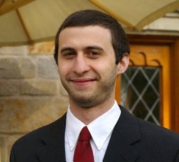 DeSalvo Matthew N合作论文数Harvard Medical School, Boston, MA 02115, USA. matthew_desalvo@hms.harvard.edu16Abhijeet Gummadavelli合作论文数Cincinnati Children's Hospital Medical Center, Cincinnati Children's Hospital Medical Center15
 Jason Gerrard合作论文数Semmes Murphey Clinic & UTenn Health Sciences Center14
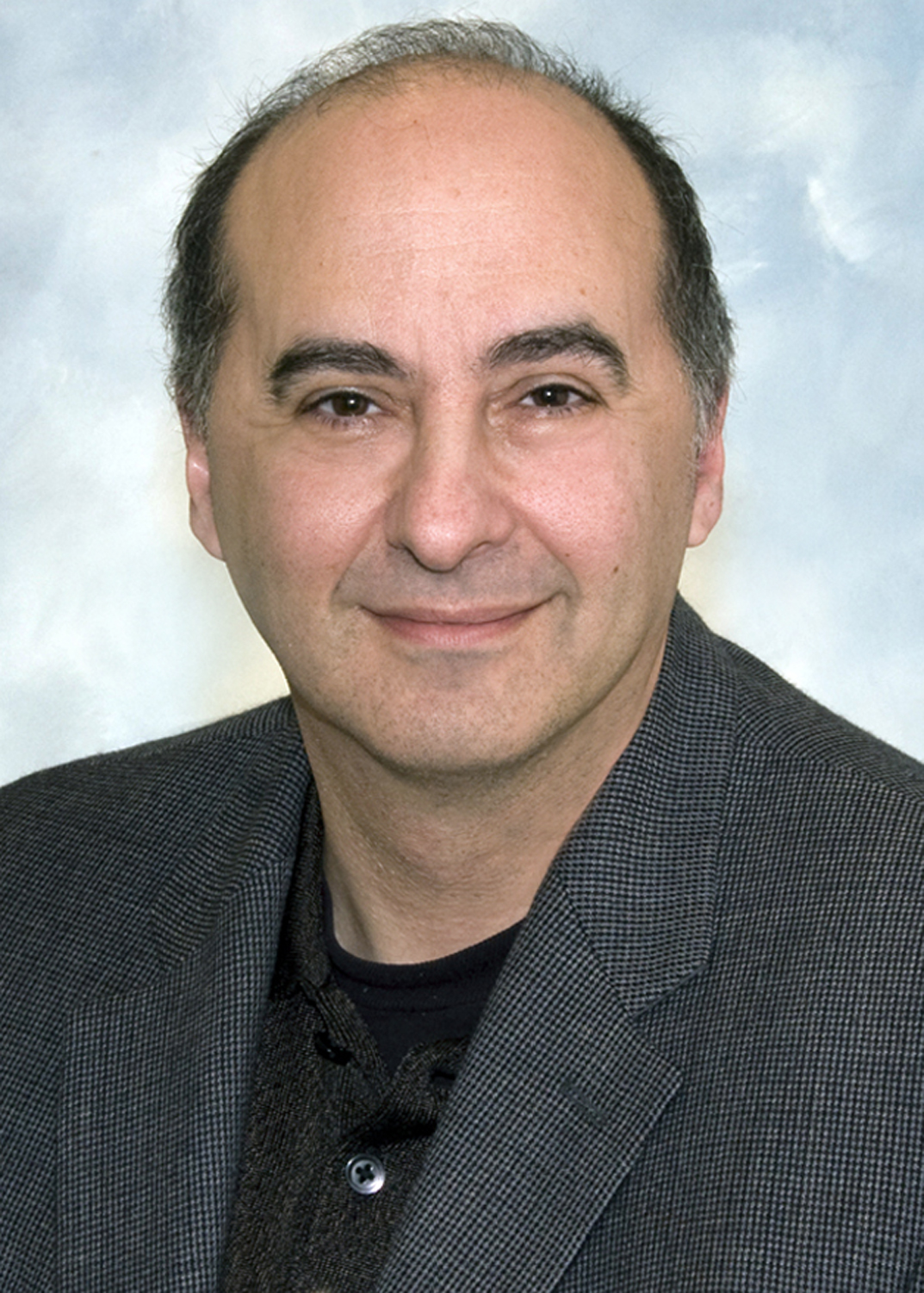 Douglas Rothman合作论文数Magnetic Resonance Research Center, Yale School of Medicine14Michael J. Purcaro合作论文数Department of Neurology, Yale University School of Medicine14
 Jun Hwan Ryu合作论文数Department of Neurology, Yale School of Medicine13
 Pue Farooque合作论文数Department of Neurology, Yale School of Medicine, Yale University13
 McCafferty Cian P合作论文数13
 I. George Zubal合作论文数National Institutes of Health;Z-Concepts LLC13
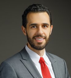 Kronemer Sharif I合作论文数David O. Robbins Neuroscience Program;Ohio Wesleyan University;David O. Robbins Neuroscience Program, Ohio Wesleyan University12
 Xiaoxiao Bai合作论文数12M. Negishi合作论文数11
 William (Chien-Wen) Chen合作论文数Departments of Radiation Oncology and Neurological Surgery, University of California San Francisco11
 Susan Spencer合作论文数Department of Neurology, Yale School of Medicine;Department of Neurosurgery, Yale School of Medicine11Nathan Danielson合作论文数Dept Neurosci, Columbia Univ10
 Michiro Negishi合作论文数Neuroverb, LLC10
 Christison-Lagay Kate L合作论文数Perelman School of Medicine, University of Pennsylvania9
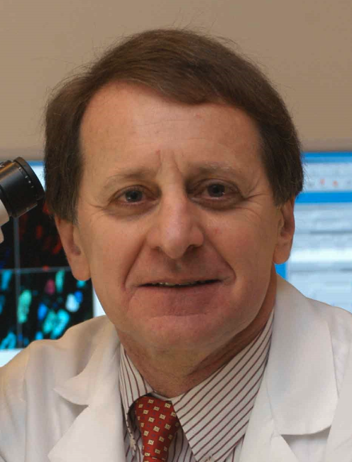 Stephen Waxman合作论文数Department of Neurology, School of Medicine, Yale University;Department of Neuroscience, School of Medicine, Yale University;OliPass BioTherapeutics9
 Robert Duckrow合作论文数9
 Matthew Vestal合作论文数9KA McNally合作论文数8Jennifer Guo合作论文数NIH, NIH Undiagnosed Dis Program8
 Michael Crowley合作论文数Child Study Center, Yale School of Medicine, Yale University8
 Marisa N. Spann合作论文数School of Medicine, Yale University8
 Rafeed Alkawadri合作论文数Department of Neurology, Yale Comprehensive Epilepsy Center8
 Andrews John合作论文数Department of Surgery, Columbia University Medical Center8
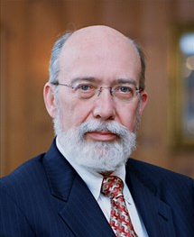 Dennis D. Spencer合作论文数Department of Neurosurgery8Ulrich Schridde合作论文数8Damien J. Ellens合作论文数7R. Berman合作论文数7Peter Vincent合作论文数Department of Neurology, Yale School of Medicine7
 Adithya Sivaraju合作论文数Department of Neurology, Yale School of Medicine, Yale University7
 Christopher Benjamin合作论文数Department of Neurology, Yale School of Medicine, Yale University7
 Xenophon Papademetris合作论文数Department of Biomedical Informatics and Data Science, School of Medicine, Yale University;Department of Radiology and Biomedical Imaging, School of Medicine, Yale University7
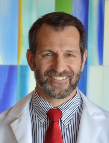 Joseph T. Giacino合作论文数Center for Bioethics, Harvard Medical School;Department of Physical Medicine and Rehabilitation, Harvard Medical School;Neurorehabilitation Laboratory, Spaulding Rehabilitation Hospital;Department of Psychiatry, Massachusetts General Hospital;Spaulding Rehabilitation Outcomes Center7Quraishi Imran H合作论文数Department of Bioengineering, Rice University7
 Hunki Kwon合作论文数Harvard Medical School;Department of Neurology, Massachusetts General Hospital7




