Cryo-electron tomography provides 3D images of macromolecules in their cellular context. To detect macromolecules in tomograms, template matching (TM) is often used, which uses 3D models that are often reliable for substantial parts of the macromolecules. However, the extent of rotational searches in particle detection has not been investigated due to computational limitations. Here, we provide a GPU implementation of TM as part of the PyTOM software package, which drastically speeds up the orientational search and allows for sampling beyond the Crowther criterion within a feasible timeframe. We quantify the improvements in sensitivity and false-discovery rate for the examples of ribosome identification and detection. Sampling at the Crowther criterion, which was effectively impossible with CPU implementations due to the extensive computation times, allows for automated extraction with high sensitivity. Consequently, we also show that an extensive angular sample renders 3D TM sensitive to the local alignment of tilt series and damage induced by focused ion beam milling. With this new release of PyTOM, we focused on integration with other software packages that support more refined subtomogram-averaging workflows. The automated classification of ribosomes by TM with appropriate angular sampling on locally corrected tomograms has a sufficiently low false-discovery rate, allowing for it to be directly used for high-resolution averaging and adequate sensitivity to reveal polysome organization.
Cryo-electron tomography (cryo-ET) is an imaging technique that allows three-dimensional visualization of macro-molecular assemblies under near-native conditions. Cryo-ET comes with a number of challenges, mainly low signal-to-noise and inability to obtain images from all angles. Computational methods are key to analyze cryo-electron tomograms. To promote innovation in computational methods, we generate a novel simulated dataset to benchmark different methods of localization and classification of biological macromolecules in tomograms. Our publicly available dataset contains ten tomographic reconstructions of simulated cell-like volumes. Each volume contains twelve different types of complexes, varying in size, function and structure. In this paper, we have evaluated seven different methods of finding and classifying proteins. Seven research groups present results obtained with learning-based methods and trained on the simulated dataset, as well as a baseline template matching (TM), a traditional method widely used in cryo-ET research. We show that learning-based approaches can achieve notably better localization and classification performance than TM. We also experimentally confirm that there is a negative relationship between particle size and performance for all methods.
Cryo-electron tomography (cryo-ET) is an imaging technique that allows us to three-dimensionally visualize both the structural details of macro-molecular assemblies under near-native conditions and its cellular context. Electrons strongly interact with biological samples, limiting electron dose. The latter limits the signal-to-noise ratio and hence resolution of an individual tomogram to about 50 (5 nm). Biological molecules can be obtained by averaging volumes, each depicting copies of the molecule, allowing for resolutions beyond 4 (0.4 nm). To this end, the ability to localize and classify components is crucial, but challenging due to the low signal-to-noise ratio. Computational innovation is key to mine biological information from cryo-electron tomography. To promote such innovation, we provide a novel simulated dataset to benchmark different methods of localization and classification of biological macromolecules in cryo-electron tomograms. Our publicly available dataset contains ten tomographic reconstructions of simulated cell-like volumes. Each volume contains twelve different types of complexes, varying in size, function and structure. In this paper, we have evaluated seven different methods of finding and classifying proteins. Six research groups present results obtained with learning-based methods and trained on the simulated dataset, as well as a baseline template matching, a traditional method widely used in cryo-ET research. We find that method performance correlates with particle size, especially noticeable for template matching which performance degrades rapidly as the size decreases. We learn that neural networks can achieve significantly better localization and classification performance, in particular convolutional networks with focus on high-resolution details such as those based on U-Net architecture. (C) 2020 Elsevier Ltd. All rights reserved.
We study the structural dynamics of liquid water by time-resolved anisotropic x-ray scattering under the optical Kerr effect condition. In this way, we can separate the anisotropic scattering decay of 160 fs from the delayed temperature increase of similar to 0.1 K occurring at 1 ps and quantify transient changes in the O-O pair distribution function. Polarizable molecular dynamics simulations reproduce well the experiment, indicating transient alignment of molecules along the electric field, which shortens the nearest-neighbor distances. In addition, analysis of the simulated water local structure provides evidence that two hypothesized fluctuating water configurations exhibit different polarizability.
Different imaging techniques allow us to study the organization of life at different scales. Cryo-electron tomography (cryo-ET) has the ability to three-dimensionally visualize the cellular architecture as well as the structural details of macro-molecular assemblies under near-native conditions. Due to beam sensitivity of biological samples, an inidividual tomogram has a maximal resolution of 5 nanometers. By averaging volumes, each depicting copies of the same type of a molecule, resolutions beyond 4 A have been achieved. Key in this process is the ability to localize and classify the components of interest, which is challenging due to the low signal-to-noise ratio. Innovation in computational methods remains key to mine biological information from the tomograms. To promote such innovation, we organize this SHREC track and provide a simulated dataset with the goal of establishing a benchmark in localization and classification of biological particles in cryo-electron tomograms. The publicly available dataset contains ten reconstructed tomograms obtained from a simulated cell-like volume. Each volume contains twelve different types of proteins, varying in size and structure. Participants had access to 9 out of 10 of the cell-like ground-truth volumes for learning-based methods, and had to predict protein class and location in the test tomogram. Five groups submitted eight sets of results, using seven different methods. While our sample size gives only an anecdotal overview of current approaches in cryo-ET classification, we believe it shows trends and highlights interesting future work areas. The results show that learning-based approaches is the current trend in cryo-ET classification research and specifically end-to-end 3D learning-based approaches achieve the best performance.
Authors: Tais Gorkhover, Anatoli Ulmer, Ken Ferguson, Max Bucher, Filipe R. N. C. Maia, Johan Bielecki, Tomas Ekeberg, Max F. Hantke, Benedikt J. Daurer, Carl Nettelblad, Jakob Andreasson, Anton Barty, Petr Bruza, Sebastian Carron, Dirk Hasse, Jacek Krzywinski, Daniel S. D. Larsson, Andrew Morgan, Kerstin Mühlig, Kenta Okamoto, Alberto Pietrini, Daniela Rupp, Mario Sauppe, Gijs van der Schot, Marcin Siebert, Jonas A. Selberg, Martin Svenda, Michelle Swiggers, Nicusor Timneanu, Daniel Westphal, Garth Williams, Alessando Zani, Henry N. Chapman, Gyula Faigel, Thomas Möller, Janos Hajdu and Christoph Bostedt
Different imaging techniques allow us to study the organization of life at different scales. Cryo-electron tomography (cryo-ET) has the ability to three-dimensionally visualize the cellular architecture as well as the structural details of macro-molecular assemblies under near-native conditions. Due to beam sensitivity of biological samples, an inidividual tomogram has a maximal resolution of 5 nanometers. By averaging volumes, each depicting copies of the same type of a molecule, resolutions beyond 4 Å have been achieved. Key in this process is the ability to localize and classify the components of interest, which is challenging due to the low signal-to-noise ratio. Innovation in computational methods remains key to mine biological information from the tomograms. To promote such innovation, we organize this SHREC track and provide a simulated dataset with the goal of establishing a benchmark in localization and classification of biological particles in cryo-electron tomograms. The publicly available dataset contains ten reconstructed tomograms obtained from a simulated cell-like volume. Each volume contains twelve different types of proteins, varying in size and structure. Participants had access to 9 out of 10 of the cell-like ground-truth volumes for learning-based methods, and had to predict protein class and location in the test tomogram. Five groups submitted eight sets of results, using seven different methods. While our sample size gives only an anecdotal overview of current approaches in cryo-ET classification, we believe it shows trends and highlights interesting future work areas. The results show that learning-based approaches is the current trend in cryo-ET classification research and specifically end-to-end 3D learning-based approaches achieve the best performance. CCS Concepts •Information systems → Evaluation of retrieval results; Specialized information retrieval; Multimedia and multimodal retrieval; Retrieval models and ranking;
Corrections to the article by Daurer et al.[IUCrJ (2017). 4, 251–262] are given.
The aim of this tutorial is to introduce you to the computational workflow in cryo-electron tomography (Cryo-ET). Cryo-ET is an imaging technique used to obtain high-resolution threedimensional images of biological objects such as macromolecules and cells in their near native environment. Samples are tilted as they are imaged, resulting is a set of 2D images (tilt series), that can be combined to form a three-dimensional (3D) reconstruction. In Cryo-ET samples are immobilized in non-crystalline ice (imaged at temperatures below -150 °C) allowing them to be imaged without dehydration or chemical fixation, processes which could disrupt or distort biological structures.
The possibility of imaging single proteins constitutes an exciting challenge for x-ray lasers. Despite encouraging results on large particles, imaging small particles has proven to be difficult for two reasons: not quite high enough pulse intensity from currently available x-ray lasers and, as we demonstrate here, contamination of the aerosolized molecules by nonvolatile contaminants in the solution. The amount of contamination on the sample depends on the initial droplet size during aerosolization. Here, we show that, with our electrospray injector, we can decrease the size of aerosol droplets and demonstrate virtually contaminant-free sample delivery of organelles, small virions, and proteins. The results presented here, together with the increased performance of next-generation x-ray lasers, constitute an important stepping stone toward the ultimate goal of protein structure determination from imaging at room temperature and high temporal resolution.
Current Flash X-ray single-particle diffraction Imaging (FXI) experiments, which operate on modern X-ray Free Electron Lasers (XFELs), can record millions of interpretable diffraction patterns from individual biomolecules per day. Due to the practical limitations with the FXI technology, those patterns will to a varying degree include scatterings from contaminated samples. Also, the heterogeneity of the sample biomolecules is unavoidable and complicates data processing. Reducing the data volumes and selecting high-quality single-molecule patterns are therefore critical steps in the experimental setup. In this paper, we present two supervised template-based learning methods for classifying FXI patterns. Our Eigen-Image and Log-Likelihood classifier can find the best-matched template for a single-molecule pattern within a few milliseconds. It is also straightforward to parallelize them so as to match the XFEL repetition rate fully, thereby enabling processing at site. The methods perform in a stable way on various kinds of synthetic data. As a practical example we tested our methods on a real mimivirus dataset, obtaining a convincing classification accuracy of 0.9.
Ultrafast X-ray imaging on individual fragile specimens such as aerosols 1 , metastable particles 2 , superfluid quantum systems 3 and live biospecimens 4 provides high-resolution information that is inaccessible with conventional imaging techniques. Coherent X-ray diffractive imaging, however, suffers from intrinsic loss of phase, and therefore structure recovery is often complicated and not always uniquely defined 4 , 5 . Here, we introduce the method of in-flight holography, where we use nanoclusters as reference X-ray scatterers to encode relative phase information into diffraction patterns of a virus. The resulting hologram contains an unambiguous three-dimensional map of a virus and two nanoclusters with the highest lateral resolution so far achieved via single shot X-ray holography. Our approach unlocks the benefits of holography for ultrafast X-ray imaging of nanoscale, non-periodic systems and paves the way to direct observation of complex electron dynamics down to the attosecond timescale.
Diffraction before destruction using X-ray free-electron lasers (XFELs) has the potential to determine radiation-damage-free structures without the need for crystallization. This article presents the three-dimensional reconstruction of the Melbournevirus from single-particle X-ray diffraction patterns collected at the LINAC Coherent Light Source (LCLS) as well as reconstructions from simulated data exploring the consequences of different kinds of experimental sources of noise. The reconstruction from experimental data suffers from a strong artifact in the center of the particle. This could be reproduced with simulated data by adding experimental background to the diffraction patterns. In those simulations, the relative density of the artifact increases linearly with background strength. This suggests that the artifact originates from the Fourier transform of the relatively flat background, concentrating all power in a central feature of limited extent. We support these findings by significantly reducing the artifact through background removal before the phase-retrieval step. Large amounts of blurring in the diffraction patterns were also found to introduce diffuse artifacts, which could easily be mistaken as biologically relevant features. Other sources of noise such as sample heterogeneity and variation of pulse energy did not significantly degrade the quality of the reconstructions. Larger data volumes, made possible by the recent inauguration of high repetition-rate XFELs, allow for increased signal-to-background ratio and provide a way to minimize these artifacts. The anticipated development of three-dimensional Fourier-volume-assembly algorithms which are background aware is an alternative and complementary solution, which maximizes the use of data.
Imaging live cells at a resolution higher than achieved using optical microscopy is a challenge. Ultra-fast coherent diffractive imaging with X-ray free-electron lasers (XFELs) has the potential to achieve sub-nanometer resolution on micron-sized living cells. Our container-free injection method can introduce a beam of live cyanobacteria into the micron-sized focus of the Linac Coherent Light Source (LCLS) to record of diffraction patterns from individual cells, with very low noise at high hit rates (millions of cells/day). We used iterative phase retrieval to derive two-dimensional projection images directly from the diffraction patterns. In a first experiment, we collected diffraction patterns to 33-46 nm full-period resolution, and reconstructed the exit wave front to 76 nm full-period resolution. In a second experiment, we demonstrate that it is indeed possible to record diffraction data to nanometer resolution on live cells with an intense, ultra-short X-ray pulse as predicted earlier. These results are encouraging, and future developments to the XFELs and improvements to the X-ray area-detectors will bring sub-nanometer resolution reconstructions of living cells within reach. Utilizing this type of diffraction data will require the development of new analysis methods and algorithms for studying structure and structural variability in large populations of cells and to create abstract models. Such studies will allow us to understand living cells and populations of cells in new ways.
Single-particle diffraction from X-ray Free Electron Lasers offers the potential for molecular structure determination without the need for crystallization. In an effort to further develop the technique, we present a dataset of coherent soft X-ray diffraction images of Coliphage PR772 virus, collected at the Atomic Molecular Optics (AMO) beamline with pnCCD detectors in the LAMP instrument at the Linac Coherent Light Source. The diameter of PR772 ranges from 65–70 nm, which is considerably smaller than the previously reported ~600 nm diameter Mimivirus. This reflects continued progress in XFEL-based singleparticle imaging towards the single molecular imaging regime. The data set contains significantly more single particle hits than collected in previous experiments, enabling the development of improved statistical analysis, reconstruction algorithms, and quantitative metrics to determine resolution and self-consistency.
We use extremely bright and ultrashort pulses from an x-ray free-electron laser (XFEL) to measure correlations in x rays scattered from individual bioparticles. This allows us to go beyond the traditional crystallography and single-particle imaging approaches for structure investigations. We employ angular correlations to recover the three-dimensional (3D) structure of nanoscale viruses from x-ray diffraction data measured at the Linac Coherent Light Source. Correlations provide us with a comprehensive structural fingerprint of a 3D virus, which we use both for model-based and ab initio structure recovery. The analyses reveal a clear indication that the structure of the viruses deviates from the expected perfect icosahedral symmetry. Our results anticipate exciting opportunities for XFEL studies of the structure and dynamics of nanoscale objects by means of angular correlations.
This study explores the capabilities of the Coherent X-ray Imaging Instrument at the Linac Coherent Light Source to image small biological samples. The weak signal from small samples puts a significant demand on the experiment. Aerosolized Omono River virus particles of ∼40 nm in diameter were injected into the submicrometre X-ray focus at a reduced pressure. Diffraction patterns were recorded on two area detectors. The statistical nature of the measurements from many individual particles provided information about the intensity profile of the X-ray beam, phase variations in the wavefront and the size distribution of the injected particles. The results point to a wider than expected size distribution (from ∼35 to ∼300 nm in diameter). This is likely to be owing to nonvolatile contaminants from larger droplets during aerosolization and droplet evaporation. The results suggest that the concentration of nonvolatile contaminants and the ratio between the volumes of the initial droplet and the sample particles is critical in such studies. The maximum beam intensity in the focus was found to be 1.9 × 10 12 photons per µm 2 per pulse. The full-width of the focus at half-maximum was estimated to be 500 nm (assuming 20% beamline transmission), and this width is larger than expected. Under these conditions, the diffraction signal from a sample-sized particle remained above the average background to a resolution of 4.25 nm. The results suggest that reducing the size of the initial droplets during aerosolization is necessary to bring small particles into the scope of detailed structural studies with X-ray lasers.
Single particle diffractive imaging data from Rice Dwarf Virus (RDV) were recorded using the Coherent X-ray Imaging (CXI) instrument at the Linac Coherent Light Source (LCLS). RDV was chosen as it is a well-characterized model system, useful for proof-of-principle experiments, system optimization and algorithm development. RDV, an icosahedral virus of about 70 nm in diameter, was aerosolized and injected into the approximately 0.1 μm diameter focused hard X-ray beam at the CXI instrument of LCLS. Diffraction patterns from RDV with signal to 5.9 Ångström were recorded. The diffraction data are available through the Coherent X-ray Imaging Data Bank (CXIDB) as a resource for algorithm development, the contents of which are described here.
Gijs van der Schot, Martin Svenda, Filipe R.N.C. Maia, Max F. Hantke, Daniel P. DePonte, M. Marvin Seibert, Andrew Aquila, Joachim Schulz, Richard A. Kirian, Mengning Liang, Francesco Stellato, Sadia Bari, Bianca Iwan, Jakob Andreasson, Nicusor Timneanu, Johan Bielecki, Daniel Westphal, Francisca Nunes de Almeida, Duško Odić, Dirk Hasse, Gunilla H. Carlsson, Daniel S.D. Larsson, Anton Barty, Andrew V. Martin, Sebastian Schorb, Christoph Bostedt, John D. Bozek, Sebastian Carron, Ken Ferguson, Daniel Rolles, Artem Rudenko, Sascha W. Epp, Lutz Foucar, Benedikt Rudek, Benjamin Erk, Robert Hartmann, Nils Kimmel, Peter Holl, Lars Englert, N. Duane Loh, Henry N. Chapman, Inger Andersson, Janos Hajdu & Tomas Ekeberg
Structural studies on living cells by conventional methods are limited to low resolution because radiation damage kills cells long before the necessary dose for high resolution can be delivered. X-ray free-electron lasers circumvent this problem by outrunning key damage processes with an ultra-short and extremely bright coherent X-ray pulse. Diffraction-before-destruction experiments provide high-resolution data from cells that are alive when the femtosecond X-ray pulse traverses the sample. This paper presents two data sets from micron-sized cyanobacteria obtained at the Linac Coherent Light Source, containing a total of 199,000 diffraction patterns. Utilizing this type of diffraction data will require the development of new analysis methods and algorithms for studying structure and structural variability in large populations of cells and to create abstract models. Such studies will allow us to understand living cells and populations of cells in new ways. New X-ray lasers, like the European XFEL, will produce billions of pulses per day, and could open new areas in structural sciences.
 Martin Svenda合作论文数Division of Biomedical and X-ray Physics, Department of Applied Physics, School of Engineering Sciences, KTH Royal Institute of Technology17
 filipe r n c maia合作论文数Uppsala Univ, Dept Cell & Mol Biol, Lab Mol Biophys, SE-75124 Uppsala, Sweden16
 Max Hantke合作论文数Laboratory of Molecular Biophysics, Uppsala University16
 Janos Hajdu合作论文数Department of Cell and Molecular Biology, Molecular Biophysics, Uppsala Universitet16M. Marvin Seibert合作论文数Department of Cell and Molecular Biology, Uppsala University15Ekeberg Tomas合作论文数14
 Anton Barty合作论文数Center for Data and Computing in Natural Sciences, Universitat Hamburg14
 Daniel Westphal合作论文数Laboratory of Molecular Biophysics, Uppsala University13Bielecki Johan合作论文数Lab Mol Biophys, Uppsala Univ13
 Duane Loh合作论文数Department of Physics, Faculty of Science, National University of Singapore13
 Daniel Larsson合作论文数Department of Cell and Molecular Biology, Uppsala University13
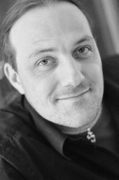 Jakob Andreasson合作论文数Uppsala Univ, Dept Cell & Mol Biol, Lab Mol Biophys, SE-75124 Uppsala, Sweden12
 Nicusor Timneanu合作论文数High Energy Physics;Uppsala University;Uppsala University12
 Henry Chapman合作论文数Coherent X-Ray Imaging Division, Center for Free-Electron Laser Science, Deutsches Elektronen-Synchrotron;University of Hamburg12
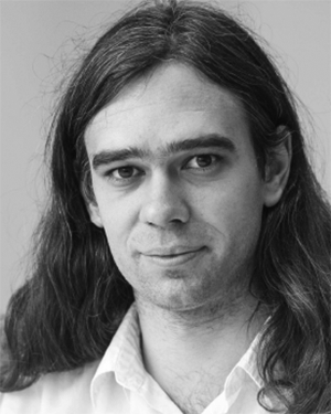 Carl Nettelblad合作论文数Uppsala University, University of Uppsala12
 Christoph Bostedt合作论文数Laboratory for Ultrafast X-ray Sciences, Ecole Polytechnique Federale de Lausanne;Laboratory for Synchrotron Radiation and Femtochemistry, Paul Scherrer Institut11
 Benedikt J. Daurer合作论文数Uppsala Univ, Dept Cell & Mol Biol, Lab Mol Biophys, Husargatan 3,Box 596, SE-75124 Uppsala, Sweden10Dirk Hasse合作论文数10Andersson Inger合作论文数10
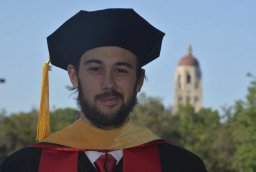 Ken R. Ferguson合作论文数Linac Coherent Light Source (LCLS), SLAC National Accelerator Laboratory, 2575 Sand Hill Road, Menlo Park, CA 94025, USA.10
 Jonas Sellberg合作论文数Division of Biomedical and X-ray Physics, Department of Applied Physics, School of Engineering Sciences, KTH Royal Institute of Technology10Mühlig Kerstin合作论文数10
 Richard Kirian合作论文数Center for Biological Physics, Arizona State University;Biodesign Center for Applied Structural Discovery, Arizona State University9
 Gunilla H Carlsson合作论文数8
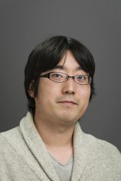 Okamoto Kenta合作论文数Department of Infectious Medicine, Kurume University School of Medicine;Institute of Tropical Medicine, Nagasaki University;Institute of Tropical Medicine, Nagasaki University;Fukuoka Tokushukai Medical Center;Laboratory of Hygienic Chemistry, Daiichi University of Pharmacy;Experimental Therapeutics Institute, Mount Sinai School of Medicine;Houston Methodist Research Institute, Weill Cornell Medical College;Nagasaki University, Kurume University School of Medicine8Garth J. Williams合作论文数Australian Research Council Centre of Excellence for Coherent X-Ray Science8
 Sebastian Carron Montero合作论文数California Lutheran University7Mengning Liang合作论文数7
 Bianca Iwan合作论文数Physics Department, Royal Institute of Technology7
 Artem Rudenko合作论文数Department of Physics, Kansas State University7
 Daniel Rolles合作论文数Department of Physics, College of Arts and Sciences, Kansas State University7Robert Hartmann合作论文数7s w epp合作论文数6
 Pietrini Alberto合作论文数6Andrew Aquila合作论文数SLAC Natl Accelerator Lab6Nils Kimmel合作论文数6
 F.G. (Friedrich) Förster合作论文数Situ Structural Biology Lab, Utrecht University;Bijvoet Center for Biomolecular Research, Utrecht University6Daniel P. DePonte合作论文数SLAC National Accelerator Laboratory6Hemanth K N Reddy合作论文数5
 Ilja Gubins合作论文数Corresponding author.5
 Anna Munke合作论文数Uppsala Univ, Dept Cell & Mol Biol, Lab Mol Biophys, Husargatan 3,Box 596, SE-75124 Uppsala, Sweden5
 Salah Awel合作论文数DESY, Ctr Free Electron Laser Sci, Notkestr 85, D-22607 Hamburg, Germany5
 Francesco Stellato合作论文数Center for Free-Electron Laser Science (CFEL), DESY, Notkestrasse 85, 22607 Hamburg, Germany.5Sebastian Schorb合作论文数Linac Coherent Light Source (LCLS), SLAC National Accelerator Laboratory, 2575 Sand Hill Road, Menlo Park, CA 94025, USA.5
 R.C. (Remco) Veltkamp合作论文数Department of Game and Media Technology, Faculty of Science, Utrecht University5
 Andrew Martin合作论文数Division of Heat and Power Technology, Department of Energy Technology, School of Industrial Engineering and Management, KTH Royal Institute of Technology5
 Wim Vranken合作论文数ULB VUB, Interuniv Inst Bioinformat Brussels5
 Alexandre Bonvin合作论文数Bijvoet Center for Biomolecular Research, Utrecht University;Faculty of Science, Utrecht University;Faculty of Chemistry, Utrecht University5J. Krzywinski合作论文数Institute of Physics, the Polish Academy of Sciences5




