Current immunotherapies often fail in immunologically "cold", macrophage-rich tumor microenvironments (TMEs). Multi-targeting approaches that modulate innate-adaptive immune activation represent a promising frontier in cancer immunotherapy. Here, we developed phagocytic synapse enhancers (PSEs), a class of modular immune engagers comprising a high-affinity PD-L1 binder coupled to a macrophage-stimulating peptide, tuftsin. PSEs strengthened effector-target cell interactions and enhanced tumor phagocytosis by bridging tumor PD-L1 to macrophage neuropilin-1 (NRP1), bypassing the classical FcγR-dependent pathways. The PSEs also acted in cis on PD-L1+ macrophages, accelerating the endocytosis and lysosomal compartmentalization of surface PD-L1, therefore stripping the immunosuppressive checkpoint from the local microenvironment. Beyond physical clearance, PSEs reprogrammed macrophage phenotype and triggered a robust pro-inflammatory cytokine and chemokine response. The lead molecule, longPSE, and its half-life-extended variant fused to the albumin binding domain, ABD- longPSE, showcased superior efficacy than the macrophage enhancer magrolimab in a syngeneic tumor model of colorectal cancer and an orthotopic model of pancreatic cancer. PSE treatment remodeled the TME by inducing phenotypic changes in the lymphoid and myeloid compartments, together with a reduction of tumor-associated macrophages and regulatory T cells. These findings establish PSEs as bifunctional molecules that complement innate and adaptive immune modulation. The bifunctional design offers a versatile approach for next-generation immunotherapies and provides a blueprint for a plug-and-play platform of immune engagers targeting diverse cancer-associated pathways.
Glioblastoma (GBM) is the most aggressive primary brain tumour and is frequently accompanied by severe neurological symptoms, including epilepsy and cognitive impairment. Neurological symptoms often persist after surgical resection, indicating that GBM induces durable and self-sustaining changes in the surrounding neuronal networks. However, the mechanisms by which GBM reshapes network structure and function in the tumour periphery remain poorly understood. We present a compartmentalised in vitro platform enabling long-term coculture of iPSC-derived neurons and primary GBM cells to investigate these changes. Placed on high-density microelectrode arrays, the platform permits longitudinal electrophysiological recordings at single-neuron resolution. Using effective network inference, we find that GBM drives a reproducible structural progression: first toward a hyperconnected, hub-dominated architecture, then a collapse of community structure accompanied by a widespread neuron loss. This evolving structure shapes population dynamics, constraining features such as network burst rate and instantaneous synchrony. The reorganisation also carries computational consequences: signal propagation becomes progressively redundant and synergistic rather than unique. As a result, neurons lose the capacity to encode distinct input combinations independently, and the repertoire of accessible network states contracts. Together, these findings reframe GBM as a driver of neuronal network reorganisation rather than uniform hyperexcitability, and establish a compartmentalised, single-neuron-resolution platform for the longitudinal observation, dissection, and ultimately targeting of the network processes that underlie disease progression.
Glioblastoma remains one of the most aggressive primary brain tumors in adults, with a survival rarely exceeding 15 months despite multimodal therapy. Novel immunotherapeutic strategies, particularly chimeric antigen receptor T-cell therapy, have emerged as promising approaches to overcome the limitations of conventional treatments. This review summarizes recent early-phase clinical trials investigating locoregional chimeric antigen receptor T-cell delivery in recurrent glioblastoma and highlights key considerations for multidisciplinary neuro-oncology teams involved in this evolving therapeutic paradigm. Phase I studies of intratumoral, intracavitary, intraventricular, or combined delivery routes have demonstrated technical feasibility and safety, with most adverse events being manageable. Dual-route delivery may enhance chimeric antigen receptor T-cell distribution and produce early radiographic and clinical responses in selected patients. However, therapeutic durability remains limited by tumor heterogeneity, antigen loss, and the immunosuppressive tumor microenvironment. Multidisciplinary care teams play a critical role in catheter and reservoir placement, infusion planning, and management of neuroinflammatory toxicities. Although current findings are preliminary, ongoing optimization of target selection, dosing strategies, and combination therapies may expand treatment options for recurrent glioblastoma and further integrate immunotherapy into contemporary neuro-oncology care.
Microglia are regarded as self-maintaining brain parenchymal macrophages without contribution from adult hematopoiesis. While peripheral macrophage engraftment into the brain has been reported, the biological variables governing central nervous system (CNS) macrophage niche access remain unclear. We show that CNS macrophage engraftment depends on (1) the balance between tissue-resident macrophage (TRM) self-renewal, (2) the temporal alignment of niche opening with the availability of engraftment-competent cells and (3) proximity to the vacated niche, rather than prolonged niche vacancy. Fate mapping revealed that monocytes entering the subdural space undergo border-associated macrophage (BAM)-like differentiation, clonal expansion, and transpial migration into the parenchyma, whereas mature BAMs directly repopulate selectively vacated parenchymal niches. We further identified a parenchymal macrophage population with a peripheral BAM-like transcriptional program in aged and neurodegenerative human brains, challenging the dogma of MG-exclusivity. Together, these discoveries provide a predictive framework for understanding macrophage maintenance and replacement, and for developing macrophage-based cellular therapies.
Although direct biological factors underlying the progression of Glioblastoma (GBM), an aggressive form of brain cancer, have been extensively studied, emerging evidence suggests that indirect biological triggers, such as traumatic brain injury (TBI), may also have a role. Since proteoglycans, secreted by reactive astrocytes and astroglial cells contribute to biophysical characteristics (stochastic topography, stiffness) of the brain, we postulated a role for stochastic nanoroughness in the induction of glioma following brain trauma. Using a model system to emulate such physical cues that manifest following traumatic injury, we demonstrate that human cortical astrocytes undergo spontaneous organization into spheroids in response to nanoroughness and retain the spheroid phenotype even upon withdrawal of the physical cues. Furthermore, spheroids serve as aggregation foci for naïve astrocytes, express activated MMP2, and disseminate upon implantation in the mouse brain. RNA-seq analysis revealed that astrocytes within spheroids differentially express genes, including p53, ADAMTS proteases, and NOTCH3, and adopt a transcriptional program enriched for GBM proneural signatures, with reactome analysis pointing toward astrocytes with GBM-associated transcriptional traits. Moreover, nanoroughness mediates a cross-talk between cancer cells and astrocytes through induced senescence. These findings implicate a role for stochastic biophysical cues in driving a potential malignant transformation of astrocytes.
Chimeric antigen receptor (CAR) T-cell therapy has demonstrated remarkable efficacy in hematological malignancies, but its success in solid tumors remains limited. Conventional CAR-T designs do not integrate into the T-cell receptor (TCR) and rely on non-natural CD3ζ signaling. They thus often suffer from tonic signaling, rapid T-cell exhaustion, and antigen escape due to reduced sensitivity. To address these limitations, we explored alternative CAR architectures that take into account the evolutionary optimized TCR signaling machinery. Specifically, we employed T-cell receptor fusion constructs (TRuCs), where a single-chain variable fragment (scFv) targeting the Sialyl-Thomson-Nouveau antigen (STn) is attached to the CD3ε subunit. We then systematically screened a library of costimulatory domains and their combinations, fused to the TRuC, using a novel in vitro and in vivo screening approach. This screen identified a potent TRuC variant incorporating both CD28 and CD27 costimulatory domains. This optimized construct exhibited increased in vitro and in vivo proliferation and enhanced 4-1BB and IFNγ expression upon repeated antigen stimulation. It further showed improved antitumor efficacy compared to conventional second-generation CAR-T cells in a mouse melanoma model. Moreover, we validated the adaptability of this approach by targeting B7H3 in a sarcoma model. The TRuC with costimulatory domains again outperformed other CAR-variants. Our findings highlight the potential of incorporating combined CD28 and CD27 costimulatory domains into TCR-based CAR architectures to overcome limitations associated with conventional CAR-T therapy and improve efficacy against solid tumors. ### Competing Interest Statement A. Zingg reports grants from Bristol Myers Squibb during the conduct of the study. H. Laubli reports grants from Fondaction and Bristol Myers Squibb during the conduct of the study, as well as grants and nonfinancial support from Bristol Myers Squibb, nonfinancial support from Merck Sharp Dohme, grants and personal fees from GlycoEra, and grants from Palleon Pharmaceuticals outside the submitted work. No disclosures were reported by the other authors. H. Laubli is a co-founder of Glycocalyx Therapeutics and OmnilinX Therapeutics. Swiss National Science Foundation, https://ror.org/00yjd3n13, 310030_184720/1
Expression of inhibitory Siglec receptors has been described on T cells of patients with viral infection and cancer. However, the exact function and their role in T-cell activation and control of infections have not been well investigated. Here, we show that Siglec-9 levels increase on T cells in patients with acute severe acute respiratory syndrome coronavirus 2 infection relative to levels on healthy controls. T cells of these patients are inhibited by Siglec-9, and specific blocking with antibodies released this inhibition. To further test the role of Siglec-9 in acute viral infections, we characterized a murine model with T-cell intrinsic Siglec-9 overexpression. We found that Siglec-9 restricted T-cell immunity after acute infection with lymphocytic choriomeningitis virus, thereby dampening T-cell-mediated immune pathology. These results show that inhibitory Siglec receptors including Siglec-9 T cell intrinsically modulate and fine-tune antiviral T-cell activation, proliferation, and effector function.
Immunotherapy profoundly impacted cancer treatments by harnessing the patient’s immune system. Phagocytosis, the process whereby immune cells engulf and destroy foreign particles or cells, plays a critical role in tumour cell clearance. Herein, we introduce a novel concept termed “ENPHASYS” – Enhancement of Phagocytic Synapses – designed to direct and amplify phagocytosis of cancer cells using heterobifunctional molecules named phagocytic synapse enhancers (PSEs). By engineering a de novo PD-L1 binder linked to a natural phagocytosis promoting peptide, tuftsin, the resulting PSE combines PD-L1 blockade with enhanced tumour cell phagocytosis; in addition, the PSEs induce macrophages to internalize a membrane or extracellular target. Intratumoural treatment of colorectal carcinoma- or glioblastoma-burdened immunocompetent animals resulted in beneficial overall survival, delayed tumour growth and a potent antitumor response driven by T-cell activation and TAM reprogramming, underpinning the translational relevance of ENPHASYS.
Purpose: Proteomics of glioma have not yet provided biomarkers and pathways that would clearly discriminate glioma subgroups. Methods: 82 glioma biopsies were prospectively collected and classified into six subgroups defined by methylomic classification: two low-grade glioma (LGG) and four high-grade glioma (HGG) subgroups. Proteins were extracted and processed for liquid chromatography-mass spectrometry (LC-MS). Differentially expressed proteins (DEPs) between subgroups were annotated, and functional validation was performed using inhibitor response assays in subtype-positive, patient-derived glioblastoma single-cell suspensions. Results: 5057 proteins were quantified for each sample. Tumor grading and IDH mutation status were the strongest discriminators for differential expression patterns. The glioblastoma IDH-wildtype subgroups showed diverse patterns of functions enriched with overexpressed DEPs: translation and cell cycle/telomere regulation in proneural glioblastoma (linked to cell proliferation), actin cytoskeleton, cell adhesion, and apoptosis regulation in classical glioblastoma (migration and invasion), and mitochondrial ATP synthesis in mesenchymal glioblastoma (metabolism). The most overexpressed proteins were correlated with survival and mRNA expression data. In vitro, inhibition of these proteins led to reduced cell viability that differed among subgroups, albeit in a small patient-derived exploratory cohort. Conclusion: This mainly descriptive study on proteomics in glioma provides insights into subgroup metabolism and potential biomarkers for further experimental testing.
Glioblastoma (GBM) is widely considered unresponsive to immunotherapy due to its immunosuppressive microenvironment and limited immune cell infiltration. However, recent single-cell transcriptomic studies reveal the presence of exhausted, potentially antigen-specific T cells in human GBM. We have developed a locally administered, brain-confined cytokine therapy with minimal systemic exposure using an engineered, neonatal Fc receptor (FcRn)-silenced, Compartment-Locked (CL) IL-12Fc fusion protein. This approach thus relies on presence and reactivation of tumor-resident, IL-12-responsive cells. In syngeneic, preclinical GBM models, intratumorally administered CL IL-12Fc induces robust antitumor efficacy, also under conditions of peripheral lymphopenia (induced via systemic temozolomide) or when T cell influx was pharmacologically inhibited. In 3D-perfused, patient-derived GBM explant cultures—with a largely preserved native tumor microenvironment and without addition of autologous peripheral immune cells—CL IL-12Fc triggered pro-inflammatory reprogramming. Proteomics-based secretome analysis revealed significant upregulation of interferon-gamma (IFNγ) pathway signatures. Single-cell RNA sequencing of GBM tissues prior to culture setup confirmed the presence of resident memory T cells in most patients. Notably, single cell transcriptome analysis of post-treatment explants demonstrated a robust IFNγ response in both tumor-associated macrophages, microglia and tumor cells. Our findings demonstrate that CL IL-12Fc can activate local immune responses within human GBM samples, independent of peripheral immune cell recruitment. This localized immune activation is an important prerequisite for the clinical translation of locally confined cytokine therapy. We aim to validate these findings using spatially resolved multiplex immunofluorescence histology. In summary we provide evidence that CL IL-12Fc can facilitate local immune activation without peripheral immune cell requirement.
Glioblastoma is the most common malignant primary brain tumor in adults and associated with a poor prognosis and a median survival of approximately 15 months, largely due to the lack of effective therapies and inevitable recurrence. A key challenge is the spatial and cellular heterogeneity, evident on MRI as a necrotic center, a contrast-enhancing tumor core, and a non-enhancing, T2/FLAIR-hyperintense infiltration zone. The diffuse infiltration of tumor cells into surrounding brain tissue impedes complete surgical resection and drives relapse from persisting residual cells. While regional molecular differences have been well characterized, their functional implications - particularly drug sensitivities - remain insufficiently understood. To address this, we performed MRI- and 5-aminolevulinic acid (5-ALA)-guided tissue sampling from the tumor core and from the infiltration zone of patients with glioblastoma. We utilized pharmacoscopy—a single-cell, microscopy-based drug screening platform—to evaluate region- and tumor cell type-specific drug responses. Cancer cell types were defined based on differential expression of nestin (marking stem-like cells) and S100B (marking more differentiated tumor cells). Profiling a library of 59 drugs across region-specific tumor samples from 23 patients revealed pronounced heterogeneity in drug responses across patients, tumor regions and tumor cell states. To overcome this heterogeneity, we computationally predicted complementary drug pairs and validated 20 drug combinations in an independent cohort. The combinations demonstrated enhanced efficacy compared to single agents and effectively addressed glioblastoma’s spatial, cellular, and inter-patient heterogeneity. Notably, drug combinations of oncology and neuroactive drugs outperformed those involving agents from the same drug class (oncology + oncology or neuroactive + neuroactive). Ongoing mechanistical explorations reveal first insights into mechanisms of the complementary drug efficacy. Our findings underscore the limitations of monotherapies for patients with glioblastoma and highlight the potential of rationally designed drug combinations tailored to the tumor’s spatial and functional complexity.
Glioblastoma remains a challenging indication for immunotherapy: the blood-brain barrier hampers accessibility for systemic treatments and the immunosuppressive microenvironment impedes immune attack. Intratumoral therapy with the proinflammatory cytokine interleukin-12 (IL-12) can revert immunosuppression but leakage into the circulation causes treatment-limiting toxicity. Here we engineer an IL-12Fc fusion cytokine with reduced binding to the neonatal Fc receptor FcRn. FcRn-silenced IL-12Fc avoids FcRn-mediated brain export, thus exhibits prolonged brain retention and reduced blood levels, which prevents toxicity. In murine glioblastoma, FcRn-silenced IL-12Fc induces more durable responses with negligible systemic cytokine exposure and boosts the efficacy of radio- and chemotherapy. It triggers anti-tumor responses independently of peripheral T cell influx or lymphopenia and leads to inflammatory polarization of the tumor microenvironment in patient-derived glioblastoma explants. FcRn-silencing of IL-12Fc may unlock the full potential of IL-12 for brain cancer therapy and could be further applied to containing the activity of other therapeutics targeting neurological diseases.
Glioblastoma (GBM) is a lethal brain tumor without effective treatment options. This study aimed to characterize longitudinal tumor changes in order to find potentially actionable targets to prevent GBM relapse. We extracted RNA and proteins from fresh frozen tumor samples from patient-matched IDHwt WHO grade 4 primary (pGBM) and recurrent (rGBM) tumors for transcriptomics and proteomics analysis. A tissue microarray containing paired tumor samples was processed for spatial transcriptomics analysis. Differentially expressed genes and proteins between pGBM and rGBM were involved in synapse development and myelination. By categorizing patients into short (STTR) and long (LTTR) time-to-lapse, we identified genes/proteins whose expression levels positively or negatively correlated with TTR. In rGBM, expressions of Fcγ receptors (FCGRs) and complement system genes were negatively correlated with TTR, whereas expression of genes involved in DNA methylation was positively correlated with TTR. Spatial transcriptomics of the tumor cells showed enrichment of oligodendrocytes in rGBM. Besides, we observed changes in the myeloid compartment such as a switch from quiescent to activated microglia and an enrichment in B and T cells in rGBM with STTR. Our results uncover a role for activated microglia/macrophages in GBM recurrence and suggest that interfering with these cells may hinder GBM relapse.
The treatment of solid tumors faces substantial hurdles because of inadequate drug delivery and the immunosuppressive tumor microenvironment. To address these challenges, we developed a therapeutic platform using macrophages loaded with ferritin-drug conjugates, referred to as macrophage-drug conjugates (MDC), and applied it to glioblastoma, an immunologically cold solid tumor. MDC loaded with ferritin-conjugated monomethyl auristatin E enabled efficient, cell contact-dependent transfer of the payload by a mechanism involving transfer of iron-binding proteins, from either mouse or human macrophages preferentially into glioma cells. This targeted delivery and therapeutic efficacy was demonstrated across in vitro coculture systems, ex vivo assays using dissociated glioblastoma patient tumor samples, and in vivo using orthotopic glioblastoma mouse models, all while maintaining a favorable preclinical safety profile evidenced by minimal systemic toxicity and localized drug biodistribution. Beyond direct tumor cell killing leading to significant tumor regression and prolonged survival in these models, MDC therapy reprogrammed the immunosuppressive tumor microenvironment. Immune profiling by spectral flow cytometry revealed enhanced infiltration and activation of cytotoxic T lymphocytes and B lymphocytes while reducing immunosuppressive regulatory T cells. This culminated in a robust, durable, T cell-dependent antitumor immune response, the necessity of which was confirmed through studies in immunodeficient mouse models and by lymphocyte depletion, and which conferred protection against tumor rechallenge. The combined cytotoxic and immunomodulatory effects highlight the potential of MDC therapy as a promising strategy for glioblastoma treatment and support its further clinical development.
Recent findings show that immune cells from the skull bone marrow and peripheral blood can migrate through the meninges and engraft as long-lived, monocyte-derived microglia—challenging traditional views of central nervous system (CNS) immune privilege. While the impact of such engraftment remains unclear, it opens therapeutic opportunities for microglial replacement, CAR-macrophages, or, if harmful, blockade of monocyte entry. Understanding the dynamics of the CNS macrophage niche is critical to guide these approaches. We assessed CNS macrophage repopulation across distinct depletion models (CSF1R inhibition, Cx3cr1CreERT2/wtCsf1rfl/fl, Sall1CreERT2/wtCsf1rfl/fl) using immunofluorescence and high-dimensional single-cell analyses (scRNA-seq, spectral flow cytometry) of dissected CNS compartments, and lineage tracing (ZMAN-FACS, skull bone marrow transplantation). Using a published scRNA-seq atlas, we derived and validated a high-dimensional FACS panel that distinguishes all CNS macrophage subsets in CNS macrophage reporter lines (Sall1GFP, Cx3cr1GFP,Pf4-Cre;Ai14). Pharmacological CSF1R inhibition depleted all subsets, including choroid plexus MHCII-high BAMs, but spared MHCII-high BAMs in the dura and leptomeninges, likely due to ongoing monocyte input from nearby skull bone marrow. Sall1CreERT2/wt; Ai14-lineage tracing showed that microglia repopulation post-BLZ945 originates from residual Sall1⁺ cells. In contrast, permanent CSF1R deletion (Cx3cr1CreERT2/wtCsf1rfl/fl) delayed repopulation and led to engraftment of CD49d⁺Clec12a⁺non-microglial macrophages. Microglia-selective CSF1R deletion (Sall1CreERT2/wtCsf1rfl/fl)) allowed rapid niche reconstitution by CSF1R-sufficient peripheral macrophages. scRNA-seq and flow cytometry identified repopulating cells with BAM-like signatures within the brain. Blood monocyte lineage tracing confirmed contribution, while skull marrow grafts did not contribute under rapid-repopulation conditions. Monocyte engraftment into the brain requires failed microglial self-renewal and the ontogeny is defined by the presence of CSF1R-sufficient cells at niche opening. In rapid, microglia-specific depletion, only blood and border-associated macrophages—not skull marrow monocytes—repopulate the niche, revealing tight spatial and temporal constraints on CNS access. These insights redefine the rules of monocyte engraftment in the brain.
Glioblastoma (GBM), the most common malignant primary brain tumor in adults, is resistant to Standard of Care (SOC) therapy and almost all available therapies. Clinical trials have not been successful in improving patients' survival, which underscores an urgent need for developing new effective therapies for this treatment-refractory disease. Considering the significant inter- and intratumoral heterogeneity existing in GBM, assessment of therapies in a personalized manner would allow for assigning patients to effective targeted therapies suitable for each patient's unique tumor signature.Patient-tailored ex vivo drug response platform for GBM allows for therapy planning in a personalized manner and enables the discovery of biomarkers of response. Advanced 3D methodologies have shown high efficacy in assessing the effect of therapeutics on intact tumor tissues immediately following tumor resection, allowing for studying the effect of therapies on tumor as a whole followed by detailed analysis of interactions between tumor cells and their microenvironment pre- and post-treatment.Here we describe the use of 3D perfusion bioreactors for translational assessment of therapeutics on regionally annotated patient-derived GBM explants. This model system provides the possibility of generating a 3D dynamic culture with continuous exposure and delivery of drug candidates to an intact tumor tissue material providing crucial insights on treatment-induced mechanisms and facilitating the identification of biomarkers of response, paving the way toward designing and generating more effective therapeutic interventions for GBM patients.
Abstract BACKGROUND Glioblastoma is the most common malignant primary brain tumor in adults with a poor survival and an unmet need for more effective therapies. Macroscopically, as observed through MR imaging and microscopy, these tumors are characterized by distinct tumor regions: a contrast-enhancing tumor core with a high cancer cell density and an infiltration zone that appears T2/FLAIR hyperintense without the uptake of contrast agent and consists of infiltrating tumor cells as well as non-malignant cells of the microenvironment. Previous genomic and transcriptomic studies revealed molecular differences between the tumor core and the infiltration zone. However, functional differences, such as drug sensitivity, between tumor regions are still largely unknown. MATERIAL AND METHODS To address this gap, we collected region-specific glioblastoma patient tissue samples from both the contrast-enhancing tumor core and the non-contrast-enhanced but 5-aminolevulinic acid (5-ALA) fluorescent infiltration zone directly from surgery. We utilized pharmacoscopy, a microscopy-based single-cell drug screening approach, to identify drug responses across regions and patients. RESULTS By profiling a library of 60 drugs across regions in a cohort of 23 patients, we identified region-specific drug responses and developed a complementarity score to pinpoint drug combinations capable of targeting both the tumor core and the infiltration zone. These combination treatments exhibited highly promising anti-tumor activity across regions and patients. CONCLUSION Given the high levels of functional spatial intra- and inter-tumor heterogeneity, relying on a single drug across patients may not be feasible for effective patient treatment. Thus, there is a strong rationale to administer drug combinations that not only consider regional heterogeneity but also account for patient-specific variations.
 Marie-Francoise Ritz合作论文数Neurosurgery Laboratory, Cantonal Hospital Basel34
 Tala Shekarian合作论文数22
 Tomas A. Martins合作论文数Brain Tumor Immunotherapy Lab, University of Basel20Philip Schmassmann合作论文数16
 Manina Maja Etter合作论文数12
 Sabrina Hogan合作论文数12
 Jean-Louis Boulay合作论文数12
 Nazanin Tatari合作论文数11
 Irving Weissman合作论文数Department of Developmental Biology, Stanford University;Department of Biology and Neurosurgery, Stanford University;Department of Pathology, Stanford University11
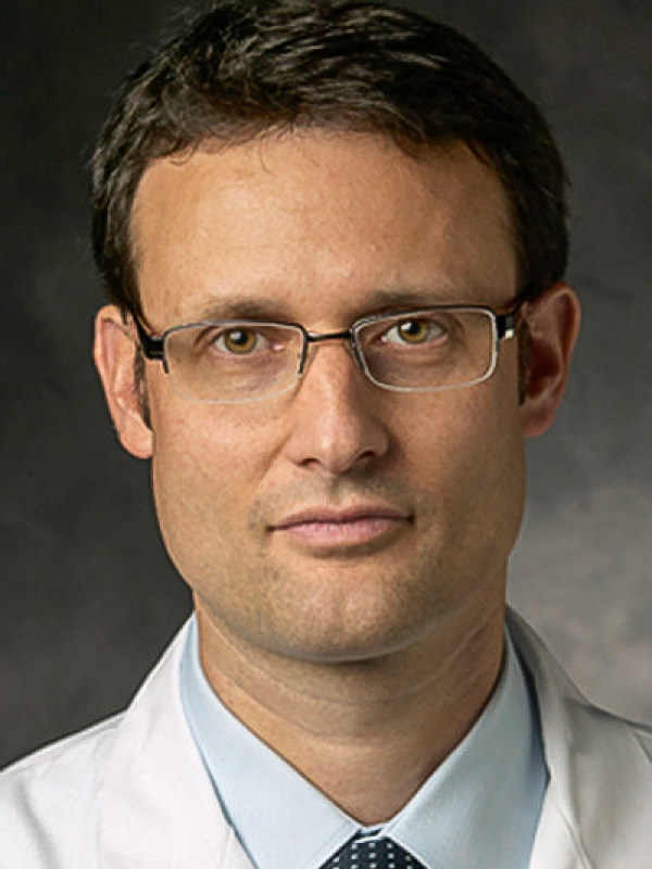 Raphael Guzman合作论文数Department of Neurosurgery, University Hospital Basel;Department of Biomedicine, Medizinische Fakultät, University of Basel10
 Michael Weller合作论文数Department of Neurology, University Hospital Zurich10
 Siddhartha Mitra合作论文数USGS10
 Heinz Läubli合作论文数Department of Biomedicine, University of Basel;University Hospital Basel9Deniz Kaymak合作论文数8Weiss Tobias合作论文数8Stephan Frank合作论文数Institute of Pathology, University Hospital Basel8
 Adriano Aguzzi合作论文数Institute of Neuropathology, University Hospital Zurich8
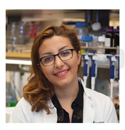 Sharareh Gholamin合作论文数Shahid Modarres Hospital, Shahid Beheshti University (MC),8
 Suzana Assad Kahn合作论文数Instituto de Ciências Biomédicas - INNT/INCT/MCT, Universidade Federal do Rio de Janeiro (UFRJ),8
 Berend Snijder合作论文数Department of Biology, Eidgenössische Technische Hochschule Zürich7
 Roux Julien合作论文数7vom Berg Johannes合作论文数Institute of Experimental Immunology, University of Zurich7
 Alexandra L Gerber合作论文数7
 Severina Leu合作论文数7Samuel Cheshier合作论文数Department of Health Services, California;Human Stem Cell Research7Alicia Buck合作论文数6
 Steven Zanganeh合作论文数Department of Bioengineering, University of Massachusetts Dartmouth6
 Marios Psychogios合作论文数Clin Radiol & Nucl Med, Univ Hosp Basel6
 Alexandar Tzankov合作论文数Institute of Medical Genetics and Pathology, University of Basel6Martin Sailer合作论文数Ludwig-Maximilians-University of Munich6
 Matthias S Matter合作论文数Institut für Pathologie, Universitätsspital Basel6Emilie Le Rhun合作论文数5C. Tostado合作论文数5
 Johanna Lieb合作论文数5Jim Q. Ho合作论文数5Flavio Vasella合作论文数Lab Mol Neurooncol, Univ Hosp Zurich5
 Johanna Theruvath合作论文数Stanford Univ, Stanford, CA 94305 USA5Wandrille Duchemin合作论文数5
 Tej D. Azad合作论文数Johns Hopkins Medicine5Bartoszek Ewelina M合作论文数5
 Neidert Marian C合作论文数Department of Neurosurgery, University Hospital Zurich5
 Gretel Sanabria-Diaz合作论文数5
 Cristina Granziera合作论文数Department of Biomedical Engineering, University of Basel5P. Krebs合作论文数4T Shekarian合作论文数4Marta McDaid合作论文数4
 Claus Moritz Graef合作论文数4K. Schulze合作论文数4
 Stefanie von Felten合作论文数4




