Abstract Diffuse Midline Glioma (DMG) remains one of the most lethal pediatric brain tumors, characterized by near-universal therapeutic resistance. Tumor-associated macrophages and resident microglia comprise the dominant immune population in DMG, yet the functional consequences of their engagement with tumor cells following therapy remain poorly defined. Here, we investigate how distinct modes of tumor cell clearance shape macrophage activation states and immune function, with direct relevance to pediatric DMG. Using complementary in vivo glioma models, ex vivo phagocytosis assays, and single-cell transcriptomic profiling, we demonstrate that macrophage responses are not uniformly pro-inflammatory following tumor cell uptake. Instead, the qualitative nature of phagocytosis, particularly the cellular context in which tumor cells are engulfed, drives divergent and durable macrophage activation programs. Phagocytosis of therapy-damaged tumor cells, such as those induced by radiotherapy, preferentially activates innate immune signaling pathways including cGAS–STING and NF-κB, promoting antigen presentation, interferon signaling, and cross-priming potential. In contrast, engulfment of apoptotic or metabolically intact tumor cells induces a transcriptional program enriched for scavenger receptors, lipid metabolism, oxidative stress responses, and immune suppressive pathways characteristic of dysfunctional tumor-associated macrophages. Single-cell RNA sequencing reveals that these phagocytosis-dependent programs coexist spatially and temporally within the tumor microenvironment, suggesting that macrophage heterogeneity in DMG may be driven by differential access to distinct tumor cell substrates. Importantly, blockade of the CD47–SIRPα axis amplifies phagocytic flux but does not uniformly reprogram macrophages toward immunostimulatory states, highlighting the need to consider phagocytic context rather than magnitude alone. Collectively, these findings establish phagocytosis as a fate-determining event for macrophages in pediatric DMG and identify tumor cell clearance as a critical regulator of myeloid-driven immune outcomes. Notably, the act of phagocytosis itself appears to directly drive an immunosuppressive scavenger signature under specific tumor and therapy contexts, with important implications for the rational design of macrophage-targeted immunotherapies in DMG.
Traditional cell culture models are often infeasible and fail to adequately capture the tumor microenvironment of pediatric brain tumors. To address this gap, we developed a live bio-bank of patient-derived tumor-oids (PDTs) without single-cell dissociation to maintain the 3D architecture and microenvironment of pediatric brain tumors. Within multiple sites at the Children’s Brain Tumor Network (CBTN), we successfully established 125 PDTs from over 30 pediatric brain tumor types that underwent tumor resection, biopsy, or autopsy. The vast majority (96%) of PDTs from surgical resections demonstrated feasibility, defined as rounded viable organoids after 4 weeks, including previously difficult or impossible to culture low grade brain tumor types. Feasibility was optimized for: (1) tissue direct from OR, (2) storage in cold transport media for up to 48 hours, and (3) freeze/thaw with subsequent re-growth. Assessment of low-grade glioma PDTs demonstrated CD3 positive T cells in early passage PDTs (<4 weeks) and non-tumor neurons (MAP2+) and microglia (IBA1+) in late passage PDTs (4-8 weeks+). We performed personalized panel drug screens to rapidly identify tumor-specific drug sensitivities. To date, 15 PDTs have undergone individualized drug screens within 4 weeks of resection, revealing actionable vulnerabilities that correlated with molecular findings. In one case, a H3K27M-DMG PDT showed unique low nM sensitivity to selinexor; the patient was independently treated with selinexor for 12 months+ with ongoing remission. Ongoing work includes: (1) multi-omic comparisons of tumor tissue, early and late passage PDTs, and subsequent (tumor only) cell lines in DMG, CNS sarcoma, ependymoma and LGG/DNET models, and (2) integration of PDT-drug screen across homogenously treated target validation cohorts of patients in multi-site clinical trials (e.g. PNOC041). In summary, our work establishes the technical and biological feasibility of a precision medicine pipeline, allowing unprecedented access to living tumor tissue across the entire spectrum of pediatric brain tumors.
Ultraviolet (UV) radiation can damage DNA and kill cells. We use laboratory and observational studies of the harmful effect of UV radiation on marine photosynthesizers to inform the implementation of a UV radiation damage function for phytoplankton photosynthesis in a modified version of the Community Earth System Model version 2 (CESM2-UVphyto). CESM2-UVphyto is capable of simulating UV inhibition of photosynthesis among modeled phytoplankton and ocean column penetration of UV-A, UV-B, and UV-C radiation. We conduct a series of simulations with CESM2-UVphyto using the Marine Biogeochemistry Library (MARBL) ecosystem model to understand the sensitivity of phytoplankton productivity to UV radiation. Results from the simulations indicate that increased UV radiation shifts the vertical distribution of phytoplankton biomass and productivity deeper into the column, causes a moderate decline in total global productivity, and changes phytoplankton community structure. Our new CESM2-UVphyto model configuration can be used to quantify the potential ocean biogeochemical and ecosystem impacts resulting from events that disturb the stratospheric ozone layer, such as an asteroid impact, a volcanic eruption, a nuclear war, and stratospheric-aerosol-injection-based geoengineering.
Atypical teratoid/rhabdoid tumor (AT/RT) is a highly malignant embryonal brain tumor driven by genetic alterations inactivating the SMARCB1 or, less commonly, the SMARCA4 gene. Large-scale molecular profiling studies have identified distinct molecular subtypes termed AT/RT-TYR, -SHH and -MYC. Despite the increasing knowledge of AT/RT biology, curative treatment options are still lacking for certain risk groups and outcomes of these patients remain poor. We performed an in vitro high-throughput drug screen of 768 small molecule drugs covering conventional chemotherapeutic agents and late-stage developmental drugs in 13 AT/RT cell lines and determined intra- and inter-entity differential responses to unravel specific vulnerabilities. Our data demonstrated in vitro preferential activity of mitogen-activated protein kinase kinase (MEK) and mouse double minute 2 homolog (MDM2) inhibitors in AT/RT cell lines compared to other high-grade brain tumor cell lines including medulloblastoma and malignant glioma models. Moreover, we were able to link distinct drug response patterns to AT/RT molecular subtypes through integration of drug response data with large-scale DNA methylation and RNASeq-based expression profiles. Subtype-dependent drug response profiles demonstrated sensitivity of AT/RT-SHH cell lines to B-cell lymphoma 2 (BCL2) and heat shock protein 90 (HSP90) inhibitors, and increased activity of microtubule inhibitors, kinesin spindle protein (KSP) inhibitors, and the eukaryotic translation initiation factor 4E (eIF4E) inhibitor briciclib in a subset of AT/RT-MYC cell lines. In summary, our in vitro pharmacogenomic approach revealed preclinical evidence of tumor type- and subtype-specific therapeutic vulnerabilities in AT/RT cell lines that may inform future in vivo and clinical evaluations of novel pharmacological strategies.
M1/M2 macrophages convey differential survival outcomes. M1/M2 gating strategy. CD68 gating of monocytes in culture system.
BACKGROUND:Cellular senescence can have positive and negative effects on the body, including aiding in damage repair and facilitating tumor growth. Adamantinomatous craniopharyngioma (ACP), the most common pediatric sellar/suprasellar brain tumor, poses significant treatment challenges. Recent studies suggest that senescent cells in ACP tumors may contribute to tumor growth and invasion by releasing a senesecence-associated secretory phenotype. However, a detailed analysis of these characteristics has yet to be completed. METHODS:We analyzed primary tissue samples from ACP patients using single-cell, single-nuclei, and spatial RNA sequencing. We performed various analyses, including gene expression clustering, inferred senescence cells from gene expression, and conducted cytokine signaling inference. We utilized LASSO to select essential gene expression pathways associated with senescence. Finally, we validated our findings through immunostaining. RESULTS:We observed significant diversity in gene expression and tissue structure. Key factors such as NFKB, RELA, and SP1 are essential in regulating gene expression, while senescence markers are present throughout the tissue. SPP1 is the most significant cytokine signaling network among ACP cells, while the Wnt signaling pathway predominantly occurs between epithelial and glial cells. Our research has identified links between senescence-associated features and pathways, such as PI3K/Akt/mTOR, MYC, FZD, and Hedgehog, with increased P53 expression associated with senescence in these cells. CONCLUSIONS:A complex interplay between cellular senescence, cytokine signaling, and gene expression pathways underlies ACP development. Further research is crucial to understand how these elements interact to create novel therapeutic approaches for patients with ACP.
Diffuse midline gliomas (DMGs) are devastating brain tumors that occur primarily in children. The salient feature of these tumors is the presence of a H3K27M mutation (K27M), associated with the worst prognosis. We identified the cell surface antigen CD99 as notably expressed in DMGs, particularly in K27M+DMGs. We found that the increased expression of CD99 in K27M+DMGs was a result of the onco-histone K27M mutation. In K27M+DMG cells, CD99 inactivation impaired tumor growth by inducing cell differentiation, indicating an oncogenic role of CD99 enabled by blocking differentiation. We then developed a novel therapeutic anti-CD99 chimeric antibody, 10D1, with a membrane-proximal binding epitope, and evaluated its antitumor efficacy in preclinical models of K27M+DMG. 10D1 suppressed DMG growth in vitro and in vivo by inducing apoptosis. When combined with radiation treatment, 10D1 exhibited improved antitumor efficiency and xenograft survival, providing a strong justification for its clinical development as a therapy for DMGs.Statement of Significance This study emphasizes that CD99 overexpression occurs due to the H3K27M mutation in Diffuse Midline Gliomas (DMGs). This heightened expression suppresses apoptosis, inhibits differentiation, and induces radio-resistance in DMGs. This research justifies using a novel CD99 antibody alone or combined with radiation therapy in human pediatric clinical trials.### Competing Interest StatementIB, KM, DW, JM, ND, SLC, JD, BB, ZN, KJ, ND, AG, AP, and AD report no affiliations with or involvement in any organization or entity with any financial interest in the subject matter or materials discussed in this manuscript. SV and RV are patent holders for 10D1 and co-founders of Vinasa Oncology.
Abstract Chromobox 2 (CBX2), an epigenetic reader and component of polycomb repressor complex 1, is highly expressed in >75% of high-grade serous carcinoma. Increased CBX2 expression is associated with poorer survival, whereas CBX2 knockdown leads to improved chemotherapy sensitivity. In a high-grade serous carcinoma immune-competent murine model, knockdown of CBX2 decreased tumor progression. We sought to explore the impact of modulation of CBX2 on the tumor immune microenvironment (TIME), understanding that the TIME plays a critical role in disease progression and development of therapy resistance. Exploration of existing datasets demonstrated that elevated CBX2 expression significantly correlated with specific immune cell types in the TIME. RNA sequencing and pathway analysis of differentially expressed genes demonstrated immune signature enrichment. Confocal microscopy and co-culture experiments found that modulation of CBX2 leads to increased recruitment and infiltration of macrophages. Flow cytometry of macrophages cultured with CBX2-overexpressing cells showed increased M2-like macrophages and decreased phagocytosis activity. Cbx2 knockdown in the Trp53-null, Brca2-null ID8 syngeneic murine model (ID8 Trp53−/−Brca2−/−) led to decreased tumor progression compared with the control. NanoString immuno-oncology panel analysis suggested that knockdown in Cbx2 shifts immune cell composition, with an increase in macrophages. Multispectral immunohistochemistry (mIHC) further confirmed an increase in macrophage infiltration. Increased CBX2 expression leads to recruitment and polarization of protumor macrophages, and targeting CBX2 may serve to modulate the TIME to enhance the efficacy of immune therapies. Significance: CBX2 expression correlates with the TIME. CBX2 modulation shifts the macrophage population, potentially leading to an immunosuppressive microenvironment, highlighting CBX2 as a target to improve efficacy of immunotherapy.
While major advances have been made in improving the quality of life and survival of children with most forms of medulloblastoma (MB), those with MYC-driven tumors (Grp3-MB) still suffer significant morbidity and mortality. Here, we report a cell surface proteome analysis of Group3-MB cell lines after direct MYC inhibition to identify changes in surface proteins and sensitivity to macrophage-mediated phagocytosis. We had previously demonstrated the preferential activity of HDACi (CI994) in MYC-driven medulloblastoma. CI-994, showed significant cell viability reduction mediated by reduction in the MYC (mRNA and protein), induction of apoptosis in MYC-driven medulloblastoma. In this study, we directly inhibited MYC using MYCi975 and OMO-MYC and carried out a cell surface proteomic analysis on Group3-MB cell lines to reveal CD24 and CD59 as potential immune evasion markers. While CD24 is a potential don’t eat me signal, CD59 is a ubiquitously expressed cell-surface glycosylphosphatidylinositol-anchored protein that acts as an inhibitor to the membrane attack complex and protects cells from CDC. In certain tumors, CD59 expression is enhanced, posing a significant obstacle to treatment by hindering effective monoclonal antibody-induced CDC. We use inhibitors to CD24 and CD59 to confirm their role as innate immune evasion proteins in Group 3MB. Meta-analysis of published datasets reveals CD24 and CD59 as strong prognostic indicators in Group3-MB as well as within the Group3g-subtype. Inhibition of MYC reduces surface levels of CD24, increasing macrophage-mediated phagocytosis. The use of a blocking anti-CD24 mAb significantly enhanced macrophage-mediated phagocytosis. Furthermore, a significant reduction in surface expression was observed in CD59. Blocking CD59 mAb significantly enhanced anti-CD47 mAb mediated phagocytosis. Combined treatment of anti-CD24 and anti-CD47 significantly enhanced the survival of Grp3-MB tumor-bearing mice.
Adamantinomatous craniopharyngiomas (ACPs) are the most challenging pituitary tumors in children. Despite their histological classification as benign, ACPs exhibit an aggressive clinical demeanor, posing challenges in management and conferring a suboptimal quality of life. The anatomical intricacies of ACPs often preclude complete surgical excision. The greatest morbidity of ACP is associated with damage to the hypothalamus - iatrogenically or through direct invasion, which may be dependent upon extracellular matrix (ECM) remodeling. We endeavored to describe the ECM components of ACPs and the protein milieu that may alter the ECM. High throughput imaging mass cytometry (IMC) alongside conventional histological staining methods. IMC offers unparalleled resolution in detecting ECM constituents, which may play pivotal roles in tumor pathophysiology and could represent novel therapeutic targets. We also employed SomaScan, an innovative, high-throughput, aptamer-based protein quantification platform. SomaScan utilizes Slow Off-Rate Modified Aptamers (SOMAmer Reagents) for the measurement of a broad spectrum of proteins within various biological fluids, including plasma and cyst fluid. We identified prominent collagen tracts within ACP, which are further interspersed with areas of increased fibrosis surrounding wet keratin, suggestive of focal inflammatory response to cyst fluid. SomaScan identified elevated levels of proteins implicated in neutrophil degranulation. Neutrophil degranulation plays a significant role in ECM remodeling, a process critical to tumor progression and invasion. The association of neutrophils with ECM remodeling in ACPs suggests a potential mechanism through which these tumors might facilitate their own growth and invasion. The elevated protein markers of neutrophil degranulation may also serve as potential biomarkers or therapeutic targets, offering a novel approach to managing these tumors. By integrating the data from IMC and SomaScan, our findings reveal a complex interplay between ACPs and the immune system, particularly highlighting the role of neutrophils in the tumor microenvironment.
Abstract Atypical teratoid rhabdoid tumor (ATRT) is an uncommon, aggressive tumor affecting the brain and spinal cord primarily in children. Despite significant strides in understanding cancer biology through DNA and RNA sequencing, nearly 90% of ATRTs exhibit a crucial deletion that triggers rampant tumor growth. The absence of easily targetable genetic mutations in ATRT complicates the development of effective treatments, necessitating innovative strategies for therapy. Evading immune system detection is a key feature of cancer cells. In our study, we conducted an exhaustive, unbiased examination of 350 surface receptors through high-throughput multicolor flow cytometry on samples from ATRT surgeries, cell lines, and xenograft models derived from patients. By using a combination of antibodies targeting CD31, CD45, CD11b, CCR2, Cx3CR1, CD4, and CD8, we could identify immune cell populations within the tumors. Our findings revealed a heightened presence of CD44, CD146, CD59, CD151, and CD276 on the surface. Using qPCR we identified CD44V6 a variant isoform of the CD44 molecule, which is a cell surface glycoprotein involved in cell-cell interactions, cell adhesion, and migration. CD44 has multiple isoforms generated through alternative splicing, with CD44V6 being one of these variants. This specific isoform includes an additional variable exon (exon v6) in its extracellular domain. CD44v6-directed CAR-T cells effectively controlled tumor growth in multiple myeloma and solid tumors such as HNSCC, lung and ovarian adenocarcinomas. We developed CD44V6-directed CAR-T cells and investigated the capacity of Anti-CD44v6 CAR-T cells to execute cytotoxicity against ATRT tumor cells. We see significant cell killing by Anti-CD44v6 CAR-T cells in vitro and a significant increase in the survival of ATRT tumor-bearing mice.
Supplemental Figure 4. Free BTK levels at baseline and trough TIRA concentration for CLL patients treated with 80 mg TIRA
At Abu Hureyra, a well-studied archeological site in Syria, the onset boundary of the Younger Dryas climatic episode ~12,800 years ago has previously been proposed to contain evidence supporting a near-surface cosmic airburst impact that generated temperatures >2000°C. Here, we present a wide range of potential impact-related proxies representing the catastrophic effects of this cosmic impact that destroyed the village. These proxies include nanodiamonds (cubic diamonds, n-diamonds, i-carbon, and lonsdaleite-like crystals); silica-rich and iron-rich micro-spherules; and melted chromite, quartz, and zircon grains. Another proxy, meltglass, at a concentration of 1.6 wt% of bulk sediment, appears to have formed from terrestrial sediments and was found to partially coat toolmaking debitage, bones, and clay building plaster, suggesting that village life was adversely affected. Abundant meltglass fragments examined display remarkably detailed imprints of plant structures, including those of reeds. The nanodiamonds are proposed to have formed under anoxic conditions from the incineration of plant materials during high-temperature, impact-related fires, while geochemical evidence indicates that the micro-spherules formed from the melting of terrestrial sediments. Broad archeological and geochemical evidence supports the hypothesis that Abu Hureyra is the oldest known archeological site catastrophically destroyed by cosmic impact, thus revealing the potential dangers of such events.
Supplemental Figure 3. Mean (SD) TIRA plasma concentration. Cycle 1 Day 8 data are shown in patients receiving TIRA monotherapy with A) TIRA/IDELA, and B) TIRA/ENTO*
At Abu Hureyra, a well-studied archeological site in Syria, the onset boundary of the Younger Dryas climatic episode ~12,800 years ago has previously been proposed to contain evidence supporting a near-surface cosmic airburst impact that generated temperatures >2000°C. Here, we present a wide range of potential impact-related proxies representing the catastrophic effects of this cosmic impact that destroyed the village. These proxies include nanodiamonds (cubic diamonds, n -diamonds, i -carbon, and lonsdaleite-like crystals); silica-rich and iron-rich micro-spherules; and melted chromite, quartz, and zircon grains. Another proxy, meltglass, at a concentration of 1.6 wt% of bulk sediment, appears to have formed from terrestrial sediments and was found to partially coat toolmaking debitage, bones, and clay building plaster, suggesting that village life was adversely affected. Abundant meltglass fragments examined display remarkably detailed imprints of plant structures, including those of reeds. The nanodiamonds are proposed to have formed under anoxic conditions from the incineration of plant materials during high-temperature, impact-related fires, while geochemical evidence indicates that the micro-spherules formed from the melting of terrestrial sediments. Broad archeological and geochemical evidence supports the hypothesis that Abu Hureyra is the oldest known archeological site catastrophically destroyed by cosmic impact, thus revealing the potential dangers of such events.
PDF file - 386K, A, Cell viability relative to day 0 is shown following the indicated treatments in the indicated cell lines. Error bars represent the mean plus-minus SD of three replicates per condition. B, qPCR assessment of MYC mRNA expression in three MYC-amplified patient derived medulloblastoma cell lines treated with JQ1R or JQ1S at doses shown. Values represent mean plus-minus SD of six replicate measurements.
 Samuel Cheshier合作论文数Department of Health Services, California;Human Stem Cell Research61
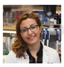 Sharareh Gholamin合作论文数Shahid Modarres Hospital, Shahid Beheshti University (MC),45
 Irving Weissman合作论文数Department of Developmental Biology, Stanford University;Department of Biology and Neurosurgery, Stanford University;Department of Pathology, Stanford University38
 Yoon-Jae Cho合作论文数Dept Neurol & Neurol Sci, Stanford Univ22
 Suzana Assad Kahn合作论文数Instituto de Ciências Biomédicas - INNT/INCT/MCT, Universidade Federal do Rio de Janeiro (UFRJ),22
 Johanna Theruvath合作论文数Stanford Univ, Stanford, CA 94305 USA18
 Rajeev Vibhakar合作论文数Anschutz Med Campus, Univ Colorado18
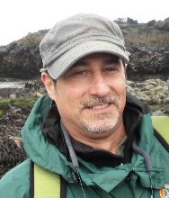 Andrew R. Zimmerman合作论文数Department of Geological Sciences, College of Liberal Arts and Sciences, University of Florida18
 Albert J. Wong合作论文数Department of Neurosurgery, School of Medicine, Stanford University;Cancer Institute, School of Medicine, Stanford University18
 Michelle Monje合作论文数Department of Psychiatry and Behavioral Sciences, School of Medicine, Stanford University18
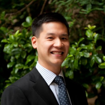 Gordon Li合作论文数Department of Neurosurgery, School of medicine, Stanford University;Department of Neurology, School of medicine, Stanford University;Department of Otolaryngology - Head & Neck Surgery, School of medicine, Stanford University15Abdullah H. Feroze合作论文数14
 Claus Moritz Graef合作论文数12Donson Andrew M合作论文数Dept Pediat, Univ Colorado Denver12
 Hannes Vogel合作论文数Department of Neurosurgery, School of Medicine, Stanford University;Department of Pathology, School of Medicine, Stanford University12Sujatha Venkataraman合作论文数Department of Radiation Oncology, The University of Iowa;b12
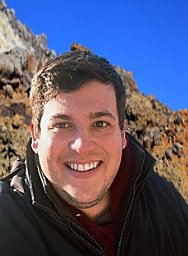 Kyle W. Bostick合作论文数Fugro Marine USA Inc11
 Steven Finkbeiner合作论文数Department of Neurology, University of California, San Francisco11
 Christopher R. Moore合作论文数11Esparza Rogelio合作论文数11
 Senthilnath Lakshmanachetty合作论文数11
 Michael Zhang合作论文数Stanford Artificial Intelligence Laboratory, Stanford University;Together AI11
 James Kennett合作论文数Department of Earth Sciences, University of California, Santa Barbara11
 Christopher Fegan合作论文数Institute of Cancer & Genetics, Cardiff University11
 Martin Dyer合作论文数Department of Cancer Studies and Molecular Medicine, University of Leicester;Ernest and Helen Scott Haematological Research Institute, University of Leicester;Department of Cancer Studies and Molecular Medicine, University of Leicester10
 Gregor Hutter合作论文数Institute of Neuropathology, University Hospital Zurich10
 Marc Remke合作论文数9Ryan Nitta合作论文数9
 Michael Taylor合作论文数Department of Medical Biophysics, Temerty Faculty of Medicine, University of Toronto;Departments of Pediatrics, Baylor College of Medicine;Texas Children’s Hospital9Picard Daniel合作论文数9
 Tej D. Azad合作论文数Johns Hopkins Medicine9Simone Schubert合作论文数9
 Wendy S. Wolbach合作论文数Dept Chem, De Paul Univ9
 Allen West合作论文数9
 Michael S. B. Edwards合作论文数Department of Neurosurgery, Lucile Packard Children’s Hospital, Stanford University Medical Center;Department of Neurosurgery, Stanford University School of Medicine9
 Sabran Masoud合作论文数8
 Allison Cole合作论文数8Nujsaubnusi Vue合作论文数8
 Aleksandar I. Goranov合作论文数Dept Chem & Biochem, Old Dominion Univ8Chase Richard合作论文数8
 Viktoria Marquardt合作论文数Inst Neuropathol, Heinrich Heine Univ8Furong Yu合作论文数8
 Achal Singh Achrol合作论文数8
 Kun-Wei Liu合作论文数NCI-Designated Cancer Center, Sanford Burnham Prebys Medical Discovery Institute8Brian Nguyen合作论文数8Harriet S Walter合作论文数University Hospitals of Leicester, University of Leicester8
 Hankinson Todd C合作论文数School of Medicine, University of Colorado Anschutz Medical Campus8
 Jun Qi合作论文数Dana-Farber Cancer Institute8
 Vijay Ramaswamy合作论文数Department of Medical Biophysics, Temerty Faculty of Medicine, University of Toronto8




