The brain vasculature comprises diverse specialized cells that are essential for brain function, yet their spatial organization remains poorly understood. Here, we construct a comprehensive cerebrovascular cell atlas encompassing 314,535 transcriptomes that captures the arteriovenous axis and defines consensus cell states. We then perform spatial transcriptomics to map 1,529,740 cells across the human temporal cortex and hippocampus, uncovering stereotyped micro-communities termed vascular cell ensembles. These ensembles comprise specialized subsets of endothelial cells, mural cells, fibroblasts, and perivascular macrophages that align with the arteriovenous architecture to coordinate segment-specific functions, such as neurovascular coupling, blood-brain barrier transport, and immune surveillance. By overlaying genetic risk and pharmacologic reactivity, we identify ensemble-specific susceptibilities and candidate therapeutic targets across neurological diseases, including small vessel disease and stroke. This study provides a resource to dissect the spatial and functional logic underlying human cerebrovascular biology and establishes a blueprint for decoding neurological disease susceptibility and therapeutic response.
BACKGROUND AND OBJECTIVES:Extent of resection (EOR) predicts local freedom from recurrence (LFFR) for meningiomas and is a key clinical trial design parameter. Simpson grade (SG) defines EOR based on intraoperative assessment of tumor removal, but MRI-based methods represent promising alternatives. The aim of this study was to compare the prognostic performance of SG vs MRI-based EOR paradigms for predicting recurrence and survival across histomolecular subgroups. METHODS:International multicenter, retrospective cohort study included 475 meningiomas, resected between 1983 and 2024, which were classified by World Health Organization grade and molecular subgroups (DNA methylation, gene expression, and integrated grade). Area under the curve (AUC) was calculated for LFFR and overall survival (OS) from Cox models with a histomolecular subgroup and an EOR paradigm. Delta AUC (ΔAUC) compared EOR predictive performance within each subgroup, and log-rank comparisons of LFFR and OS were performed. RESULTS:MRI-defined gross total resection was associated with significantly longer LFFR and OS when compared with subtotal resection across most histomolecular subgroups. SG1-3 vs 4 distinguished differences in LFFR across several subgroups, but there were no consistent differences in outcomes when comparing degrees of dural treatment. Multivariable Cox including gene expression groups revealed that volumetric EOR (%) had a significantly higher AUC than SG (ΔAUC 0.07, P = .036) for 5-year OS; otherwise, there were no other differences between MRI-based or SG EOR paradigms for 5-year LFFR or OS. Additional significant differences for predicting 10-year LFFR all favored MRI-based EOR paradigms. CONCLUSION:Although SG and MRI-based EOR paradigms provide similar prognostic performance for predicting LFFR and OS in the era of molecular classification, MRI-based definitions may be preferred for future clinical trial inclusion criteria.
Glioblastoma is a lethal brain tumor that is resistant to conventional therapies. Here we present a non-lytic replicating retrovirus (RRV) that delivers an IL-15-receptor-linked fusion protein (RLI) superagonist directly into glioblastoma cells, creating localized immunotherapy biofactories. In orthotopic mouse models, RRV-RLI dramatically suppresses tumor growth, prolongs survival, and induces lasting remission with immunologic memory. Mechanistically, we observe increased CD8⁺ T cell and natural killer cell infiltration and activation, alongside elevated antigen presentation pathways. Combining RRV-RLI with temozolomide, which is standard-of-care chemotherapy for glioblastoma, enhances antitumor immunity. T cell receptor sequencing reveals a polyclonal repertoire of T cells, enhanced by combining RRV-RLI with temozolomide. Analysis of the T-cell repertoire suggests it to be directed against tumor rather than viral antigens, supporting the specificity and re-applicability of our approach. These findings illustrate that RRV-RLI reprograms glioblastoma into an immunostimulatory hub, offering a viral immunotherapy against glioblastoma and potentially other therapy-resistant solid tumors.
Objective Blood-based biomarkers are becoming increasingly relevant for the diagnosis of acute traumatic brain injury (TBI) in adults, but they remain underutilized in children. There exists the need for high-quality evidence to support their use in defined clinical contexts. Methods This review synthesizes recent evidence on three structural biomarkers cleared for clinical use in adult TBI: glial fibrillary acidic protein (GFAP), ubiquitin C-terminal hydroxylase L1 (UCH-L1), and S100 calcium-binding protein B (S100B) for the diagnosis, management, and prognostication in pediatric TBI. Results Notably, plasma/serum biomarker levels in non-injured children were highest in infancy and inversely associated with age, and reference levels for GFAP and S100B across pediatric age groups were higher than those of non-injured adults. Biomarker levels <24 hours post-injury showed acceptable diagnostic performance for clinically-important TBI (area under the receiver-operating characteristic curve (AUC): GFAP=0.75, UCH-L1=0.76, S100B=0.77), and GFAP and UCH-L1 showed strong performance for traumatic intracranial lesions on computed tomography (CT) (overall AUC: GFAP=0.85, UCH-L1=0.83, S100B=0.67; patients aged <5-years: GFAP=1.00, UCH-L1=0.79, S100B=0.62). In European interventional studies, clinical management aided by S100B was associated with reductions in CT utilization by 67-70% and hospitalizations from 54%, underscoring its utility for improving diagnostic efficacy and safety (i.e., decreasing ionizing radiation and time in-hospital). Structural biomarkers predicted death/severe functional disability at 12 months post-injury (AUC: GFAP=1.00, UCH-L1=0.98, S100B=0.96). Conclusions Blood-based biomarkers hold promise for improving pediatric TBI care. Prospective studies incorporating protocolized assessments are needed to validate their diagnostic and prognostic performance and support clinical qualification and implementation.
Importance Glioblastoma (GBM) remains a disease with high mortality despite treatment with maximal safe surgical resection, radiation, and temozolomide. Standard of care for GBM has remained unchanged for decades. Gene therapies using both viral and nonviral platforms have the potential to significantly improve outcomes but have faced challenges related to delivery, intratumoral heterogeneity, and immunologic limitations. Observations This review discusses the most recent progress in genetic therapies for gliomas, with a focus on clinical platforms and those nearing clinical translation. Oncolytic and nonlytic replicating viral therapies remain the most clinically advanced, with adeno-associated virus and nonviral systems approaching clinical translation. Treatment efficacy has been variable across trials, platforms, and payloads. Oncolytic herpes simplex virus (G47Δ, teserpaturev) has been conditionally approved for the treatment of gliomas in Japan, while next-generation constructs show a potential association between immunologic activation and survival. Common challenges include the verification and quantification of delivery efficiency, the balance between antivector and antitumor immunity, appropriately designed clinical trials, relevant modeling of vector behavior in the preclinical setting, regulatory clearance, and manufacturing. Conclusions and Relevance Understanding the potential and limitations of gene therapies for gliomas is vital in navigating the path forward for these promising therapies. The future of these treatments will focus on improved clinical trial design with rapid confirmation and tracking of delivery, biomarker feedback and assay optimization, and combination strategies. A focus on delivery platform development can compress iteration cycles and lead to therapeutic benefit for patients with gliomas and other solid tumors.
Intraoperative hemostasis and dural closure are critical components of neurosurgical procedures, directly affecting operative time, blood loss, neurological outcomes, and the risk of cerebrospinal fluid leak, infection, and reoperation. Over the past 2 decades, there has been rapid expansion in the availability of hemostatic adjuncts, dural substitutes, and liquid sealants. Despite widespread adoption, product selection is frequently guided by surgeon preference and institutional practice rather than a detailed understanding of material composition, mechanism of action, and product-specific limitations. This qualitative review provides a neurosurgery-focused, product-level comparison of commonly used intraoperative hemostatic agents, dural substitutes, and dural sealants, with emphasis on composition, mechanism of action, indications, advantages, limitations, safety considerations, and cost. A comprehensive literature search was conducted using PubMed, Embase, United States Food and Drug Administration device summaries, manufacturer instructions for use, and key clinical trials and observational studies. Products were categorized into 3 groups: intraoperative hemostatic adjuncts, dural substitutes and graft materials, and liquid dural sealants and adhesives. The review encompasses gelatin-based products, oxidized regenerated cellulose, collagen-based agents, flowable gelatin-thrombin matrices, topical thrombin preparations, autologous and nonautologous dural grafts, and fibrin- and polyethylene glycol-based sealants. Selection of these materials should be guided by anatomic considerations, patient-specific factors, and best available evidence. This review serves as a practical reference to support informed intraoperative decision-making in neurosurgical practice.
INTRODUCTION:External ventricular drain (EVD) placement is a life-saving neurosurgical procedure used to divert cerebrospinal fluid and reduce intracranial pressure in conditions such as subarachnoid hemorrhage, intraparenchymal hemorrhage, and intraventricular hemorrhage. While known complications include infection and hemorrhage, the formation of iatrogenic dural arteriovenous fistulas (dAVFs) following ventriculostomy is under-reported. METHODS:We conducted a retrospective review of patients at our institution from 2002 to 2023 who developed dAVFs after EVD placement. Inclusion criteria required angiographic confirmation of a new dAVF near the EVD site. Demographics, primary pathology, EVD characteristics, angiographic findings, treatment approach, and outcomes were recorded. RESULTS:Sixteen patients met the inclusion criteria. Subarachnoid hemorrhage was the most common indication for EVD placement (75.0%). All drains were placed at Kocher's point, most commonly on the right (75.0%). All dAVFs involved the middle meningeal artery, with venous drainage documented in 93.8%-most frequently into the middle meningeal vein (80.0%). Eight patients (50.0%) underwent treatment, primarily with ethanol embolization (62.5%). All treated cases achieved radiographic cure without complications. Two patients experienced spontaneous resolution. Three patients were managed conservatively for low-risk fistulas and five were lost to follow-up. CONCLUSION:This study presents the largest institutional series to date of iatrogenic dAVFs following ventriculostomy. While rare, iatrogenic dAVFs have the potential for significant morbidity if they develop complications from venous hypertension and may require additional treatments. Our findings highlight the need for awareness of identifying and appropriately managing ventriculostomy-related dAVFs.
Intraoperative functional brain mapping is an essential and intricate technique in modern-day glioma surgery. This article is not a review of the literature but of the technical protocol at our institution that has evolved over the recent decades to the current time and is intended to highlight details that enable us to perform maximal safe resection of gliomas. Prior to surgery, anatomical and functional imaging protocols are obtained to determine the tumor to be resected within its anatomical and functional environment. Preoperative assessments are used to determine which mapping procedures and tasks are most appropriate. Cortical and subcortical motor and language mapping using low and high frequency stimulation paradigms are applied when appropriate during resection. Methods to interpret findings and troubleshoot issues are reviewed herein. All preoperative imaging including magnetic resonance imaging, magnetoencephalography of functional cortex, and diffusion tensor imaging of subcortical tracts are uploaded into the neuronavigation station and used throughout surgery for guidance. The decision to continue with tumor resection is based on constant feedback from the mapping paradigms as functional pathways are approached in real time. Both awake and asleep anesthesia regimens are utilized depending on the type of testing required to assess and preserve functional areas during tumor resection. Postoperatively, deficits are assessed using MRI along with clinical exam to predict whether they will be temporary or permanent. The standard of care for all gliomas is maximal safe resection. In this review, we describe brain mapping methods that have been developed, refined, and utilized over decades at a single institution, which have allowed us to achieve this goal safely.
BACKGROUND AND OBJECTIVES:Traumatic brain injury (TBI) often involves alcohol intoxication, yet the effects of elevated blood alcohol content (BAC) on the Glasgow Coma Scale (GCS) are poorly understood. This study investigates the effect of alcohol intoxication on the initial GCS evaluation of patients with TBI. METHODS:Data from a subset of patients enrolled in the Transforming Research and Clinical Knowledge in Traumatic Brain Injury (TRACK-TBI) prospective cohort study were analyzed through Mann-Whitney and Fisher exact tests, and regression analyses as appropriate. Clinical and demographic data were reviewed, and patients were grouped by presenting BAC. RESULTS:In the cohort of 2032 patients, presenting BAC was available for 1448 patients; 25.6% (n = 370) of whom had a BAC ≥0.08 (+BAC). Intoxicated patients exhibited higher rates of recreational drug use (35% vs 19%; P < .001). Despite no difference in injury severity scores between the groups, +BAC patients were more likely to have a positive head computerized tomography (66% vs 53%; P < .001), a lower GCS at presentation (12 vs 13; P < .001), and be categorized as severe TBI (GCS 3-8) at emergency department presentation (26% vs 17%; P < .001). Importantly, the effect of BAC on the GCS remained significant after the use of multivariable regression analysis to control for Marshall score, nonhead injury severity scores, age, sex, and cannabinoid and hard drug use ( P = .007). Multivariable regression analysis also demonstrated a dose-dependent effect with only the most intoxicated patients (BACs 0.16-0.24 and ≥0.24) significantly affecting the GCS. CONCLUSION:Patients with elevated BACs scored approximately 1 point lower on the GCS than patients with BACs under 0.08. BAC's effect on the GCS persisted even after controlling for other covariates by multivariable regression analysis. Providers should be aware of this when initially evaluating intoxicated patients with TBI as it can affect their clinical management.
BACKGROUND AND OBJECTIVES:Extent of resection (EOR) is prognostic for meningioma outcomes. DNA methylation profiling can shed light on biological drivers and therapeutic vulnerabilities. The goal of this study was to re-evaluate the impact of EOR on clinical outcomes across meningioma DNA methylation groups. METHODS:Patients with sporadic meningiomas who underwent resection from a multicenter, international cohort were retrospectively reviewed. Gross vs subtotal resection (GTR vs STR, respectively) was determined based on postoperative MRI. The Kaplan-Meier method, log-rank statistics, and multivariable Cox proportional hazard analyses were performed to evaluate the impact of EOR on local freedom from recurrence (LFFR) and overall survival (OS). RESULTS:In total, 587 patients (Male: 195, Female: 392) underwent 644 surgeries for intracranial meningioma (GTR: 438, STR: 206), with 124 surgeries (19.3%) for recurrent intracranial meningiomas. The cohort included 375 (58.2%) World Health Organization (WHO) Grade 1, 202 (31.4%) WHO Grade 2, and 67 (10.4%) WHO Grade 3 meningiomas based on histological criteria. DNA methylation profiling was used to categorize meningiomas as Merlin-intact (N = 214, 33.2%), Immune-enriched (N = 236, 36.6%), or Hypermitotic (N = 194, 30.1%). GTR was associated with longer LFFR across all meningioma DNA methylation groups (Merlin-intact P < .0001; Immune-enriched P = .013; Hypermitotic P = .001) and was associated with longer OS for Hypermitotic meningiomas ( P = .0022). In multivariable Cox proportional hazard analyses, EOR was significantly associated with LFFR across all DNA methylation groups and WHO grades but was significantly associated with OS only for Hypermitotic meningiomas (hazard ratio [GTR vs STR] 0.64, 95% CI 0.43-0.97, P = .034). CONCLUSION:MRI-defined GTR is associated with improved LFFR across all meningioma DNA methylation groups and improved OS for patients with Hypermitotic meningiomas. These data continue to support maximal safe resection when feasible and demonstrate how molecular classification systems complement rather than supersede the prognostic impact of surgery.
BACKGROUND:Preoperative embolization is hypothesized to reduce blood loss and operative time for meningioma resection, but the impact of preoperative embolization on long-term oncological outcomes and molecular features of meningiomas is incompletely understood. Here, we investigate how preoperative embolization influences perioperative and long-term outcomes as well as molecular features of atypical WHO grade 2 meningiomas. METHODS:Patients who underwent resection of WHO grade 2 meningiomas from 1997 to 2021 were retrospectively identified from an institutional database. Univariate and multivariate Cox proportional hazards modeling and propensity matching were used for clinical analyses. Available DNA methylation profiling, bulk RNA sequencing, and targeted gene expression profiling data were used to elucidate how preoperative embolization influences the molecular architecture of atypical WHO grade 2 meningiomas. RESULTS:A total of 319 patients with atypical WHO grade 2 meningiomas were identified, of which 106 (33.2%) underwent preoperative embolization without significant changes in perioperative outcomes, such as blood loss or operative time, in comparison to patients who did not undergo preoperative embolization. In propensity matched multivariate analyses, preoperative embolization was independently associated with longer recurrence-free survival (RFS, HR 0.55, 95% CI 0.31-0.96, P = .037), particularly in patients with subtotal resection (median RFS 16.2 years vs 5.9 years, P = .045; HR 0.32, 95% CI 0.14-0.70, P = .005). Bioinformatic analyses demonstrated that preoperative embolization led to enrichment of pathways linked to cellular differentiation and hypoxia, and suppression of pathways implicated in mitosis and cell cycle progression, suggesting that improved long-term oncological outcomes may occur through inhibition of the cell cycle in atypical WHO grade 2 meningiomas. CONCLUSIONS:Preoperative embolization improves local control and modulates gene expression in atypical WHO grade 2 meningiomas, a subgroup of meningiomas that have intermediate clinical outcomes with standard interventions.
Background: Publicly reported hospital risk-adjusted mortality rates (RAMRs) reflect real-world outcomes and may be used to understand the impact of advances in medical evidence. Our study presents an analysis of RAMRs in California hospitals across the time period of publication of major trials in stroke intervention, to interrogate the effect of these trials upon population-level mortality from stroke. Methods: Stroke (total acute, ischemic, hemorrhagic, subarachnoid hemorrhage) RAMR data from 2010 to 2020 was extracted from the California Hospital Inpatient Mortality Rates and Quality Ratings dataset. Hospitals were categorized by county population, size and type (academic/community). ANOVA with Tukey-Kramer and Bonferroni-corrected t-tests, and independent t-tests were used for statistical comparison of RAMRs across different population groups and hospital types. Results: There was a statewide decline in acute stroke mortality from 11.4 % to 8.6 %, with ischemic stroke mortality decreasing from 24.9 % to 21.6 %. RAMRs decreased from 5.7 % to 5.0 % in community hospitals (p = 0.006), a trend not mirrored in academic settings. Hemorrhagic stroke RAMRs fluctuated, while subarachnoid hemorrhage RAMRs increased, except in academic institutions. Hospitals in the >2M population group had significantly lower RAMRs (p < 0.005) than the 0-500k group. There were no significant RAMR differences between academic and community hospitals across all stroke types. Conclusions: Despite the publication of paradigm-shifting trials, California in-patient stroke mortality only modestly changed, reflecting the complexity of replicating clinical trial outcomes in real-world data. Consistent, longitudinal quality and outcome metrics at state and national levels remain essential for understanding the impact of clinical research and innovation.
While preoperative embolization is hypothesized to reduce blood loss and surgical time for resection of meningiomas, the impact of embolization on long-term outcomes and molecular profiling of meningiomas is incompletely understood. A series of patients with grade 2 meningiomas who underwent resection at our institution from 1997 to 2021 were identified and reviewed. Available DNA methylation profiling and bulk RNA sequencing data were analyzed (n= 200, WHO grade 1 or 2). Univariate and multivariate analyses, Cox proportional hazards modeling, and propensity matching (sex, age at surgery, previous surgery, extent of resection, postop radiation) were utilized. A total of 357 WHO grade 2 meningioma patients were included, 129 (36.1%) underwent embolization and 228 (63.9%) did not undergo embolization. Mean age at diagnosis was 53.5 years and 61.4% of patients were female. There was no difference in time to recurrence with or without embolization for cases with gross total resection (229/357). Embolization was independently associated with improved local control for cases with subtotal resection even after accounting for postoperative radiotherapy (HR 0.49, 95% CI 0.32-2.20 p=0.028). Average time to recurrence of atypical WHO grade 2 meningioma was 3.3 years after subtotal resection and 6.7 years after embolization and subtotal resection (p<0.001, propensity matched). The distribution of Merlin-intact vs Immune-enriched vs Hypermitotic meningioma DNA methylation groups was equivalent with versus without embolization, but differential gene expression analysis of RNA sequencing data revealed enrichment of hypoxia related pathways after embolization that were unique to each DNA methylation group. These data show that embolization improves local control and reprograms gene expression in WHO grade 2 meningiomas. Additional investigation into the impact of embolization on clinical outcomes and gene expression for meningiomas, especially for higher-grade tumors, is warranted.
OBJECTIVE There is persistent debate in the literature surrounding the true predictors of biochemical remission after resection of somatotroph adenoma. A multimodal analysis of a large number of patients is needed to better understand which patients may be at higher or lower risk for remission failure after surgery. METHODS A retrospective review was performed on patients undergoing somatotroph adenoma resection. Biochemical remission was defined as age- and sex-adjusted normalization of serum insulin growth factor-1 (IGF-1) levels at least 6 months after surgery. Patient case characteristics and clinicopathologic variables were tested for statistical associations with remission and were included in a random forest machine learning model to assess for their importance in determining remission status. Preoperative variables found to be significant remission predictors on statistical testing and important in the random forest model were subsequently assessed via receiver operating characteristic (ROC) analysis to determine numeric thresholds that optimally predicted preoperative likelihood of remission success or failure. RESULTS Eighty patients were identified with somatotroph adenoma who underwent transsphenoidal resection, with 60 patients (75%) achieving biochemical remission. Statistical testing found that patients with failed remission were more likely to have larger tumors (1.9 vs 1.6 cm by the largest axis, p = 0.014; and 3.61 vs 2.66 cm3 by 3D volume, p = 0.013) that invaded the cavernous sinus more frequently (70% vs 22% of patients, p < 0.001) and have higher preoperative IGF-1 level (860 vs 660 ng/ml, p = 0.044). An optimized random forest machine learning model with 10,000 iterations found that tumor size, preoperative growth hormone and IGF-1 levels, and cavernous sinus invasion were important preoperative predictors of remission status. ROC analysis revealed that 96% of patients with preoperative 3D tumor volume less than 1.51 cm3 (area under the curve [AUC] 0.691, p = 0.003) and 100% with nonadjusted preoperative IGF-1 level less than 718.5 ng/ml (AUC 0.736, p = 0.002) achieved remission. CONCLUSIONS Important preoperative predictors of postoperative remission for somatotroph adenoma resection include serum IGF-1 level, cavernous sinus invasion, and tumor size. Ninety-five percent of patients who achieved postoperative remission had preoperative 3D tumor volume less than 1.51 cm3.
OBJECTIVE:Maximal safe resection of gliomas near motor pathways is facilitated by intraoperative mapping. Here, the authors review their results with triple-modality asleep motor mapping with motor evoked potentials and bipolar and monopolar stimulation for cortical and subcortical mapping during glioma surgery in an expanded cohort. METHODS:This was a retrospective analysis of patients who underwent resection of a perirolandic glioma near motor pathways. Clinical and neuromonitoring data were extracted from the electronic medical records for review. All patients with new or worsened postoperative motor deficits were followed for at least 6 months. Regression analyses were performed to assess factors associated with a persistent motor deficit. RESULTS:Between January 2018 and December 2021, 160 operations were performed in 151 patients with perirolandic glioma. Sixty-four patients (40%) had preoperative motor deficits, and the median extent of resection was 98%. Overall, patients in 38 cases (23.8%) had new or worse immediate postoperative deficits by discharge, and persistent deficits by 6 months were seen in 6 cases (3.8%), all in patients with high-grade gliomas. There were no new persistent deficits in low-grade glioma patients (0%). The risk factors for a persistent deficit included an insular tumor component (OR 8.6, p = 0.01), preoperative motor weakness (OR 8.1, p = 0.03), intraoperative motor evoked potential (MEP) changes (OR 36.5, p < 0.0001), and peri-resection cavity ischemia (OR 7.5, p = 0.04). Most persistent deficits were attributable to ischemic injury despite structural preservation of the descending motor tracts. For patients with persistent motor deficits, there were 3 cases (50%) in which a change in MEP was noted but subsequent subcortical monopolar stimulation still elicited a response in the corresponding muscle groups, suggesting axonal activation distal to a point of injury. CONCLUSIONS:Asleep triple motor mapping results in a low rate of permanent deficits, especially for low-grade gliomas. Peri-resection cavity ischemia continues to be a significant risk factor for permanent deficit despite maintaining appropriate distance for subcortical tracts based on monopolar feedback.
BACKGROUND AND IMPORTANCE: Acute intracerebral hematomas are known to induce significant mass effects within the brain, leading to critical complications such as cerebral midline shift, herniation, and increased intracranial pressure. The timing and efficacy of intracerebral hematoma evacuation remain subjects of ongoing debate in current literature. CLINICAL PRESENTATION: In our case report, we present a 74-year-old female patient diagnosed with basal ganglia hematoma. The resultant mass effect from the intracerebral hematoma led to middle cerebral artery (MCA) stenosis. Notably, early-stage minimally invasive hematoma evacuation was pivotal in facilitating successful revascularization of the MCA. CONCLUSION: Our case underscores the significance of prompt identification and management of MCA stenosis arising from intracerebral hematoma. Early intervention through minimally invasive hematoma evacuation proved instrumental in achieving successful MCA revascularization. These findings emphasize the critical role of timely interventions in mitigating potential complications associated with intracerebral hematoma.
 Manish Aghi合作论文数Department of Neurological Surgery, School of Medicine, University of California, San Francisco40
 Jacob Young合作论文数33
 Morshed Ramin A合作论文数The Brain Tumor Center, The University of Chicago Pritzker School of Medicine24Rushikesh S Joshi合作论文数22
 Christopher P. Ames合作论文数School of Medicine, University of California, San Francisco;Helen Diller Family Comprehensive Cancer Center, University of California, San Francisco;Weill Institute for Neurosciences, University of California, San Francisco;UCSF Medical Center20
 Vedat Deviren合作论文数University of California17
 Saket Jain合作论文数15Darryl Lau合作论文数Department of Neurosurgery, University of California at San Francisco15Matheus P. Pereira合作论文数14
 Philip Theodosopoulos合作论文数Mayfield Clinic14
 Mitchel Berger合作论文数Brain Tumor Center, University of California, San Francisco;School of Medicine, University of California, San Francisco13Praveen V. Mummaneni合作论文数Dept. of Neurosurgery, Emory University13Sabraj Gill合作论文数12Angad Beniwal合作论文数12
 Taemin Oh合作论文数12
 Lewis Stuart Blevins合作论文数12Sweta Sudhir合作论文数11Zain Peeran合作论文数11Sandeep Kunwar合作论文数11
 Anthony M DiGiorgio合作论文数Institute for Health Policy Studies, University of California11
 Elaina Wang合作论文数Warren Alpert Med Sch, Brown Univ11Robert C. Osorio合作论文数11
 Jia-Shu Chen合作论文数The Warren Alpert Medical School, Brown University11
 Shawn Hervey-Jumper合作论文数School of Medicine, University of California, San Francisco;Weill Institute for Neurosciences, University of California, San Francisco;Medical School, University of Michigan11Spatz Jordan M合作论文数Harvard-MIT Institute for Medical Engineering and Science, Massachusetts Institute of Technology10
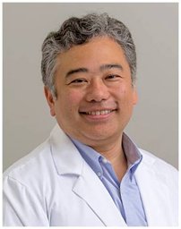 Noriyuki Kasahara合作论文数Brain Tumor Center, University of California, San Francisco;Department of Neurological Surgery, University of California, San Francisco;Department of Radiation Oncology,University of California, San Francisco10Dean Chou合作论文数10Phiroz Tarapore合作论文数9
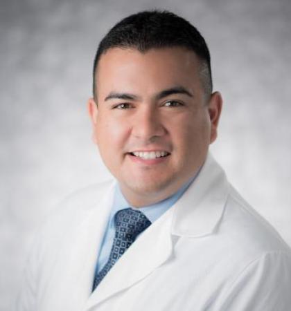 Jose Gurrola合作论文数School of Medicine, University of California San Francisco;Department of Otolaryngology – Head and Neck Surgery, University of California San Francisco;University of California San Francisco Medical Center9Ivan H. El-Sayed合作论文数9
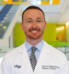 David R Raleigh合作论文数9Kaitlyn M Pereira合作论文数8
 Megan Montoya合作论文数8Kevin C Donohue合作论文数8
 Geoffrey Manley合作论文数Department of Neurological Surgery, School of Medicine, University of California, San Francisco;Brain And Spinal Injury Center;Zuckerberg San Francisco General Hospital8
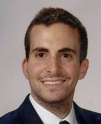 Safaee Michael合作论文数Departments of Neurological Surgery and.8Savastano Luis E合作论文数Departments of 1 Neurosurgery and.7Sara D. Collins合作论文数Washington Hosp Ctr, Washington, DC 20010 USA7
 Vijay Letchuman合作论文数Department of Neurological Surgery, University of California6
 Yoshua Esquenazi合作论文数Department Of Neurosurgery, University of Texas Health Science Center at Houston6
 Michael Weller合作论文数Department of Neurology, University Hospital Zurich6
 Michael Vogelbaum合作论文数Moffitt Cancer Center;Research Institute, Inc6
 Susan M. Chang合作论文数Weill Institute for Neurosciences, University of California, San Francisco;School of Medicine, University of California, San Francisco6
 Nitin Tandon合作论文数The Vivian L. Smith Department of Neurosurgery, The University of Texas Medical School at Houston6
 Ethan Winkler合作论文数Weill Institute for Neurosciences, University of California, San Francisco;School of Medicine, University of California, San Francisco6
 Bello Lorenzo合作论文数Department of Oncology and Hematology-Oncology, Universita degli Studi di Milano6
 Annette Molinaro合作论文数Helen Diller Family Comprehensive Cancer Center, University of California, San Francisco;Department of Neurological Surgery, School of Medicine, University of California, San Francisco;Brain Tumor Research Center, University of California, San Francisco6Aymen Kabir合作论文数5Atul Saha合作论文数5




