Background:Glioblastoma multiforme (GBM) is an aggressive brain tumor with abysmal prognosis because cancer cell growth in the tumor microenvironment (TME) is orchestrated by complex interplay between aerobic glycolysis (AG) and endothelial dysfunction (ED). AG acidifies extracellular pH (pHe) to promote tumor invasion and suppress immune response, whereas ED leads to leaky blood vessels which hampers perfusion and stimulates hypoxia. Since metabolism generates heat and perfusion removes heat, we hypothesized that temperature could reflect both metabolic and vascular reprogramming in the TME mediated by AG and ED. Methods:We used multiple magnetic resonance methods and bioheat modeling to dissect temperature contributions from metabolic and vascular sources in rat gliomas. Results:Upregulated AG in the TME results from enhanced glycolysis (∼4.2× higher) and reduced glucose oxidation (∼4.8× lower), which leads to more acidic pHe (6.9 ± 0.1 vs 7.3 ± 0.1). Since TME is hypoperfused (∼40% lower) and glycolysis is less exothermic compared to glucose oxidation, simulations predict a cooler TME as in vivo measurements clearly demonstrate (0.5-1.5 °C). Moreover, temperature and pHe are correlated both inside and outside the TME for untreated and treated rats (r > 0.6). Conclusions:Since TME is more glycolytic, acidic, hypoperfused, and cooler than neighboring milieu, thermal mapping can represent combined effects of AG and ED for early GBM detection and therapy optimization.
We hypothesized that vascular maladaptations resulting from chronic hypoxemia worsen end-organ function via insidious positive feedback. To test this hypothesis in the systemic circulation, we quantified brain perfusion and left heart function and then biomechanically phenotyped the left common carotid artery (LCCA) in adult female mice under normoxic (21% oxygen) and chronic hypoxic (10%) conditions. Functional MRI revealed that hypoxia impaired cerebrovascular reactivity during transient hypercapnia (pCO2 5%) while echocardiography revealed altered left ventricular diastolic, but not systolic, function. These end organ changes associated with an 18.8% decrease in distensibility of the LCCA due to a 34.4% increase in circumferential material stiffness without an increase in wall thickness. This finding suggested a change in the state (e.g., collagen microstructure), not amount, of the LCCA extracellular matrix. Active biaxial testing of the LCCA further revealed up to an 11% reduction of SMC contractility in response to vasoactive agents. Treatment with the mTOR inhibitor rapamycin improved LCCA properties in hypoxic mice, as reflected by a 53.37% increase in distensibility and a 43% improvement in contractility relative to the hypoxic group. We conclude that chronic hypoxia causes multiorgan effects: stiffer carotid arteries having impaired vascular reactivity associate with impaired cerebral and cardiac function. Maladaptive LCCA changes are prevented with mTOR inhibition during hypoxia, suggesting mTOR as a potential target to break the insidious feedback loop that leads to hypoxia-induced maladaptive remodeling and to mitigate associated end-organ dysfunction, with possible advantages to single ventricle patients who experience long periods of hypoxemia.
Extracellular acidosis is a biologically important feature of the tumor microenvironment in the liver, promoting immune evasion, angiogenesis, and resistance to therapy, and representing a mechanistically important and potentially targetable axis in liver cancer. Imaging extracellular pH (pHe) at high resolution is needed to better understand the immuno-metabolic interplay, especially at the transition regions between the tumor core, tumor margin, and background liver, which is critical for any pharmacological or image-guided intervention. Yet, there is a paucity of imaging techniques capable of providing pHe mapping at high resolution. Here, we demonstrate high-resolution pHe imaging in a mouse Hepa1-6 liver tumor model using 1H Biosensor Imaging of Redundant Deviation in Shifts (BIRDS) with REduced Spherical Encoding with GAussian Weighting (RESEGAW). Eight tumor-bearing C57BL/6J mice were used to demonstrate pHe imaging with RESEGAW using the macrocyclic agent TmDOTP5- at 0.6 mm isotropic resolution on a 9.4 T scanner, which was validated using 31P-MRSI with 3-aminopropylphosphonate (3-APP). pHe imaging with 1H-BIRDS-RESEGAW consistently showed acidic tumor regions (pHe = 6.77 ± 0.14) relative to adjacent normal liver (pHe = 7.14 ± 0.07). Mean pHe values measured by 31P-MRSI with 3-APP and 1H-BIRDS-RESEGAW with TmDOTP5- show no significant differences in tumors (pHe = 6.81 ± 0.13) and normal liver (pHe = 7.14 ± 0.06). Voxelwise comparison after co-registration of 31P-MRSI with 3-APP to 1H-BIRDS-RESEGAW using Bland-Altman analysis demonstrated excellent agreement between the two methods, with minimal mean bias (-0.005 pH units) and variance of less than 0.1 pH units. These results demonstrate the feasibility and quantitative reliability of 1H-BIRDS-RESEGAW for imaging extracellular acidosis in liver tumors at submillimeter resolution, establishing a technical foundation for studying the immuno-metabolic interplay in liver cancer and its response to therapy.
Childhood neglect and deprivation are the most common forms of early adversity, yet their biological impact on cognitive development—and how enrichment mitigates these effects—remains poorly understood. Using limited bedding (LB) as a mouse model of deprivation, we previously showed that abnormal microglia-mediated synaptic pruning during the second and third postnatal weeks impairs synaptic connectivity and hippocampal function, particularly in males. However, the molecular basis of this microglial dysfunction is unclear. Here, we demonstrate that LB reduces expression of Triggering Receptor Expressed on Myeloid cells 2 (TREM2) across multiple mouse strains and that TREM2 deficiency accounts for roughly half of the phagocytic deficit. Overexpressing TREM2 restores microglial phagocytic function and rescues deficits in hippocampal connectivity and fear learning later in life. Brief postnatal enrichment normalizes synaptic pruning in a TREM2-dependent manner and restores contextual fear conditioning in adolescent LB male mice. Together, these findings identify TREM2 activity during early development as a key mediator of the long-term impact of deprivation and enrichment on synaptic connectivity and cognitive function.
Introduction: The failure to translate promising preclinical stroke therapies into clinical success is largely attributed to a lack of rigorous, reproducible outcome measures. While magnetic resonance imaging (MRI) offers a translational alternative to traditional histology, its use in large, multi-site trials is challenged by data heterogeneity and the need for scalable analysis. To address this, we developed and validated a fully automated, open-source image analysis pipeline for the Stroke Preclinical Assessment Network (SPAN), a six-center preclinical trial. Methods: T2 and ADC MRI scans were acquired from 2443 mice and rats (including aged and obese cohorts) at 6 centers 2 and 30 days after middle cerebral artery occlusion (MCAO). Our open-source pipeline performs a complete workflow ( Figure 1 ): 1) preprocessing (image reconstruction, denoising, parameter estimation and quality assessment measures); 2) intensity harmonization to reduce inter-site variability; 3) brain extraction using either traditional rule-based segmentation (rats) or U-net deep learning model (mice); 4) rule-based lesion segmentation via thresholding of harmonized T2 and ADC maps ( Figure 2A-G ); and 5) quantification of midline shift as a proxy for swelling and atrophy ( Figure 3A ). Validation was performed against expert manual tracing on both MRI and 2,3,5-Triphenyltetrazolium chloride (TTC)-stained tissue. Results: The pipeline successfully processed thousands of scans from a heterogeneous collection of scanners. The U-net brain extraction model was highly accurate (Dice score=0.964) and successfully segmented cases where traditional methods failed. Automated lesion volumes correlated strongly with manual expert MRI tracing (R=0.957) and with TTC staining in optimal preparations (R=0.86; Figure 2H ). Harmonization significantly reduced site-specific differences in MRI values. Our geometric midline estimation consistently demonstrated a midline shift towards the contralesional side indicative of swelling on Day 2 and a midline shift towards the ipsilesional side indicative of atrophy on Day 30 ( Figure 3B ). Conclusion: We have developed a validated, end-to-end automated pipeline for quantifying stroke injury in large, multi-site preclinical trials. This work delivers a scalable, objective, and reproducible framework as a shareable, open-source tool that enhances the rigor of preclinical research to help bridge the translational gap in stroke.
Alzheimer’s disease (AD) poses a significant global health challenge, being the most prominent cause of dementia with prevalence increasing as the population ages. While the majority of AD cases are late-onset (LOAD), current animal models predominantly represent the more aggressive, faster progressing early-onset AD (EOAD), limiting their ability in assessing early biomarkers and gaining deeper understanding of LOAD progression. This study explores a promising translatable model, the APOE4.TREM2 mouse, which combines the APOE4 allele and the Trem2 p.R47H mutation, both linked to increased AD risk in the human population. We performed behavioral phenotyping and measured hemodynamics and neurovascular coupling in dorsal olfactory bulbs (dOB) during odor stimulation of the APOE4.TREM2 mouse line. Experimental evidence of olfactory dysfunction prior to clinical symptoms suggests the opportunity of utilizing smell testing and fMRI as tools for screening of AD, both for preclinical and clinical studies. Here we assess and confirm the translatability of the APOE4.TREM2 mouse LOAD model, reporting exacerbated anxiety, deficits in odor-based foraging and spatial memory, and exacerbated odor-evoked dOB neural and intrinsic responses, but stable neurovascular coupling, in an age-dependent manner.
Background: Alzheimer’s disease (AD) is a progressive neurodegenerative disorder marked by memory loss and cognitive decline, reflecting widespread brain dysfunction across multiple neural systems. Early detection of pathological changes is critical for enabling timely intervention, improved management, and better therapeutic outcomes. Methods: Using non-transgenic AD rats (Samaritan) and sham rats (Long–Evans), we explored structural and functional differences with multimodal MRI and multi-unit activity (MUA). Results: Diffusion tensor imaging (DTI) revealed no significant changes in mean diffusivity of water, but AD-related microstructural alterations of fractional anisotropy were confined to subcortical regions with cortical areas and white matter tracts remaining intact. We used functional MRI (fMRI) with blood oxygenation level-dependent (BOLD) contrast in rest-state (R-fMRI) and task-based (T-fMRI) paradigms. R-fMRI revealed much stronger functional connectivity in subcortical vs. cortical areas in AD rats, implicating AD-related functional changes in subcortical areas in agreement with DTI data. T-fMRI with sensory stimulation revealed reproducible fMRI responses in both groups; however, AD rats exhibited reduced BOLD response amplitude and spatial activation extent, which was accompanied by attenuated stimulus-evoked MUA responses. These suggest that attenuated evoked BOLD response reflects diminished neuronal activity in AD, rather than impaired neurovascular and/or neurometabolic coupling. Conclusions: Together these findings suggest that AD-induced anatomical and functional changes in subcortical areas are related to altered cortical responses, highlighting multimodal MRI as a sensitive tool for early AD-related brain changes.
The failure to translate promising preclinical stroke therapies into clinical success is a multi-faceted problem; however, a critical contributing factor is the lack of rigorous, reproducible preclinical outcome measures. While magnetic resonance imaging (MRI) offers a translational alternative to traditional histology, its use in large, multi-site trials is challenged by data heterogeneity and the need for scalable analysis. To address this, we developed and validated a fully automated, open-source image analysis pipeline for the Stroke Preclinical Assessment Network (SPAN), a six-center preclinical trial network. The pipeline processed T2-weighted and apparent diffusion coefficient (ADC) maps from over 2,000 mice and rats, incorporating steps for cross-site data harmonization, deep learning-based brain extraction, and rule-based segmentation to quantify infarct volume, brain swelling, and atrophy. The pipeline demonstrated high accuracy, as automated lesion volumes strongly correlated with manual expert tracing on both MRI (R = 0.96) and 2,3,5-triphenyl-tetrazolium chloride (TTC)-stained tissue (R = 0.86). The U-net model for brain extraction achieved a Dice score of 0.96, and our harmonization method successfully reduced inter-site variability in quantitative MRI parameters. This robust and reproducible pipeline provides a scalable framework for standardizing tissue outcome assessment, enhancing the rigor of multi-site preclinical studies.
Neurovascular coupling links calcium (Ca2+)-dependent neuronal activity to cerebral blood volume changes, whereas neurometabolic coupling describes alterations of neuronal activity and glucose uptake. While mesoscale optical imaging of neurovascular coupling is prevalent, neurometabolic coupling has been explored much less. We describe a multiplexed optical system with a closed cranial window setup for longitudinal studies in Thy1-jRGECO1a mice where neuronal activity is measured with Ca2+-dependent red fluorescence, glucose uptake with bolus injections of 2NBDG with green fluorescence, and cerebral blood volume (CBV) with near-infrared spectroscopy (NIRS). Genetically encoded calcium indicators (GECIs) provide strong fluorescent signals for assessing Ca2+-dependent neuronal activity. Thy1-jRGECO1a, a novel GECI with red fluorescence emission that penetrates deeper into tissue, allows for simultaneous imaging of metabolic activity using a green-fluorescent glucose analog, 2-(N-(7-nitrobenz-2-oxa-1,3-diazol-4-yl)amino)-2-deoxyglucose (2NBDG), which is taken up like glucose and then phosphorylated. Dual-fluorescent (red, green) and NIRS recordings confirm strong neurovascular coupling during hindpaw stimuli (Ca2+-CBV; P = 0.0033, r(2) = 0.91), whereas neurometabolic coupling (Ca2+-2NBDG; P < 0.001) was three times stronger during stimulation (r(2) = 0.75; slope = 0.6) compared to rest (r(2) = 0.49; slope = 0.23). In summary, multiplexed optical imaging can be used to reveal mechanisms of neurovascular and neurometabolic (un)couplings during ischemia, traumatic brain injury, aging, and Alzheimer's disease.
Background & Aims: Increasing enthusiasm around integrating locoregional therapy with systemic immunotherapy in primary liver cancer underscores the need for non-invasive imaging biomarkers. In this study, we aimed to establish advanced molecular MRI tools for monitoring T-cell responses to cryoablation in murine models, distinguishing between immunologically "hot" and "cold" hepatocellular carcinoma (HCC). Methods: Immunocompetent 7-10-week-old C57BL/6J and BALB/cJ mice (n = 18 each) received carbon tetrachloride for 12 weeks to induce cirrhosis. Intrinsically immunogenic Hepa1-6 ("hot") and non-immunogenic TiB75 ("cold") cells were orthotopically implanted into C57BL/6 or BALB/c mice, respectively, to generate focal HCC lesions. After one week, animals were randomly assigned to (A) partial cryoablation (pCryo) (1.2 mm cryoprobe, -40 degrees C) or (B) no treatment (n = 8 per group and tumor type). Gadolinium 160 (Gd-160)-labeled CD8(+) antibody was administered intravenously either 1 week after tumor induction (control) or 1-week post (pCryo) (treatment). T1-weighted MRI scans were performed using a 9.4 T MRI scanner. Radiological-pathological correlation included imaging mass cytometry and immunohistochemistry. Results: pCryo-treated Hepa1-6 tumors displayed peritumoral ring enhancement on T1-weighted MRI with Gd-160-CD8, correlating with imaging mass cytometry signal patterns. Untreated Hepa1-6 tumors lacked such enhancement. Radiological-pathological correlation confirmed significantly increased tumor-infiltrating CD8(+) T lymphocytes in pCryo Hepa1-6 tumors compared with untreated tumors (p <0.001), and a stronger local response compared with systemic lymph nodes (p = 0.0415). Increased T-lymphocyte infiltration was not observed in TiB75 tumors, as indicated by MRI and histopathology. Conclusion: pCryo induced increased T-cell infiltration in Hepa1-6 tumors compared to TiB75 tumors. T1-weighted MRI, following Gd-160-CD8 antibody administration, reproducibly detected the ablation-induced changes. These findings encourage further investigation of MRI-based molecular imaging biomarkers to assess immune responses to local tumor therapies. (c) 2024 The Authors. Published by Elsevier B.V. on behalf of European Association for the Study of the Liver (EASL). This is an open access article under the CC BY-NC-ND license (http://creativecommons.org/licenses/by-nc-nd/4.0/).
Leukemia Inhibitory Factor (LIF) is an injury-induced cytokine that peaks 48 hours after a traumatic brain injury (TBI). Juvenile LIF haplodeficient mice exhibit desynchronized glial responses, increased neurodegeneration, decreased axonal conductivity and behavioral deficits after a concussive head injury. Given the necessity of LIF during the acute recovery phase after injury, we hypothesized that intranasal LIF (IN-LIF) treatment would prevent neurodegeneration when administered during the chronic recovery period from a mild TBI (mTBI). Young adult male CD1 mice were subjected to a midline, closed-head frontal cortex injury using a flat metal impactor with a 3mm tip to induce a mTBI. In the 6-8 weeks post-mTBI, known to precede axonal atrophy in this mTBI model, two doses of 40 ng and 100 ng of LIF were administered twice daily, 5 days/week for two consecutive weeks. Sensorimotor functions were assessed at 4 and 8 weeks post mTBI, followed by ex-vivo brain magnetic resonance imaging at 9.4T and histopathology. mTBI mice showed sensorimotor deficits at 4 weeks, which worsened by 8 weeks post-injury. IN-LIF treatment prevented the progressive sensorimotor loss seen in the vehicle-treated controls. Increased mean diffusivity and decreased fractional anisotropy were observed in the corpus callosum and prefrontal cortex of mTBI brains. In a dose-dependent manner, IN-LIF prevented the mTBI-induced mean diffusivity increase and fractional anisotropy decrease. Histologically, there was significantly less astrogliosis, microgliosis and axonal injury in the IN-LIF treated mice vs. controls. These results support the therapeutic potential of IN-LIF to reduce delayed neurodegeneration and improve neurological outcomes after mTBIs.
Solid tumors, including hepatocellular carcinoma (HCC), arises most often in cirrhotic livers, where immune exclusion and metabolic reprogramming drive extracellular acidosis (the "Warburg effect") and create an immunosuppressive tumor microenvironment (TME). This study applied a non-invasive MR Spectroscopic Imaging method called Biosensor Imaging of Redundant Deviation in Shifts (BIRDS) to quantify extracellular pH (pHe) dynamics in a mouse model of cirrhosis-associated HCC. Forty-two Mdr2-/- mice received chronic carbon tetrachloride (CCl4), inducing cirrhosis and HCC, confirmed by contrast-enhanced MR and histology. BIRDS revealed significantly lower tumor pHe in untreated tumors (6.78 ± 0.3) compared with liver parenchyma (7.17 ± 0.02). Cryoablation induced tumor pHe normalization (7.08 ± 0.03), coinciding with downregulation of metabolic markers and increased T-cell and macrophage infiltration. These results demonstrate that BIRDS enables non-invasive monitoring of the metabolic and immunologic response to cryoablation in HCC within cirrhotic livers. Cryoablation-induced re-normalization of tumor acidity, coupled with enhanced immune activity, suggests a favorable therapeutic outcome and establishes pHe imaging as a tool for assessing treatment efficacy in acidic TMEs.
Preclinical stroke research faces a critical translational gap, with animal studies failing to reliably predict clinical efficacy. To address this, the field is moving toward rigorous, multicenter preclinical randomized controlled trials (mpRCTs) that mimic phase 3 clinical trials in several key components. This collective statement, derived from experts involved in mpRCTs, outlines considerations for designing and executing such trials. mpRCTs offer advantages such as increased sample sizes, robust statistical design, incorporation of heterogeneity, and standardized protocols, but they face challenges in finding the right balance between standardization and heterogeneity, appropriate stroke model selection, and outcome measures, as well as the implementation of complex network infrastructure. We discuss the importance of rigorous study design, including appropriate stroke models, representation of biological variables and comorbidities, functional outcome readouts, and handling of attrition and mortality. Statistical considerations such as adaptive sequential designs, covariate adjustments, and appropriate handling of missing data are also addressed. The integration of machine learning, the implementation of common data elements, and the selection of appropriate therapeutic candidates are crucial for maximizing the efficiency and utility of mpRCTs. Furthermore, the transition toward mpRCT platforms, akin to clinical trial platforms, holds promise for facilitating continuous evaluation of therapies. Finally, we discuss data-sharing practices and the collateral benefits of mpRCTs, emphasizing their potential to improve preclinical stroke research and bridge the translational gap. Altogether, we hope that this article will serve as a starting point for a lasting debate on the future of stroke mpRCTs and their evolution toward a universally accepted set of principles.
In autism spectrum disorder (ASD), a neurodevelopmental disorder with social-cognitive deficits, macrocephaly occurs in 20% of patients with severe symptoms. However, the role of macrocephaly in ASD pathogenesis remains unclear. Here, we address the mechanistic link between macrocephaly and ASD by investigating a novel ASD-associated gain-of-function A1877T mutation in ASPM ( abnormal spindle-like microcephaly-associated ). ASPM is a key regulator of cortical size and cell proliferation expressed in both excitatory and inhibitory neuronal progenitors but not in differentiated neurons. We found that Aspm gain-of-function knock-in mice exhibit macrocephaly, excessive embryonic neurogenesis with expanded outer radial glia, an increased excitatory-inhibitory (E-I) ratio, brain hyperconnectivity, and social-cognitive deficits with male specificity. Our results suggest that macrocephaly in ASD is not a proportional expansion of excitatory and inhibitory neurons, but a shift in the E-I ratio, independent of the expression patterns of the causative gene. Thus, macrocephaly alone can cause a subset of ASD-like symptoms.
Brain's high energy demands require abundant production of ATP from glucose oxidation, mandating coupling between neural activity and nutrient supply. Understanding how neural activity augments blood flow (CBF) to support metabolism of glucose (CMRglc) and oxygen (CMRO2) can help unravel mysteries of neurovascular and neurometabolic couplings underlying functional MRI (fMRI) with blood oxygenation level-dependent (BOLD) contrast. Key to this enigma is oxygen extraction fraction (OEF). Fundamentally, OEF is defined by flow-metabolism (i.e., CBF-CMRO2) coupling generating mitochondrial ATP to signify limits of hypoxia and ischemia. However, to fully account for observed CBF-CMRO2 coupling, the OEF must include a term for oxygen diffusivity (DO2) that is regulated by rheological properties of blood. BOLD contrast depends on intravoxel spin dephasing of tissue water protons due to paramagnetic fields generated by deoxyhemoglobin. During augmented neural activity, if CBF increases more than CMRO2, then deoxyhemoglobin (paramagnetic) is replaced by perfusing oxyhemoglobin (diamagnetic) to increase BOLD signal. Calibrated fMRI converts BOLD contrast into OEF according to the deoxyhemoglobin dilution model. Agreement across these OEF models (i.e., OEF trifecta) authenticates calibrated fMRI, both gas-based and gas-free methods. CMRO2 by gas-free calibrated fMRI easily and reproducibly tracks neural activity, while combining it with CMRglc can also reveal aerobic glycolysis. In summary, there is translational potential of gas-free calibrated fMRI for metabolic imaging in the resting and stimulated brain, from neurodegeneration to neurological disorders.
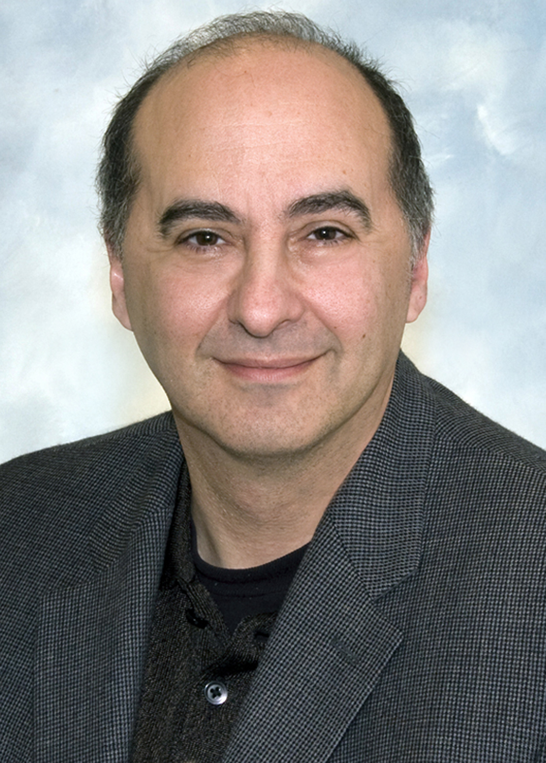 Douglas Rothman合作论文数Magnetic Resonance Research Center, Yale School of Medicine137
 Basavaraju Sanganahalli合作论文数Bioimaging Sciences, Radiology & Biomedical Imaging, Yale School of Medicine, Yale University110
 Peter Herman合作论文数Bioimaging Sciences, Radiology & Biomedical Imaging, Yale School of Medicine, Yale University110
 Daniel Coman合作论文数Bioimaging Sciences, Radiology & Biomedical Imaging, Yale School of Medicine, Yale University100
 Hal Blumenfeld合作论文数Neuroscience, Biological & Biomedical Sciences, Yale University65
 Kevin Behar合作论文数School of Medicine, Yale University45
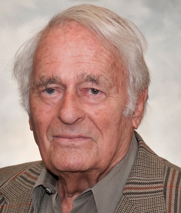 Robert Shulman合作论文数School of Medicine, Yale University39
 Ikuhiro Kida合作论文数Tokyo Metropolitan Institute of Medical Science, The Tokyo Metropolitan Institute of Medical Science22
 Stephan Kaczmarz合作论文数Tech Univ Munich, Klinikum Rechts Isar, Dept Diagnost & Intervent Neuroradiol, Munich, Germany21
 Christine Preibisch合作论文数20
 Garth J. Thompson合作论文数19Jelena Mihailovic合作论文数18
 Yiyun Huang合作论文数Department of Radiology & Biomedical Imaging, Yale School of Medicine, Yale University18
 Graeme Mason合作论文数Biomedical Engineering, School of Engineering & Applied Science, Yale University18
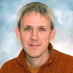 Robin A. de Graaf合作论文数Biomedical Engineering, School of Engineering & Applied Science, Yale University18
 Maxime Parent合作论文数Department of Psychology, University of Quebec in Montreal (UQAM),18
 Mishra Asht M合作论文数Department of Neurology, Yale University School of Medicine17
 Richard H. Mattson合作论文数School of Medicine, Yale University15Sridhar Kannurpatti合作论文数Department of Radiology, Rutgers New Jersey Medical School15
 Sandeep K. Mishra合作论文数Centre for Nanoscience and Technology, Pondicherry University14OAC PETROFF合作论文数13
 Xenophon Papademetris合作论文数Department of Biomedical Informatics and Data Science, School of Medicine, Yale University;Department of Radiology and Biomedical Imaging, School of Medicine, Yale University13Hubert Trubel合作论文数13
 Justus Verhagen合作论文数Neuroscience, Biological & Biomedical Sciences, Yale University12Ognen A. C. Petroff合作论文数12J. Goettler合作论文数11
 Samuel Maritim合作论文数11
 Edward Novotny合作论文数Department of Neurosurgery, Yale University11C. Zimmer合作论文数10
 Julius Chapiro合作论文数Bioimaging Sciences, Radiology & Biomedical Imaging, Yale School of Medicine, Yale University10
 Joshua E Motelow合作论文数Department of Neurology;Department of Diagnostic;Quantitative Neuroscience;Magnetic Resonance Research Center;Department of Neurology, Magnetic Resonance Research Center10
 Lauren Sansing合作论文数Department of Immunobiology, Yale University9
 Fuqiang Xu (徐富强)合作论文数Innovation Academy for Precision Measurement Science and Technology, Chinese Academy of Sciences;Shenzhen Institutes of Advanced Technology, Chinese Academy of Science;University of Chinese Academy of Sciences9
 Cenk Ayata合作论文数Massachusetts General Hospital;Mass General Research Institute;Harvard Medical School9
 Todd Constable合作论文数Departments of Diagnostic Radiology and Neurosurgery, School of Medicine, Yale University9Adil Akif合作论文数8
 Henk De Feyter合作论文数Bioimaging Sciences, Radiology & Biomedical Imaging, Yale School of Medicine, Yale University8
 DeSalvo Matthew N合作论文数Harvard Medical School, Boston, MA 02115, USA. matthew_desalvo@hms.harvard.edu8Jessica Lamb合作论文数8
 Albert H. Gjedde合作论文数Department of Clinical Medicine, University of Southern Denmark;Department of Neuroscience, University of Copenhagen;Faculty of Medicine, McGill University8Andrew M. Blamire合作论文数8
 Muhammad Khan合作论文数7R.P. Kennan合作论文数7
 Andreia Morais合作论文数Harvard Medical School, Massachusetts General Hospital7
 Helene Benveniste合作论文数Department of Anesthesiology Yale School of Medicine, Yale University7Jyotsna Upendra Rao合作论文数7
 Andras Eke合作论文数7
 Christoph Kampmann合作论文数7




