Secreted Protein Acidic and Rich in Cysteine (SPARC), also known as osteonectin or BM-40, is a prototypical matricellular protein that regulates the dynamic interplay between cells and the extracellular matrix (ECM). Unlike structural ECM components that primarily provide mechanical support, SPARC functions as a regulatory molecule that modulates cell-matrix communication, collagen fibrillogenesis, and tissue remodeling. In musculoskeletal tissues -including bone, cartilage, skeletal muscle, tendons, and intervertebral discs - SPARC plays essential roles in organizing ECM architecture, maintaining mechanical integrity, and coordinating adaptive responses to mechanical loading. Emerging evidence indicates that SPARC also acts beyond classical ECM regulation by linking ECM organization to intracellular signaling and metabolic pathways. In this review, we summarize the structural features and biological functions of SPARC, with particular emphasis on its roles in skeletal and connective tissues. We discuss its contribution to ECM assembly and cell-matrix interactions across diverse tissue environments. Finally, we highlight emerging concepts linking SPARC to tissue degeneration, inflammation, and aging, and explore its potential as a biomarker and therapeutic target in musculoskeletal disease.
Macrophages and other phagocytic cells are central regulators of tendon immunobiology, orchestrating inflammation, tissue repair, and extracellular matrix (ECM) remodeling in the tendons. They derive from circulating monocytes and resident tendon-specific populations, including tenophages. Macrophage polarization along the M1/M2 axis exerts a decisive influence on tendon healing trajectories. Activated M1 macrophages promote the early healing phase for debris clearance initiating the reparative cascade. However, their sustained activity leads to inflammation, ECM degradation, impaired healing, tendinopathy, and heterotopic ossification (HO). Conversely, a timed shift toward activated M2 macrophages promotes resolution of inflammation, angiogenesis, ECM deposition, and fibrocartilage formation, whereas excessive or prolonged M2 activity facilitates adhesion formation, fibrosis, scarring and HO. Recent single-cell and spatial profiling studies showed macrophage heterogeneity across tendon compartments, thereby extending the classical M1/M2 paradigm and underscoring the relevance of macrophages/resident tendon cell's interaction in tendon-specific local niches. Mechanobiological stimuli (depending on magnitude, frequency and duration) further modulate macrophage phenotypes and tendon healing. Emerging coculture models and human tendon-on-chip systems provide high-resolution platforms for dissecting these spatiotemporal interactions. Promising therapeutic approaches comprise the application of extracellular vesicles, controlled mechanoloading regimens, and immunomodulatory biomaterials demonstrating potential to induce regenerative macrophage signatures for improved healing outcomes. Notably, platelet-rich plasma (PRP) formulations shape macrophage responses: leukocyte-rich PRP preferentially promotes M1 activity whereas leukocyte-poor PRP supports M2 polarization. Thus, mechano- and immunomodulatory strategies can offer precise control over macrophage dynamics. Regarding the Achilles tendon pathologies, such approaches are helpful by directing macrophage-mediated inflammation towards effective tendon healing outcomes.
Tendon injuries, common in both athletic and non-athletic populations, present significant challenges due to their slow healing and the formation of scar tissue, which impairs function and potentially increases the risk of (re-)rupture. Conventional treatments often yield suboptimal functional and structural repair. This study investigates the potential of mRNA-based therapeutics to enhance tendon healing by targeting 2 distinct pathways via the delivery of chemically modified ARCA-capped mRNAs (cmRNAs) encoding Interleukin-1 receptor antagonist (IL1RA) and Platelet-Derived Growth Factor-BB (PDGF-BB) using injectable nanoparticle (NP) carriers. In vitro experiments demonstrate successful cmRNA delivery and translation, resulting in increased tendon cell proliferation, migration, and anti-inflammatory responses. In vivo, cmRNA treatment notably enhances tendon repair in a rat patellar tendon defect model, by reducing pro-inflammatory cytokines and fibrotic markers while enhancing repair tissue structure. These findings suggest that NP-based cmRNA delivery represents a promising therapeutic strategy for improving tendon healing, offering better outcomes over existing treatments by targeting both inflammatory and regenerative pathways. Statement of significance: In this study, we investigate an mRNA-based therapeutic approach aimed at enhancing tendon healing in a small animal model. Utilizing bioreducible poly(amidoamine)-based polymeric nanoparticles (PAA PNPs) for the delivery of cmRNAs encoding Interleukin-1 receptor antagonist (IL1RA) and Platelet-Derived Growth Factor-BB (PDGF-BB), we demonstrate effective delivery and protein translation in vitro and ex vivo, resulting in enhanced tendon cell proliferation, migration, and robust anti-inflammatory responses. By combining these therapeutic cmRNAs, we show improved tendon repair in vivo, with accelerated tissue regeneration, better collagen fiber organization, and signs of reduced fibrotic scarring. These findings highlight the potential of nanoparticle-mediated cmRNA delivery targeting two distinct pathways to improve tendon healing, offering a promising alternative to current treatments that often yield suboptimal results.
The repair of bony non-unions remains challenging and often requires graft material due to limited availability of autologous bone. The aim of this study was to investigate the potency of a stand-alone pedicled periosteal flap (PF) versus a ligated periosteal flap (PFx), an empty defect and a crossover group in terms of newly formed bone in a 5 mm critical-sized defect in the rat femur diaphysis. The following 4 treatment groups were formed out of a total of 36 male Sprague Dawley rats: Pedicled periosteal flap, ligated periosteal flap, crossover (each n = 10) and empty defect group (n = 6). A prospective randomized plate osteosynthesis was performed. The periosteal flap was dissected along with the supplying vessel from the medial femoral condyle with the aid of magnifying glasses and fixed to the plate and to the defect with a suture. Regular radiographic and µ-CT examinations were performed to determine bone volume inside the defect, as well as descriptive histological examinations. Newly formed bone tissue was measured by Bone Volume / Tissue Volume. The significant highest ratio to the control group was detected in the PF group after 10 weeks (18.77
Tendons are often affected by injuries or tendinopathies, resulting in serious and long-lasting impairments. The repair capacity is very low with a high risk of rerupture. Nevertheless, early, moderate and intermittent functional training adapted to the healing process has been shown to support tendon healing. The mechanosensitive tenocytes are responsible for extracellular matrix (ECM) synthesis, a process that is highly dependent on their specific and local mechanotopographical niche. The mechanical stimuli are triggered by the surrounding ECM that are then recognized by the cells via mechanosensation, transduced via activated intracellular molecular cascades to initiate the mechanoresponse, a process known as mechanotransduction. Hereby, the activation of calcium (Ca2+) dependent channels plays an essential role. Moreover, tenocyte primary cilium has been strongly suggested to participate in mechanosensation and -transduction. The cellular mechanoresponse results in processes such as ECM remodeling, collagen fiber alignment, cell proliferation and migration. Diverse approaches have been developed to recapitulate the natural mechanoenvironment and to optimize tenogenesis. It still remains difficult to identify the threshold parameters that determine optimal mechanical stimulation of tenocytes. The diverse effects of mechanical loading on tenocytes are not yet fully understood, as 2D and 3D experiments have not led to consistent conclusions. Further research is needed to fully address the mechanomics of each tendon cell population to gain a more comprehensive picture of cellular mechanoresponses and interdependencies within the tendon tissue that could help to explain possible feedback mechanisms for the regulation of the tendon ECM after mechanical loading. In turn, such efforts and subsequent achievements can help to outlining advanced therapeutic strategies and physiotherapy protocols for tendon health. Future developments in the field of mechanically assisted tendon reconstruction include 4D applications and direct in situ bioprinting.
Mechanical stabilization is crucial for bone healing, yet complex fractures, particularly osteoporotic or comminuted, remain challenging due to impaired implant osseointegration, resulting in implant loosening or non-unions. This study investigated whether co-application of small extracellular vesicles (sEVs) derived from human umbilical cord mesenchymal stromal cells (hUC-MSC-sEVs) with a low dose of recombinant human bone morphogenetic protein 2 (rhBMP-2) could enhance screw implant osseointegration. A novel small animal model was established to evaluate the effect of anatomical femur regions on screw integration. Six weeks postoperatively, bone formation and bone–implant contact were assessed by micro-computed tomography and descriptive histology. Biomechanical stability was determined using pull-out tests. Outcomes differed significantly between the proximal and distal implant locations, with no improvements in osseointegration observed in the distal region. In the proximal region, application of hUC-MSC-sEVs alone did not significantly improve osseointegration, whereas local application of 1.5 µg rhBMP-2 resulted in measurable biomechanical improvements. No additive or synergistic effects were observed when sEVs were co-administered with rhBMP-2. Descriptive histology supported these findings, demonstrating the most pronounced bone formation at the proximal site following rhBMP-2 treatment. hUC-MSC-sEVs did not enhance screw implant osseointegration and slightly reduced new bone formation. In contrast, a low dose of rhBMP-2 (1.5 µg) promoted implant integration, with no additive effect when combined with sEVs. Notably, the osteogenic effect of rhBMP-2 was observed only at the proximal femoral site, indicating that anatomical location critically influences implant osseointegration. These findings highlight the importance of considering anatomical region when evaluating osteoinductive treatments and implant materials in small animal models.
Tendinopathy is a long-lasting, debilitating disease that not only affects patients' individual lives, but also imposes a significant socioeconomic burden. Due to an insufficient understanding of the underlying pathomechanisms, treatment options remain limited. Although tendinopathy is recognized as a multifactorial disorder, known risk factors and predisposing conditions have largely been studied in isolation. In this review, it is aimed to examine the various pathomechanisms identified to date and synthesize them into a unified concept. Particular emphasis is placed on key drivers believed to be critical in the onset and progression of the disease-namely mechanical stress, inflammation, and vascular changes. By exploring molecular signaling pathways and conditions involved in tendinopathy, how these mechanisms interact and reinforce one another is illustrated, forming a complex and dynamic signaling network, referred to as the tendinopathic loop. In the future, this network is expected to become even more intricate with the inclusion of emerging concepts such as intratendinous pressure and the potential influence of metabolic factors.
Abstract Background The aim of this study was to evaluate potential synergistic effects of a single, local application of human umbilical cord MSC-derived sEVs in combination with a low dose of recombinant human rhBMP-2 to promote the regeneration of a metaphyseal femoral defect in an osteoporotic rat model. Methods 6 weeks after induction of osteoporosis by bilateral ventral ovariectomy and administration of a special diet, a total of 64 rats underwent a distal femoral metaphyseal osteotomy using a manual Gigli wire saw. Defects were stabilized with an adapted Y-shaped mini-locking plate and were subsequently treated with alginate only, or alginate loaded with hUC-MSC-sEVs (2 × 10⁹), rhBMP-2 (1.5 µg), or a combination of sEVs and rhBMP-2 (n = 16 for each group). 6 weeks post-surgery, femora were evaluated by µCT, descriptive histology, and biomechanical testing. Results Native radiographs and µCT analysis confirmed superior bony union with callus formation after treatment with hUC-MSC-sEVs in combination with a low dose of rhBMP-2. This finding was further substantiated by histology, showing robust defect consolidation 6 weeks after treatment. Torsion testing of the explanted femora revealed increased stiffness after application of both, rhBMP-2 alone, or in combination with sEVs, whereas torque was only significantly increased after treatment with rhBMP-2 together with sEVs. Conclusion The present study demonstrates that the co-application of hUC-MSC-sEVs can improve the efficacy of rhBMP-2 to promote the regeneration of osteoporotic bone defects.
Background: Cranio-maxillofacial (CMF) injuries represent a significant challenge in low- and middle-income countries (LMICs), exacerbated by inadequate infrastructure, resources, and training. This systematic review aims to evaluate the current strategies and solutions proposed in the literature to improve CMF fracture care in LMICs, focusing on education, patient transfer, and off-label solutions. Methods: A comprehensive literature search was conducted using PubMed/Medline from January 2000 to June 2023. Studies were selected based on the Preferred Reporting Items for Systematic Review and Meta-analysis Statement (PRISMA). Solutions were categorized into three main areas: education (digital and on-site teaching, fellowships abroad), patient transfer to specialized clinics, and off-label/non-operative solutions. Results: Twenty-three articles were included in the review, revealing a consensus on the necessity for enhanced education and training for local surgeons as the cornerstone for sustainable improvements in CMF care in LMICs. Digital platforms and on-site teaching were identified as key methods for delivering educational content. Furthermore, patient transfer to specialized national clinics and innovative off-label techniques were discussed as immediate solutions to provide quality care despite resource constraints. Conclusions: Effective CMF fracture care in LMICs requires a multifaceted approach, prioritizing the education and training of local healthcare professionals, facilitated patient transfer to specialized centers, and the adoption of off-label solutions to leverage available resources. Collaborative efforts between international organizations, local healthcare providers, and educational institutions are essential to implement these solutions effectively and improve patient outcomes in LMICs.
Tendinopathy is a disease associated with pain and tendon degeneration, leading to a decreased range of motion and an increased risk of tendon rupture. The etiology of this frequent disease is still unknown. In other musculoskeletal tissues like cartilage and intervertebral discs, transient receptor potential channels (TRP- channels) were shown to play a major role in the progression of degeneration. Due to their responsiveness to a wide range of stimuli like temperature, pH, osmolarity and mechanical load, they are potentially relevant factors in tendon degeneration as well. We therefore hypothesize that TRP- channels are expressed in tendon cells and respond to degeneration inducing stimuli.By immunohistochemistry, qRT-PCR and western blot analyses, we found three TRP channel members, belonging to the vanilloid (TRPV), and ankyrin (TRPA) subfamily, respectively, to be expressed in healthy human tendon tissue as well as in rodent tendon, with expression being located to cells within the dense tendon proper, as well as to endotenon resident cells. In vitro-inflammatory and ex vivo-mechanical stimulation led to a significant upregulation of TRPA1 expression in tendon cells, which correlates well with the fact that TRPA1 is considered as mechanosensitive channel being sensitized by inflammatory mediators.This is the first description of TRP- channels in human and rodent tendon. As these channels are pharmacologically targetable by both agonists and antagonists, they may represent a promising target for novel treatments of tendinopathy.
BACKGROUND:Lipedema, diagnosed most often in women, is a progressive disease characterized by the disproportionate and symmetrical distribution of adipose tissue, primarily in the extremities. Although numerous results from in vitro and in vivo studies have been published, many questions regarding the pathology and genetic background of lipedema remain unanswered. METHODS:In this study, adipose tissue-derived stromal/stem cells were isolated from lipoaspirates derived from nonobese and obese donors with or without lipedema. Growth and morphology, metabolic activity, differentiation potential, and gene expression were evaluated using quantification of lipid accumulation, metabolic activity assay, live-cell imaging, reverse transcription polymerase chain reaction, quantitative polymerase chain reaction, and immunocytochemical staining. RESULTS:The adipogenic potential of lipedema and nonlipedema adipose tissue-derived stromal/stem cells did not rise in parallel with the donors' body mass index and did not differ significantly between groups. However, in vitro differentiated adipocytes from nonobese lipedema donors showed significant upregulation of adipogenic gene expression compared with nonobese controls. All other genes tested were expressed equally in lipedema and nonlipedema adipocytes. The adiponectin/leptin ratio was significantly reduced in adipocytes from obese lipedema donors compared with their nonobese lipedema counterparts. Increased stress fiber-integrated smooth muscle actin was visible in lipedema adipocytes compared with nonlipedema controls and appeared enhanced in adipocytes from obese lipedema donors. CONCLUSIONS:Not only lipedema per se but also body mass index of donors affect adipogenic gene expression substantially in vitro. The significantly reduced adiponectin/leptin ratio and the increased occurrence of myofibroblast-like cells in obese lipedema adipocyte cultures underscores the importance of attention to the co-occurrence of lipedema and obesity. These are important findings toward accurate diagnosis of lipedema. CLINICAL RELEVANCE STATEMENT:Our study highlights not only the difficulty in lipedema diagnostics but also the tremendous need for further studies on lipedema tissue. Although lipedema might seem to be an underestimated field in plastic and reconstructive surgery, the power it holds to provide better treatment to future patients can not be promoted enough.
Introduction: Pericytes (PCs) are specialized cells located abluminal of endothelial cells on capillaries, fulfilling numerous important functions. Their potential involvement in wound healing and scar formation is achieving increasing attention since years. Thus, many studies investigated the participation of PCs following brain and spinal cord (SC) injury, however, lacking in-depth analysis of lesioned optic nerve (ON) tissue. Further, due to the lack of a unique PC marker and uniform definition of PCs, contradicting results are published.Methods: In the present study the inducible PDGFRβ-P2A-CreERT2-tdTomato lineage tracing reporter mouse was used to investigate the participation and trans-differentiation of endogenous PC-derived cells in an ON crush (ONC) injury model, analyzing five different post lesion time points up to 8 weeks post lesion.Results: PC-specific labeling of the reporter was evaluated and confirmed in the unlesioned ON of the reporter mouse. After ONC, we detected PC-derived tdTomato+ cells in the lesion, whereof the majority is not associated with vascular structures. The number of PC-derived tdTomato+ cells within the lesion increased over time, accounting for 60–90% of all PDGFRβ+ cells in the lesion. The presence of PDGFRβ+tdTomato- cells in the ON scar suggests the existence of fibrotic cell subpopulations of different origins.Discussion: Our results clearly demonstrate the presence of non-vascular associated tdTomato+ cells in the lesion core, indicating the participation of PC-derived cells in fibrotic scar formation following ONC. Thus, these PC-derived cells represent promising target cells for therapeutic treatment strategies to modulate fibrotic scar formation to improve axonal regeneration.
The aim of this current study was to establish a metaphyseal femoral non-union model in osteoporotic rats by comparing a power tool versus a manual tool for fracture creation. Twelve adult female Sprague Dawley rats were ovariectomized (OVX) and received a special diet for 6 weeks. Biweekly pQCT measurements confirmed a significant reduction in the cancellous and total bone mineral density in OVX rats compared to control (CTRL) animals. After 6 weeks, OVX rats underwent surgery creating a distal metaphyseal osteotomy, either using a piezoelectric- (n = 6) or a manual Gigli wire (n = 6) saw. Fractures were stabilized with a Y-shaped mini-locking plate. Within each group, three rats received Alginate directly into the fracture gap. OVX animals gained more weight over 8 weeks compared to CTRL animals. pQCT analysis showed a significant difference in the volumetric cancellous bone mineral density between OVX and CTRL rats. A histological examination of the osteoporotic phenotype was completed. Radiographic evaluation and Masson-Goldner trichrome staining with the piezoelectric saw failed to demonstrate bony bridging or a callus formation. New bone formation and complete healing were seen after 6 weeks in the Gigli group. For the creation of a metaphyseal atrophic non-union in the osteoporotic bone, a piezoelectric saw should be used.
Collagen rich connective tissues fulfill a variety of important functions throughout the human body, most of which having to resist mechanical challenges. This review aims to compare structural and functional aspects of tendons and sclera, two tissues with distinct location and function, but with striking similarities regarding their cellular content, their extracellular matrix and their low degree of vascularization. The description of these similarities meant to provide potential novel insight for both the fields of orthopedic research and ophthalmology.
The repair of large bone defects remains challenging and often requires graft material due to limited availability of autologous bone. In clinical settings, collagen sponges loaded with excessive amounts of bone morphogenetic protein 2 (rhBMP-2) are occasionally used for the treatment of bone non-unions, increasing the risk of adverse events. Therefore, strategies to reduce rhBMP-2 dosage are desirable. Silk scaffolds show great promise due to their favorable biocompatibility and their utility for various biofabrication methods. For this study, we generated silk scaffolds with axially aligned pores, which were subsequently treated with 10× simulated body fluid (SBF) to generate an apatitic calcium phosphate coating. Using a rat femoral critical sized defect model (CSD) we evaluated if the resulting scaffold allows the reduction of BMP-2 dosage to promote efficient bone repair by providing appropriate guidance cues. Highly porous, anisotropic silk scaffolds were produced, demonstrating good cytocompatibility in vitro and treatment with 10× SBF resulted in efficient surface coating. In vivo, the coated silk scaffolds loaded with a low dose of rhBMP-2 demonstrated significantly improved bone regeneration when compared to the unmineralized scaffold. Overall, our findings show that this simple and cost-efficient technique yields scaffolds that enhance rhBMP-2 mediated bone healing.
Vascular endothelial growth factor (VEGF) signaling is crucial for a large variety of cellular processes, not only related to angiogenesis but also in nonvascular cell types. We have previously shown that controlling angiogenesis by reducing VEGF-A signaling positively affects tendon healing. We now hypothesize that VEGF signaling in non-endothelial cells may contribute to tendon pathologies. By immunohistochemistry we show that VEGFR1, VEGFR2, and VEGFR3 are expressed in murine and human tendon cells in vivo. In a rat Achilles tendon defect model we show that VEGFR1, VEGFR3, and VEGF-D expression are increased after injury. On cultured rat tendon cells we show that VEGF-D stimulates cell proliferation in a dose-dependent manner; the specific VEGFR3 inhibitor SAR131675 reduces cell proliferation and cell migration. Furthermore, activation of VEGFR2 and -3 in tendon-derived cells affects the expression of mRNAs encoding extracellular matrix and matrix remodeling proteins. Using explant model systems, we provide evidence, that VEGFR3 inhibition prevents biomechanical deterioration in rat tail tendon fascicles cultured without load and attenuates matrix damage if exposed to dynamic overload in a bioreactor system. Together, these results suggest a strong role of tendon cell VEGF signaling in mediation of degenerative processes. These findings give novel insight into tendon cell biology and may pave the way for novel treatment options for degenerative tendon diseases.
Treatment of tendinopathies still present a major challenge, since the aetiology of the disease remains poorly understood. To determine whether the systemic inflammation accompanying predisposing factors including rheumatoid arthritis, diabetes or smoking contributes to the onset of tendinopathy, we studied the effect of a systemic inflammation induced by an allergic episode on tendon properties. To this end, we elicited an allergic response in mice by exposing them to a plant allergen and subsequently analysed both their flexor and Achilles tendons. Biomechanical testing and histological analysis revealed that tendons from allergic mice not only showed a significant reduction of both elastic modulus and tensile stress, but also alterations of the tendon matrix. Moreover, 3D tendon-like constructs treated with sera from allergic mice displayed a matrix-remodelling expression profile and the expression of macrophage-associated markers and matrix metalloproteinase 2 (MMP2) was increased in allergic Achilles tendons. Analysing data from an epidemiologic study comprising data from more than 10.000 persons, we found that persons suffering from an allergic condition appeared to have an increased propensity to develop a tendinopathy. Graphical abstract
Tendons and tendon interfaces have a very limited regenerative capacity, rendering their injuries clinically challenging to resolve. Tendons sense muscle-mediated load; however, our knowledge on how loading affects tendon structure and functional adaption remains fragmentary. Here, we provide evidence that the matricellular protein secreted protein acidic and rich in cysteine (SPARC) is critically involved in the mechanobiology of tendons and is required for tissue maturation, homeostasis, and enthesis development. We show that tendon loading at the early postnatal stage leads to tissue hypotrophy and impaired maturation of Achilles tendon enthesis in Sparc(-/-)mice. Treadmill training revealed a higher prevalence of spontaneous tendon ruptures and a net catabolic adaptation in Sparc(-/-) mice. Tendon hypoplasia was attenuated in Sparc(-/-) mice in response to muscle unloading with botulinum toxin A. In vitro culture of Sparc(-/-) three-dimensional tendon constructs showed load-dependent impairment of ribosomal S6 kinase activation, resulting in reduced type I collagen synthesis. Further, functional calcium imaging revealed that lower stresses were required to trigger mechanically induced responses in Sparc(-/-) tendon fascicles. To underscore the clinical relevance of the findings, we further demonstrate that a missense mutation (p.Cys130Gln) in the follistatin-like domain of SPARC, which causes impaired protein secretion and type I collagen fibrillogenesis, is associated with tendon and ligament injuries in patients. Together, our results demonstrate that SPARC is a key extracellular matrix protein essential for load-induced tendon tissue maturation and homeostasis.
 Renate Gehwolf合作论文数46
 Andreas Traweger合作论文数Mount Sinai Hospital, Samuel Lunenfeld Research Institute46
 Christine Lehner合作论文数45Andrea Wagner合作论文数30
 Hans-Christian Bauer合作论文数28
 Herbert Resch合作论文数Privatklinik Wehrle-Diakonissen17Henrik Bauer合作论文数Department of Molecular Medicine and Surgery, Karolinska Institutet16Hannelore Bauer合作论文数13
 Stefanie Korntner合作论文数12
 Mark Tauber合作论文数12
 Ludwig Aigner合作论文数Institute of Molecular Regenerative Medicine, Paracelsus Medical University12A Wagner合作论文数11
 Francisco Rivera合作论文数Institute of Molecular Regenerative Medicine, Paracelsus Medical University Salzburg11Nadja Weissenbacher合作论文数9
 Andrea Trost合作论文数8Nadja Kunkel合作论文数8
 Istvan A. Krizbai合作论文数Institute of Biophysics, Biological Research Centre8Florian Wichlas合作论文数7
 Sebastien Couillard-Despres合作论文数7Gabriel Spitzer合作论文数6Christian Deininger合作论文数6Kaser-Eichberger Alexandra合作论文数6Peter Augat合作论文数6Falk Schroedl合作论文数6
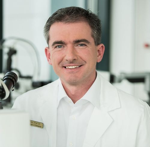 Herbert A. Reitsamer合作论文数Department of Ophthalmology, Paracelsus Medical University;Department of Ophthalmology, University Eye Clinic of Salzburg;Genumed5
 Robin Franklin合作论文数Centre for Science and Policy, University of Cambridge;Department of Veterinary Medicine, University of Cambridge;Altos Labs Cambridge Institute of Science5
 Ludwig Heindl合作论文数Department of Ophthalmology, University of Cologne;University Hospital of Cologne;Ophthalmic Oncology Center, University of Cologne5
 Amelie Deluca合作论文数4Runge Christian合作论文数4Lange Simona合作论文数4
 Heimel Patrick合作论文数4
 Alerie Guzman De la Fuente合作论文数4
 Imola Wilhelm合作论文数Biological Research Center, the Hungarian Academy of Sciences4
 Heinz Redl合作论文数Ludwig Boltzmann Institute for Experimental and Clinical Traumatology4Josef Zweimueller-Mayer合作论文数4Sebastian Toepfer合作论文数3
 Roland Nikolaus Wagner合作论文数3Lara Bieler合作论文数3W Hofmann合作论文数3
 Clemens Goegele合作论文数3Peter van Wijngaarden合作论文数Department of Surgery, Melbourne Medical School, Faculty of Medicine, Dentistry and Health Sciences, The University of Melbourne3Bogner Barbara合作论文数3
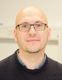 Sascha Senck合作论文数Department of Anthropology, University of Vienna3Rotheneichner Peter合作论文数3
 Christer Betsholtz合作论文数Department of Immunology, Genetics and Pathology, Uppsala University;Department of Medicine, Huddinge, Karolinska Institutet3
 Herbert Lettner合作论文数3
 Johanna Andrae合作论文数3A. Schober合作论文数3
 Eva Rohde合作论文数Laboratory for Brain Tumor Biology, Universitätskrankenhaus Eppendorf3




