Taeniolabis taoensis, the largest known multituberculate, is craniodentally one of the most well-known mammals from the early Paleocene of the Western Interior of the United States of America. Although a few postcranial elements from the Nacimiento Formation of the San Juan Basin, New Mexico, were previously referred to T. taoensis as early as the 1880s, the attribution of most of these specimens to T. taoensis has been questioned. Here we review all of the previously described postcranial remains and confirm that some attributions were indeed incorrect. However, we also place on record several new specimens recently discovered in museum collections that represent the ulna, radius, femur, and calcaneus and that we confidently assign to Taeniolabis based on morphological characters previously identified as unique to multituberculates, large size, and association with craniodental remains of Taeniolabis in the same strata. We compare these elements to those of other multituberculate mammals, particularly to those of other taeniolabidoids. Not unexpectedly considering the heavily constructed, blocky skull of Taeniolabis, all of the postcranial elements are robust, more robust than for any other known multituberculate. Although we are constrained by the poor representation of elements from critical parts of the skeleton and the incomplete nature of the available specimens, we infer that Taeniolabis was a ground-dwelling, quite possibly fossorial mammal.
Based on high-resolution computed tomography scanning, we provide new insights into the inner ear and stapedial morphology of Morganucodon from the Early Jurassic of St Brides. At the base of mammaliaforms, Morganucodon plays a pivotal role in understanding the sequence of character acquisition from basal cynodonts to mammals, including the detachment of the middle ear and the evolution of high-frequency hearing. Advancements in imaging technology enabled us to revise or newly describe crucial anatomy that was not accessible for the original description of Morganucodon. Based on 37 petrosals, we can confirm that the apex of the cochlear canal is expanded in Morganucodon, suggestive of a lagena macula. A gently raised crest along the abneural margin is reminiscent of (although much shallower than) the secondary lamina base of other Mesozoic mammaliaforms. The venous circum-promontorial plexus, which surrounded the inner ear in several basal mammaliaforms, was connected to the cochlear labyrinth in Morganucodon through numerous openings along the secondary lamina base. Two petrosals contain fragmentary stapes, which differ substantially from previously described isolated stapes attributed to Morganucodon in having peripherally placed crura and an oval and bullate footplate. Based on the revised stapedial morphology, we question the traditional view of an asymmetrical bicrural stapes as the plesiomorphic condition for Mammaliaformes.
We use synchrotron x-ray tomography of annual growth increments in the dental cementum of mammaliaforms (stem and crown fossil mammals) from three faunas across the Jurassic to map the origin of patterns of mammalian growth patterns, which are intrinsically related to mammalian endothermy. Although all fossils studied exhibited slower growth rates, longer life spans, and delayed sexual maturity relative to comparably sized extant mammals, the earliest crown mammals developed significantly faster growth rates in early life that reduced at sexual maturity, compared to stem mammaliaforms. Estimation of basal metabolic rates (BMRs) suggests that some fossil crown mammals had BMRs approaching the lowest rates of extant mammals. We suggest that mammalian growth patterns first evolved during their mid-Jurassic adaptive radiation, although growth remained slower than in extant mammals.
Postcranial remains of gondwanatherian mammals were previously known by only a single specimen, an essentially complete skull and postcranial skeleton constituting the holotype and only known specimen of Adalatherium hui (UA 9001), from the uppermost Cretaceous (Maastrichtian) Anembalemba Member of the Maevarano Formation, Mahajanga Basin, northwestern Madagascar. Here we report the discovery of a caudal vertebra (DMNH EPV.141354) that we provisionally refer to a much larger gondwanatherian, Vintana sertichi, also from the Maevarano Formation. V. sertichi, the largest known mammal from the Mesozoic of Gondwana, was previously represented only by a cranium from the Lac Kinkony Member, which represents a nearshore, peritidal paleoenvironment, but the caudal vertebra was recovered from the underlying Anembalemba Member, which represents an earlier, more fully terrestrial fluvial/flood-plain paleoenvironment. The presence of V. sertichi in the latter rock unit therefore expands the age (making it more fully contemporaneous with A. hui) and the known paleoenvironmental range for the species. Comparing various metrics of vertebral body proportions suggests that (1) gondwanatherians are unique among Mesozoic mammals in possessing mid-caudal vertebral centra that are ante-roposteriorly short; and (2) the tail of V. sertichi was relatively wider and shorter than that of A. hui.& COPY; 2023 Elsevier Ltd. All rights reserved.
Although several well-preserved crania are known for the Mesozoic Eutriconodonta, three-dimensional reconstructions of the character-rich inner ear and basicranial region based on high-resolution computed tomography scans have previously only been published for the Late Jurassic Priacodon . Here we present a description of the petrosal and inner ear morphology of a triconodontid eutriconodontan from the Lower Cretaceous Cloverly Formation, which we provisionally assign to Astroconodon . The bony labyrinth of Astroconodon is plesiomorphic for mammaliaforms in lacking a primary osseous lamina, cribriform plate, and osseous cochlear ganglion canal. However, as in Priacodon and the zhangheotheriid Origolestes , Astroconodon has a secondary osseous lamina base that extends nearly the complete length of the cochlear canal. The cochlear canal is straighter in Astroconodon and other eutriconodontans compared to several basal mammaliaform clades (e.g., morganucodontans, docodontans), that exhibit varying degrees of cochlear canal curvature. The pars cochlearis of the petrosal was well vascularized in Astroconodon , exhibiting a network of venous canals that crossed the cochlea transversely on its ventral and dorsal aspects. Of particular note are several canals that passed along the base of the secondary osseous lamina. As in Priacodon and Origolestes , those canals do not show the extensive connections to the cochlear labyrinth as seen in the basal mammaliaforms Morganucodon and Borealestes . The inner ear of Astroconodon thus highlights the complex history of the mammaliaform cochlear canal, in which different clades appear to follow independent evolutionary trajectories and various key morphological features (e.g., cochlear canal length, curvature, vascularization and osseous supports for the basilar membrane) exhibit considerable homoplasy.
Here we report on a new Early Cretaceous eutherian represented by a partial skeleton from the Jiufotang Formation at Sihedang site, Lingyuan City, Liaoning Province that fills a crucial gap between the earliest eutherians from the Yixian Formation and later Cretaceous eutherians. The new specimen reveals, to our knowledge for the first time in eutherians, that the Meckelian cartilage was ossified but reduced in size, confirming a complete detachment of the middle ear from the lower jaw. Seven hyoid elements, including paired stylohyals, epihyals and thyrohyals and the single basihyal are preserved. For the inner ear the ossified primary lamina, base of the secondary lamina, ossified cochlear ganglion and secondary crus commune are present and the cochlear canal is coiled through 360°. In addition, plesiomorphic features of the dentition include weak conules, lack of pre- and post-cingula and less expanded protocones on the upper molars and height differential between the trigonid and talonid, a large protoconid and a small paraconid on the lower molars. The new taxon displays an alternating pattern of tooth replacement with P3 being the last upper premolar to erupt similar to the basal eutherian Juramaia. Parsimony analysis places the new taxon with Montanalestes, Sinodelphys and Ambolestes as a sister group to other eutherians. This article is part of the theme issue 'The impact of Chinese palaeontology on evolutionary research'.
Newly discovered fossil evidence has led to a re-evaluation of one of the fundamental transitions in mammalian evolution: the transformation of bones of the lower jaw into those of the middle ear. Fossil evidence enables a re-evaluation of how the middle ear evolved.
Taeniolabis taoensis is an iconic multituberculate mammal of early Paleocene (Puercan 3) age from the Western Interior of North America. Here we report the discovery of significant new skull material (one nearly complete cranium, two partial crania, one nearly complete dentary) of T. taoensis in phosphatic concretions from the Corral Bluffs study area, Denver Formation (Danian portion), Denver Basin, Colorado. The new skull material provides the first record of the species from the Denver Basin, where the lowest in situ specimen occurs in river channel deposits ~730,000 years after the Cretaceous-Paleogene boundary, roughly coincident with the first appearance of legumes in the basin. The new material, in combination with several previously described and undescribed specimens from the Nacimiento Formation of the San Juan Basin, New Mexico, is the subject of detailed anatomical study, aided by micro-computed tomography. Our analyses reveal many previously unknown aspects of skull anatomy. Several regions (e.g., anterior portions of premaxilla, orbit, cranial roof, occiput) preserved in the Corral Bluffs specimens allow considerable revision of previous reconstructions of the external cranial morphology of T. taoensis. Similarly, anatomical details of the ascending process of the dentary are altered in light of the new material. Although details of internal cranial anatomy (e.g., nasal and endocranial cavities) are difficult to discern in the available specimens, we provide, based on UCMP 98083 and DMNH.EPV 95284, the best evidence to date for inner ear structure in a taeniolabidoid multituberculate. The cochlear canal of T. taoensis is elongate and gently curved and the vestibule is enlarged, although to a lesser degree than in Lambdopsalis.
The dentition in the only known specimen of Adalatherium hui, a gondwanatherian mammal from the Late Cretaceous of Madagascar, is markedly different from that of any previously known mammaliaform, extinct or extant. The two preserved upper incisors and single lower incisor of Adalatherium are each very large, open-rooted, and bear a restricted band of enamel. A shallow-rooted, rudimentary upper canine is present. The upper postcanine dentition is composed of a small, simple, two-rooted tooth mesially and four large, multiple-rooted (five roots or more) teeth distally. The latter are quadrangular in outline, each with four major cusps and three perimetric ridges bordering a central valley that opens buccally. There are four lower postcanines, also with four major cusps each. The first postcanine has two roots, but the following ones each have (or likely had) at least four. The two distal lower postcanines bear a prominent mesiobuccal basin. The cusps and other topographic features of the postcanines cannot be unambiguously homologized with those of other known mammaliaforms. Diastemata are present in the upper dentition between the incisors and canines and between the canines and postcanines, and in the lower dentition between the incisors and postcanines. Adalatherium, like several gondwanatherians from the Late Cretaceous and Paleogene of Argentina, retains relatively plesiomorphic, ‘normal’ radial enamel (i.e., single-layered schmelzmuster, non-decussating small prisms, interprismatic matrix anastomosing around prisms). This stands in contrast to the modified radial enamel found in other gondwanatherians from the Late Cretaceous of India and Madagascar, which exhibit prominent interrow sheets of interprismatic matrix.
The cranium of Adalatherium hui, as represented in the holotype and only specimen (UA 9030), is only the second known for any gondwanatherian mammal, the other being that of the sudamericid Vintana sertichi. Both Adalatherium and Vintana were recovered from the Upper Cretaceous (Maastrichtian) Maevarano Formation of northwestern Madagascar. UA 9030 is the most complete specimen of a gondwanatherian yet known and includes, in addition to the cranium, both lower jaws and a complete postcranial skeleton. Aside from Adalatherium and Vintana, gondwanatherians are otherwise represented only by isolated teeth and lower jaw fragments, belonging to eight monotypic genera from Late Cretaceous and Paleogene horizons of Madagascar, the Indian subcontinent, Africa, South America, and the Antarctic Peninsula. Although the anterior part of the cranium is very well preserved in UA 9030, the posterior part is not. Nonetheless, comparable parts of the crania of Adalatherium and Vintana indicate some level of common ancestry through possession of several synapomorphies, primarily related to the bony composition, articular relationships, and features of the snout region. Overprinted on this shared morphology are a host of autapomorphic features in each genus, some unique among mammaliaforms and some convergent upon therian mammals. The cranium of Adalatherium is compared with the crania of other mammaliamorphs, particularly those of allotherians or purported allotherians (i.e., haramiyidans, euharamiyidans, multituberculates, Cifelliodon, and Megaconus). Particular emphasis is placed on several recently described forms: the enigmatic Cifelliodon from the Early Cretaceous of Utah and several new taxa of euharamiyidans from the Late Jurassic of China.
The phylogenetic position of Gondwanatheria within Mammaliaformes has historically been controversial. The well-preserved skeleton of Adalatherium hui from the Late Cretaceous of Madagascar offers a unique opportunity to address this issue, based on morphological data from the whole skeleton. Gondwanatheria were, until recently, known only from fragmentary dental and mandibular material, as well as a single cranium. The holotype of A. hui provides the first postcranial skeleton for gondwanatherians and substantially increases the amount of character data available to score. We sampled 530 characters and 84 cynodonts (including 34 taxa historically affiliated with Allotheria) to test the phylogenetic relationships of Gondwanatheria and Allotheria using parsimony, undated Bayesian, and tip-dated Bayesian methods. We tested three lower dental formulae for Adalatherium, because its postcanines are distinctly different from those of other mammaliaforms and cannot readily be homologized with any known dental pattern. In all analyses, Adalatherium is recovered within Gondwanatheria, most frequently outside of Sudamericidae or Ferugliotheriidae, which is congruent with establishment of the family Adalatheriidae. The different dental coding schemes do not greatly impact the position of Adalatherium, although there are differences in character optimization. In all analyses, Gondwanatheria are placed within Allotheria, either as sister to Multituberculata, nested within Multituberculata, or as sister to Cifelliodon (and Euharamiyida), or in a polytomy with other allotherians. The composition of Allotheria varies in our analyses. The haramiyidans Haramiyavia and Thomasia are placed outside of Allotheria in the parsimony and tip-dated Bayesian analyses, but in a polytomy with other allotherians in the undated Bayesian analyses.
The fossil record of mammaliaforms (mammals and their closest relatives) of the Mesozoic era from the southern supercontinent Gondwana is far less extensive than that from its northern counterpart, Laurasia1,2. Among Mesozoic mammaliaforms, Gondwanatheria is one of the most poorly known clades, previously represented by only a single cranium and isolated jaws and teeth1–5. As a result, the anatomy, palaeobiology and phylogenetic relationships of gondwanatherians remain unclear. Here we report the discovery of an articulated and very well-preserved skeleton of a gondwanatherian of the latest age (72.1–66 million years ago) of the Cretaceous period from Madagascar that we assign to a new genus and species, Adalatherium hui. To our knowledge, the specimen is the most complete skeleton of a Gondwanan Mesozoic mammaliaform that has been found, and includes the only postcranial material and ascending ramus of the dentary known for any gondwanatherian. A phylogenetic analysis including the new taxon recovers Gondwanatheria as the sister group to Multituberculata. The skeleton, which represents one of the largest of the Gondwanan Mesozoic mammaliaforms, is particularly notable for exhibiting many unique features in combination with features that are convergent on those of therian mammals. This uniqueness is consistent with a lineage history for A. hui of isolation on Madagascar for more than 20 million years. Adalatherium hui, a newly discovered gondwanatherian mammal from Madagascar dated to near the end of the Cretaceous period, shows features consistent with a long evolutionary trajectory of isolation in an insular environment.
The lower jaw of the holotype of Adalatherium hui, from the Late Cretaceous of Madagascar, is the most complete yet known for a gondwanatherian mammal. It reveals for the first time the morphology of the character-rich ascending ramus of the dentary in a gondwanatherian. Each half of the lower jaw is composed of only one bone, the dentary, which is short and deep and houses only five teeth: an enlarged, procumbent incisor and four postcanine teeth. In comparable parts of its anatomy, the dentary of Adalatherium is strikingly similar to that of Sudamerica but differs slightly from that of Galulatherium (conformation anterior to first postcanine, mental foramen position), the only two other gondwanatherians represented by complete horizontal rami. Among other Mesozoic mammaliaform taxa, the dentary of Adalatherium is most similar to those of the largely Laurasian group Multituberculata, most notably in absence of postdentary trough and Meckelian sulcus; presence of short, deep dentary with sizable diastema and articulating with squamosal via mediolaterally narrow condyle that continues onto posterior surface (i.e., no distinct peduncle); possession of much reduced dentition; absence of angular process; possession of large pterygoid fossa and pterygoid shelf, ventral surface of which is flat; absence of coronoid bone; and possession of unfused mandibular symphysis. Most of these features are clearly derived and stand in stark contrast to the much more plesiomorphic morphology exhibited by the lower jaw of the haramiyaviid Haramiyavia. The lower jaws of euharamiyidans, although derived in their own right, are also relatively plesiomorphic.
 David W. Krause合作论文数Department of Earth Sciences, Denver Museum of Nature & Science;Department of Anatomical Sciences, Stony Brook University19
 John Wible合作论文数Section of Mammals, Carnegie Museum of Natural History8
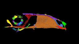 Groenke Joseph R合作论文数Department of Anatomical Sciences, Stony Brook University7
 Chris Kirk合作论文数6
 James B. Rossie合作论文数Department of Anthropology, Stony Brook University5
 Thomas Martin合作论文数Steinmann-Institut für Geologie, Universität Bonn4
 Michael D'Emic合作论文数State University of New York at Stony Brook, Stony Brook University4
 Guillermo Rougier合作论文数Department of Anatomical Science and Neurobiology, University of Louisville4
 Patrick M. O’Connor合作论文数Ignyta Inc4waymon l holloway合作论文数3Lydia J. Rahantarisoa合作论文数3Gill Pamela G合作论文数3Yaoming Hu合作论文数3
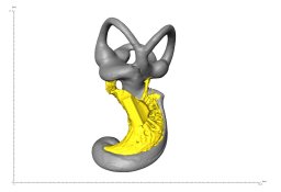 Julia A. Schultz合作论文数Universität Bonn Steinmann-Institut für Geologie3
 Raymond R Rogers合作论文数Department of Geology;Macalester College;Department of Geology, Macalester College3
 Addison D Kemp合作论文数2Dimitry Spitkovsky合作论文数2
 Rudolf J Wiesner合作论文数2
 Wighart Von Koenigswald合作论文数2
 Brady Z. Foreman合作论文数Department of Geology & Geophysics, University of Wyoming2L. R. Yohe合作论文数1Viola Henke合作论文数1
 Michael Rummel合作论文数1Isaac Sageman合作论文数1Diancan Wang合作论文数1Kai R. K. Jäger合作论文数1
 Peter Pfeiffer合作论文数Saarland Univ1
 Christoph Winkler合作论文数Hynes Institute for Entrepreneurship & Innovation, Iona College1
 Thomas L Adams合作论文数Roy M Huffington Dept Earth Sci, So Methodist Univ1Elis Newham合作论文数Sch Arts, Woodland Rd, Bristol, Avon, England1Haibing Wang合作论文数Chinese Acad Sci, Environm & Engn Res Inst, Key Lab Desert & Desertificat, Lanzhou 730000, Gansu, Peoples R China1
 Augusto Nicolas Varela合作论文数Facultad de Ciencias Naturales y Museo, Universidad Nacional de La Plata1
 Matthew Hurtgen合作论文数Department of Earth and Planetary Sciences, Weinberg College of Arts and Sciences, Northwestern University1
 Yuanqing Wang (王元青)合作论文数University of Chinese Academy of Sciences;Institute of Vertebrate Paleontology and Paleoanthropology, Chinese Academy of Sciences1Corfe Ian J合作论文数1
 Philipp Schneider合作论文数1
 Sarah Werning合作论文数Department of Integrative Biology and Museum of Paleontology, University of California1
 Jen A Bright合作论文数School of Geosciences, University of South Florida1
 Vincent Fernandez合作论文数European Synchrotron Radiation Facility, X-Ray Imaging Group1
 Liliana M. Davalos合作论文数Dept Ecol & Evolut, SUNY Stony Brook1Andriamialison Haingoson合作论文数1
 Elizabeth Dumont合作论文数School of Natural Sciences, University of California, Merced1
 Abigail Curtis合作论文数1
 Richard L. Cifelli合作论文数Department of Biology, Dodge Family College of Arts and Sciences, University of Oklahoma1
 Laurel R Yohe合作论文数Biology Department, Loyola University Chicago;Department of Ecology and Evolution, Stony Brook University;Loyola University Chicago, Stony Brook University1
 Federica Marone Welford合作论文数Paul Scherrer Institute1
 Goran Lovric合作论文数Swiss Light Source, Paul Scherrer Institute;Institute for Biomedical Engineering, University and ETH Zurich;Paul Scherrer Institute, University and ETH Zurich1
 Petermann Holger合作论文数Steinmann Institute of Geology, Mineralogy, and Paleontology, University of Bonn1
 Elsa Panciroli合作论文数Museum of Natural History, University of Oxford;National Museums Scotland1




