In stem mammals, feeding and hearing are closely linked due to jaw bones being involved in both functions. Osteological changes during the evolutionary separation of these systems are well documented in the fossil record, but changes to soft tissue anatomy are less certain. The medial pterygoid muscle is a jaw adductor that develops medial to Meckel's cartilage, inserting on the mandibular angular process in therians (placentals and marsupials). Likewise, in stem mammaliaforms, the medial pterygoid is often assumed to have passed medial to the ossified Meckel's cartilage, inserting on the dentary "angular" (i.e., pseudangular) process, a plesiomorphic pattern of muscle attachment also assumed to be present in some derived cynodonts. Thus, the traditional interpretation is that the medial pterygoid remained medial to Meckel's cartilage through the evolution from early cynodonts to therians. We highlight a pattern that contrasts with that interpretation: the medial pterygoid inserts lateral (not medial) to Meckel's cartilage in monotremes. Further, fossil evidence indicates that this pattern was also present in stem therian eutriconodontans and spalacotherioids, which lack the pseudangular process. The varying position of the medial pterygoid among mammalian groups suggests that the muscle is either (1) evolutionarily labile in terms of its relative position to Meckel's cartilage or (2) did not insert on the pseudangular process of early cynodonts. We support the latter hypothesis,which indicates that the medial pterygoid did not shift medial to Meckel's cartilage until the complete ear-jaw separation in cladotherians (therians and close relatives), with the shift linked to the evolution of the therian angular process as an insertion site.
Screen washing operations at the Busche quarry near Balve in North Rhine-Westphalia (Germany) have yielded a new paulchoffatiid multituberculate and additional teeth of the eobaatarid Cheruscodon balvensis. Angrivarodon goresi gen. et sp. nov. is represented by an upper P5 and differs from all known multituberculates of the paulchoffatiid line by a cusp formula of 4B:6L. For C. balvensis 12 new teeth are reported, representing seven formerly unknown tooth positions (dPX, P2, P3, P4, M1, M2, and m1), that allow for the reconstruction of large parts of the upper and lower premolar-molar series. A single upper P4 is attributed to Eobaataridae indet. Three upper P5s are designated as Multituberculata indet., one of which formerly had been assigned to the pinheirodontid Bructerodon alatus. After revision, B. alatus is now restricted to the holotype specimen (M1). Two isolated indeterminate multituberculate incisors have spatulate crowns with complete enamel cover. A cladistic analysis including the new Cheruscodon teeth led to an almost complete resolution of Eobaataridae under inclusion of Arginbaatar into that group. Monophyly of the genus Sinobaatar is not supported by the new cladistic analysis. The shared presence of derived cusp morphology and arrangement of cusps on the upper M2s of the Late Jurassic Teutonodon langenbergensis from the Langenberg locality (Lower Saxony) and of C. balvensis suggest the presence of eobaatarid ancestors in the Jurassic of Central Europe.
The dentition of the proviverrine hyaenodont Lesmesodon edingeri from Messel is known only from preservation in full occlusion. Moreover, only subadult individuals have been recovered so far. The mu CT-based study provides new details from lingual, buccal, and occlusal aspects of its dentition, indicating new insights on the ecology of Lesmesodon and its niche occupation in the Messel ecosystem and what dietary range it had. We analyzed the dental function of the Eocene species Lesmesodon edingeri and its close relative Proviverra typica by reconstructing the wear facets and their respective chewing cycle with the Occlusal Fingerprint Analyser (OFA) software and compared them with those of three modern carnivorous taxa. The teeth were compared by quantification of their contact areas (wear facets) and the calculation of the duration of the contact. Subsequently the masticatory path was reconstructed in detail. Differences in morphologically similar dentitions are revealed by the OFA analysis. The insectivorous species show similarities in their respective chewing cycle in terms of duration and maximum intercuspation, while species more specialized in either carnivory or omnivory show contrary trends, that can be clearly separated from the insectivores. Our study showed that the small hyaenodont Lesmesodon edingeri apparently occupied a primarily insectivorous-carnivorous dietary niche in the Messel ecosystem and how the dentitions of the insectivorous species in this study differ despite having adaptions that look similar at first glance.
The complex evolutionary history behind modern mammalian chewing performance and hearing function is a result of several changes in the entire skeletomuscular system of the skull and lower jaw. Lately, exciting multifunctional 3D analytical methods and kinematic simulations of feeding functions in both modern and fossil mammals and their cynodont relatives approach this topic, giving fresh insights into the history of mammalian masticatory behaviors and their evolutionary trends. One crucial transformation in this context is the segregation of postdentary bones (becoming the mammalian middle ear) from the lower jaw, which is posited to have led to the important functional decoupling of the hearing and feeding systems. Evolution of the middle ear is regarded as the key transition that enhanced both mammalian chewing performance and hearing capacity. Three major functional parts undergo substantial evolutionary changes in this process that are anatomically linked to each other: the lower jaw and dentition, middle ear, and inner ear. Sound, transmitted via vibrations of the bony middle ear elements to the inner ear, is converted into movements of the endolymph fluid that shift hair cells of the organ of Corti, triggering neural stimuli perceived as hearing. Structural changes in one part of the system influence the function of the other two. In this review, I highlight recent advances in research focusing on the enhancement of both chewing performance and hearing ability in mammalian history to feature the mechanisms that led to the decoupling of the hearing system (i.e., middle and inner ear) from the feeding system.
Two new vertebrate assemblages rich in elasmobranch isolated teeth and dermal denticles from the lower Cenomanian (Upper Cretaceous) of Patagonia in southern Argentina comprise ten identified taxa (†Protosqualus sp., †Heterodontus sp., †Orectoloboides sp., †Carcharias sp., †Scapanorhynchus patagonensis sp. nov., †Squalicorax sp., †Paraorthacodus sp., †Rhinobatos aff. †R. incertus, †Ptychotrygon ameghinorum, †Ptychotrygon sp.) and five unidentified lamniforms. The material comes from two closely positioned sites representing proximal palaeoenvironments (delta and lagoonal foot barrier, respectively). The new elasmobranch sites are the most diverse known from the Upper Cretaceous of Patagonia until today and indicate a remarkable elasmobranch diversity in high latitudes of the Southern Hemisphere. They are dominated by warm-water taxa demonstrating a strong Tethyan influence into the southern part of the Austral Basin, contrasting with the later Weddellian influence as indicated by the presence of cold-water taxa in that area during the Campanian and the Maastrichtian.
In non-mammalian synapsids, feeding and hearing are closely linked because some jaw bones are involved in both functions. The evolutionary decoupling of these two systems in early mammals likely catalyzed greater specialization of feeding and hearing. Although fossil osteological changes during this process are well documented, the corresponding evolutionary changes to soft tissue anatomy are less certain. The medial pterygoid muscle is a jaw adductor that is central to this evolutionary transition because in many fossil lineages it inserted near or possibly on jaw bones involved in both feeding and hearing. In therians (placentals and marsupials), the medial pterygoid muscle develops medial to Meckel’s cartilage and inserts on the mandibular angular process. Similarly, non-mammalian cynodonts are often reconstructed with a medial pterygoid muscle passing medial to the ossified Meckel’s cartilage, inserting on the dentary ‘angular’ (i.e., pseudangular) process. Thus, the traditional interpretation is that the medial pterygoid remained medial to Meckel’s cartilage through the evolutionary transitions from early cynodonts to therians. Here we highlight issues with that interpretation: the medial pterygoid muscle inserts lateral (not medial) to Meckel’s cartilage in monotremes and, presumably, early mammal groups (e.g., spalacotherioids) that lacked an angular process. This suggests at least two possible explanatory hypotheses: 1) the medial pterygoid muscle is evolutionarily labile, shifting in position relative to Meckel’s cartilage multiple times or 2) the medial pterygoid muscle did not insert on the pseudangular process of non-mammalian cynodonts and instead inserted on the mandibular medial ridge, dorsal to Meckel’s cartilage. We advocate for the latter hypothesis, proposed by Patterson (1956), which suggests that the medial pterygoid did not shift medial to Meckel’s cartilage until the complete separation of the ear and jaw in cladotherians (therians and close relatives), with the shift in position possibly triggering the evolution of the therian angular process as an insertion site. Patterson’s hypothesis is in line with a growing body of evidence that indicate concomitant evolutionary changes of muscles, ears, and jaws at the cladotherian node were important catalysts for the evolution of hearing and feeding specializations in extant mammals.
Lissamphibian assemblages of Early Cretaceous age are rare in Europe. Here, we comprehensively describe the most informative lissamphibian remains from the Lower Cretaceous (Barremian–Aptian) Balve locality in Germany. The assemblage from Balve includes the salamandroid salamanders Balveherpeton hoennetalensis and Martintriton septatus gen. et sp. nov., as well as the albanerpetontid Wesserpeton sp. and an indeterminate frog (Anura indet.). Martintriton septatus gen. et sp. nov. is characterised by the following combination of atlantal characters: anterior cotyles large and rounded with almost flat articular surfaces continuous across lateral and ventral surfaces of broad intercotylar tubercle; posterior cotyle oval and laterally compressed; prominent ventrolateral ridges; irregularly located depressions separated by bony septa and several foramina of different sizes on ventral surface of centrum; short lateral ridge on lateral surface of centrum; and large foramina between the lateral and ventrolateral ridges. Martintriton septatus gen. et sp. nov. shares similarities in atlas morphology with the Paleogene batrachosauroidid Palaeoproteus miocenicus and the Late Cretaceous scapherpetids likely indicating a close relationship. The composition of the lissamphibian assemblage from Balve (= faunal association “frogs-crown salamanders-albanerpetontids”) resembles other most diverse Early Cretaceous European lissamphibian assemblages at the family level. The transition from the Jurassic to Cretaceous lissamphibian fauna in Europe is characterised by the retention of the albanepetontid Celtedens and, possibly, by extinction of stem karaurid salamanders.
Docodon hercynicus sp. nov. from the Upper Jurassic (upper Kimmeridgian) Suntel Formation of the Langenberg Quarry in Lower Saxony is the first docodontan recorded from Central Europe. The two lower molars available are characterised by vertical enamel ridges at the distal flank of cusp a, which are typical for Docodon. This is only the second record of Docodon from Europe with the genus otherwise restricted to the Kimmeridgian-Tithonian of the Morrison Formation in western North America. A distal fragment of a much smaller lower molar from the same strata exhibits similar vertical enamel ridges and indicates the possible presence of a second Docodon species in the Langenberg Quarry. Docodon hercynicus n. sp. is further evidence for the close terrestrial faunal interrelationships between Europe and North America. It represents only the third record of a docodontan from Late Jurassic-earliest Cretaceous strata of Europe and supports a relic occurrence and late survival of mammaliaforms on a palaeo-island in the Lower Saxony Basin within the European Archipelago. Zoobank: urn:lsid:zoobank.org:pub:90E8BCB2-B4DC-49D4-B10C-115ECF60E1F3
We use synchrotron x-ray tomography of annual growth increments in the dental cementum of mammaliaforms (stem and crown fossil mammals) from three faunas across the Jurassic to map the origin of patterns of mammalian growth patterns, which are intrinsically related to mammalian endothermy. Although all fossils studied exhibited slower growth rates, longer life spans, and delayed sexual maturity relative to comparably sized extant mammals, the earliest crown mammals developed significantly faster growth rates in early life that reduced at sexual maturity, compared to stem mammaliaforms. Estimation of basal metabolic rates (BMRs) suggests that some fossil crown mammals had BMRs approaching the lowest rates of extant mammals. We suggest that mammalian growth patterns first evolved during their mid-Jurassic adaptive radiation, although growth remained slower than in extant mammals.
The primary function of the tetrapod jaw is to transmit jaw muscle forces to bite points. The routes of force transfer in the jaw have never been studied but can be quantified using load paths - the shortest, stiffest routes from regions of force application to support constraints. Here, we use load path analysis to map force transfer from muscle attachments to bite point and jaw joint, and to evaluate how different configurations of trabecular and cortical bone affect load paths. We created three models of the mandible of the Virginia opossum, Didelphis virginiana, each with a cortical bone shell, but with different material properties for the internal spaces: (1) a cortical-trabecular model, in which the interior space is modeled with bulk properties of trabecular bone; (2) a cortical-hollow model, in which trabeculae and mandibular canal are modeled as hollow; and (3) a solid-cortical model, in which the interior is modeled as cortical bone. The models were compared with published in vivo bite force and bone strain data, and the load paths calculated for each model. The trabecular model, which is preferred because it most closely approximates the actual morphology, was best validated by in vivo data. In all three models, the load path was confined to cortical bone, although its route within the cortex varied depending on the material properties of the inner model. Our analysis shows that most of the force is transferred through the cortical, rather than trabecular bone, and highlights the potential of load path analysis for understanding form-function relationships in the skeleton.
A [ACT study of two skulls of the small ruminant Hypisodus minimus from the White River Group (Late Eocene to Late Oligocene) of western North America revealed the following remarkable features: (i) downwards and inwards curved nasal bones that meet at the median line and form a bony tube, (ii) big orbitae supported by a closed orbital bar consisting almost entirely of the os frontale, (iii) a pair of indeterminate canals inside the corpus ossis basisphenoidalis, running in ventro-dorsal direction, and (iv) heavily inflated bullae tympanicae with fused paracondylar processes. The bullae are separated by a narrow slit from each other and are not in contact, as stated earlier. Large bullae enhance low-frequency hearing as seen in burrowing mammals such as the extant caviomorph rodent Dolichotis. In the inner ear, a secondary crus commune is missing and the cochlea shows 2.44 coils. In one skull the maxilloturbinalia, one undefined turbinal and the ethmoid bone (comprising two ethmoturbinalia, two frontoturbinalia, lamina semicircularis, and lamina cribrosa), as well as the anterior part of the lamina horizontalis are preserved.
The Late Jurassic docodontan Haldanodon exspectatus from the Guimarota coal mine in Portugal provides important information on the evolution of the dental replacement pattern in mammaliaforms. Haldanodon shows diphyodont replacement of antemolars and non-replacement of molars. Lower incisors are replaced in alternating order with early replacement of i2 and i4, and late replacement of i1 and i3. Upper and lower premolars were replaced sequentially from front to back. In the maxilla and mandible, four deciduous premolar positions are present (dP1-4, dp1-4), but only three permanent premolars (P1/p1, P3/p3, P4/p4) erupt with loss of the dP2/dp2 position. The anterior deciduous premolars (dP1-2/dp1-2) are small and peg-like, dP3-4/dp3-4 are much larger and molariform. The lower canine and dp4 are the last lower teeth to be replaced. The permanent lower premolars are premolariform and consist mainly of large main cusp a. The ultimate permanent lower premolar (p4) erupts at the same time as m4. After replacement of the antemolars, one or two more molars (m5-6) are added at the posterior end of the tooth row of the mandible. Growth of the ramus occurs at the anterior and posterior ends as evident from the much larger permanent canine and addition of m5-6. In the maxilla, all permanent premolars differ morphologically clearly from the molars. The replacing P1 is small and consists mainly of labial cusps A, B, and C. The P3 and P4 are increasingly larger and show a lingual extension with cusps X and Y which is shorter than in the molars. The adult dental formula of Haldanodon is 6I/4i, 1C/1c, 3P/3p, 5 M/5-6m, the deciduous dental formula is ?dI/4di, 1dC/1dc, 4dP/4dp. The tooth eruption sequence in the mandible is m1 → p1 + m2 → i2 + i4 → ?i1 + ?i3 → p3 + m3 → c + p4 + m4 → m5 → m6. Our results corroborate the earlier suggestions that “Peraiocynodon inexpectatus” is based on deciduous lower premolars (dp1-4) of Docodon victor, and that “Peraiocynodon major“ and Cyrtlatherium canei are based on deciduous lower premolars.
The relationship of food comminution and individual age in Tupaia belangeri is investigated. It is hypothesized that with increasing age the performance of the molar dentition decreases due to progressive tooth wear. While this relationship is well-documented for herbivores, age-related test series are largely lacking for insectivorous mammals. 15 individuals of Tupaia belangeri were fed exclusively with mealworms, and their faeces were analyzed for the number and size of chitin particles. The exoskeleton of a mealworm is resistant to digestive fluids in the gastrointestinal tract, and the size of individual chitin particles indicates the effectiveness of mechanical comminution that occurs in the oral cavity during mastication. It is hypothesized that a more precise occlusion of the dentition results in smaller particle size. Although individuals of all ages (juvenile, adult, and senile) were able to effectively process mealworms with their dentition prior to digestion, a larger area of very large chitin particles (98% quantile of all particles in senile animals as compared to in the same quantile in adults) in the feces of senile animals was detected. Even though the particle size of indigestible material is irrelevant for the digestive process, these findings either document somatic senescence in the functionality of the teeth, or alternatively a change in chewing behaviour with age.
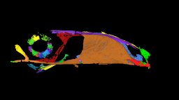
This contribution contains the 3D model described and figured in the following publication: Martin, T., Averianov, A. O., Schultz, J. A., & Schwermann, A. H. (2023). A stem therian mammal from the Lower Cretaceous of Germany. Journal of Vertebrate Paleontology, e2224848
A new extinct sclerorhynchoid sawfish, Ptychotrygon ameghinorum sp. nov., is presented here based on abundant isolated teeth and some dermal denticles, which were recovered from the Mata Amarilla Formation, belonging to the lower Upper Cretaceous of the Santa Cruz Province in the Austral Basin of Patagonia, Argentina. This new species is the first Ptychotrygon occurrence in the southern hemisphere, which so far only has been reported from northern hemisphere deposits (Europe, North Africa, and North America). The presence of P. ameghinorum sp. nov. in these southern high-latitude deposits of Patagonia, Argentina, extends the geographic range of Ptychotrygon considerably southwards. This distribution pattern in the “middle” Cretaceous seems to correlate with the South Atlantic opening at the end of the Albian. The presence of lateral cephalic dermal denticles and the simultaneous absence of rostral denticles in the abundant fossil material support the view that Ptychotrygon did not develop such rostral structures. A reinvestigation of all known species assigned to Ptychotrygon reveals that P. ellae is a junior synonym of P. boothi, P. benningensis belongs to Texatrygon, P. rugosum belongs to Asflapristis, and P. clementsi represents an unidentifiable species (Ptychotrygon? sp.). The stratigraphic distribution demonstrates that Ptychotrygon might have originated in the Albian in south-western Europe and subsequently dispersed to obtain its widest distribution during the Cenomanian. In the Coniacian, a steep diversity decline is recognizable with a subsequent distribution shift from Europe to North America.
 Thomas Martin合作论文数Steinmann-Institut für Geologie, Universität Bonn40
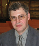 Alexander Averianov合作论文数Laboratory of Theriology, Zoological Institute of the Russian Academy of Sciences;Divisions & Labs, Institute of Earth Sciences, Department of Paleontology, St.Petersburg State University19
 Achim H Schwermann合作论文数LWL-Museum of Natural History; Sentruper Straße 285 48161 Münster Germany15
 Rico Schellhorn合作论文数14
 Zhe-Xi Luo合作论文数Department of Organismal Biology and Anatomy, The University of Chicago;Department of Geology, Center of Integrative Science and Education, Field Museum of Natural History10
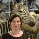 Irina Ruf合作论文数Steinmann-Institut für Geologie, Rheinische Friedrich-Wilhelms-Universität Bonn10
 Pavel Skutschas合作论文数Department of Vertebrate Zoology, Saint Petersburg State University9
 Alexander V. Lopatin合作论文数8
 Wighart Von Koenigswald合作论文数7
 Sergei Anatolyevich Krasnolutskii合作论文数6
 Sandra Engels合作论文数6
 Veniamin V. Kolchanov合作论文数4
 Stepan V. Ivantsov合作论文数Paleontology and Historical Geology Department, Tomsk State University4
 David M Grossnickle合作论文数Committee on Evolutionary Biology, The University of Chicago4
 Oliver Wings合作论文数Institut für Geowissenschaften, Universität Tübingen4
 Ottmar Kullmer合作论文数Forschungsinstitut Senckenberg, Forschungsinstitut Senckenberg4Kai R. K. Jäger合作论文数3
 Ivan Kuzmin合作论文数Dept Vertebrate Zool, Saint Petersburg State Univ3
 Hoffmann Simone合作论文数Department of Anatomical Sciences, Stony Brook University3
 Ellen Schulz-Kornas合作论文数3
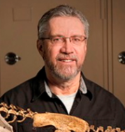 David W. Krause合作论文数Department of Earth Sciences, Denver Museum of Nature & Science;Department of Anatomical Sciences, Stony Brook University3
 Richard H. Madden合作论文数Department of Anatomy, Duke University Medical Center3
 Daniela C. Kalthoff合作论文数Institut für Paläontologie der Universiät Bonn3Ulrike Anders合作论文数3
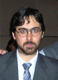 Javier N. Gelfo合作论文数Div Paleontol Vertebrados, Museo La Plata3
 Ulrike Menz合作论文数2Petr N. Kolosov合作论文数2Lydia J. Rahantarisoa合作论文数2
 Dmitrii Vitenko合作论文数2
 Arnaud Begat合作论文数2Chris Kirk合作论文数2
 Alec Wilken合作论文数2
 Dmitry Victorovich Grigoriev合作论文数Ctr Econometr & Business Analyt CEBA, St Petersburg State Univ2
 Daniela E. Winkler合作论文数Biocenter Grindel & Zoological Museum, University of Hamburg, Martin-Luther-King-Platz 3, 20146 Hamburg, Germany2
 Jurgen Kriwet合作论文数Dept Palaeontol, Univ Vienna2Corfe Ian J合作论文数2
 Karl-Heinz Südekum合作论文数Institute of Animal Science, University of Bonn2
 John Wible合作论文数Section of Mammals, Carnegie Museum of Natural History2
 Elizabeth Dumont合作论文数School of Natural Sciences, University of California, Merced2
 Groenke Joseph R合作论文数Department of Anatomical Sciences, Stony Brook University2
 Elsa Panciroli合作论文数Museum of Natural History, University of Oxford;National Museums Scotland2
 James B. Rossie合作论文数Department of Anthropology, Stony Brook University2
 Soledad Gouiric Cavalli合作论文数División Paleontología Vertebrados, Museo de La Plata2Ulrich Zeller合作论文数2
 Raymond R Rogers合作论文数Department of Geology;Macalester College;Department of Geology, Macalester College2
 Thomas M. Kaiser合作论文数Centre for Taxonomy and Morphology, Leibniz Institute for the Analysis of Biodiversity Change, University of Hamburg;Zoological Institute, University of Hamburg;Section Mammalogy & Paleoanthropology, Department of Vertebrates, University of Hamburg2
 Callum F. Ross合作论文数Department of Organismal Biology and Anatomy, The University of Chicago2
 Adam K. Huttenlocker合作论文数Dept Biol, Univ Washington2
 Michael Wuttke合作论文数2




