Equine multipotent mesenchymal stem cells (hrs-MSC) can be isolated from various tissues including Adipose Tissue (AT) and Umbilical Cord Blood (UCB). We have analyzed the effect of different oxygen tension on hrs-AT-MSC and hrs-UCB-MSC cultured and assessed proliferation, morphology, viability and immunophenotype and plasticity. Both type of equine MSC were cultured separately in normoxic (21% O2) and hypoxic (5% O2) environment
Equine multipotent mesenchymal stem cells (hrs-MSC) can be isolated from various tissues including adipose tissue (AT). We have analyzed the effect of different serum sources on hrs-AT-MSC cultured and assessed proliferation, morphology, viability and immunophenotype and plasticity. The hrs-AT-MSC was cultured separately with growth media containing three different serums: 20% FCS (Gibco), 20% horse serum (Sigma) and 20% allogenic horse serum (Belgium lab) in CO2 incubators. The hrs-AT-MSC growth and proliferation was better in cultural conditions where 20% FCS and 20% horse serum (Sigma) were used. Mesenchymal stem cell count was highest in the condition where horse serum (sigma) was used than both FCS and horse allogenic serum. The viability was more in where allogenic serum (Belgium lab) was used than both FCS (Gibco) and horse serum (Sigma). Like FCS (Gibco), horse serum (Sigma) and allogenic horse serum (Belgium lab) also showed promising /positive effects on equine adipose tissue derived mesenchymal stem cell (hrs-At-MSC) culture and proliferation. Horse serum was found as efficient as fetal calf serum in supporting proliferation and differentiation of equine mesenchymal stem cells in vitro. Further studies are needed to analyze these aspects of MSC in tissue regeneration. Stem cell biology has attracted tremendous interest recently. It is hoped that it will play a major role in the treatment of a number of incurable diseases via transplantation therapy. Several verities of stem cells have been isolated and identified in vivo and in vitro. Very broadly they comprise of two major classes: embryonic and adult mesenchymal stem cells.1 Mesenchymal stem cells (MSCs) because of their self replication, differentiate into various types of mature cells and tissues, and regeneration capabilities are regarded as an excellent source of cells for tissue engineering and for treatment of various incurable diseases and therapeutic uses in gene therapy, drug delivery, and reconstructive surgery.2,3 Recently, induced pluripotent stem cells (iPSC) and embryonic stem cells (ESCs) attracted researchers in organogenesis and cell-mediated therapy experiments, however, teratoma formation, ethical issues, and graft vs host rejection are the major limitations in development and therapeutic application of these cells.4 Due to these limitations, mesenchymal stem cells (MSCs) from adult tissues are now attractive material for and tissue engineering and cell-mediated therapy.5 Isolation of MSC derived from equine species has been reported in a number of different tissues, including bone marrow,6 peripheral blood,7 fat tissue8 and umbilical cord blood.9 Adipose tissue derived equine MSC (hrs-AT-MSC) exhibit the ability to differentiate into different types of cells and tissues in appropriate culture conditions using growth factors and specific hormones into osteoblast, chondroblat and adipocytes and a profound proliferative ability without hampering their own genetic firmness.8 Serum is an integral component for MSC culture in vitro and also it is essential for osteogenic differentiation of MSC along with other factors includes β glycerophosphate, ascorbic acid, and dexamethasone as osteogenic supplements. 10 Serum is an essential component of complete growth media for MSC culture because it provides growth factors, nutrients and extracellular matrix proteins which support MSC cells in vitro.11,12 There is also evidence that serum may act as an antioxidant for cells.13 Despite its zoogenic content, animal serum has been used since the first isolation of MSCs and remains a prime component for their culture and differentiation. Different methods for reducing animal antigens in fetal calf serum (FCS) have been suggested but none alleviate 100% percent risks,14 which leads to current researches for the development of substitute culture conditions, and a move towards the possible use of cheap, readily available as well as potentiate serum from other sources. The purpose of this study was to use three different types of serum for the culture and proliferation of equine adipose tissue derived mesenchymal stem cells (hrs-AT-MSC).
BACKGROUND/AIMS Different approaches have been considered to improve heart reconstructive medicine and direct delivery of pluripotent stem cell-derived cardiomyocytes (PSC-CMs) appears to be highly promising in this context. However, low cell persistence post-transplantation remains a bottleneck hindering the approach. Here, we present a novel strategy to overcome the low engraftment of PSC-CMs during the early post-transplantation phase into the myocardium of both healthy and cryoinjured syngeneic mice. METHODS Adult murine bone marrow mesenchymal stem cells (MSCs) and PSC-CMs were co-cultured on thermo-responsive polymers and later detached through temperature reduction, resulting in the protease-free generation of cell clusters (micro-tissues) composed of both cells types. Micro-tissues were transplanted into healthy and cryo-injured murine hearts. Short term cell retention was quantified by real-time-PCR. Longitudinal cell tracking was performed by bioluminescence imaging for four weeks. Transplanted cells were further detected by immunofluorescence staining of tissue sections. RESULTS We demonstrated that in vitro grown micro-tissues consisting of PSC-CMs and MSCs can increase cardiomyocyte retention by >10fold one day post-transplantation, but could not fully rescue a further cell loss between day 1 and day 2. Neutrophil infiltration into the transplanted area was detected in healthy hearts and could be attributed to the cellular implantation rather than tissue damage exerted by the transplantation cannula. Injected PSC-CMs were tracked and successfully detected for up to four weeks by bioluminescence imaging. CONCLUSION This approach demonstrated that in vitro grown micro-tissues might contribute to the development of cardiac cell replacement therapies.
BACKGROUND/AIMS:Embryonic stem (ES) cells have got a broad range differentiation potential. The differentiation is initiated via aggregation of non-differentiated ES cells into embryoid body (EB) capable of multi-lineage development. However experimental variables present in standard differentiation techniques lead to high EB heterogeneity, affecting development into the cells of desired lineage, and do not support the process automatization and scalability.METHODS:Here we present a novel pipe based microbioreactor (PBM) setup based on segmented flow, designed for spatial maintenance of temperature, nutrition supply, gas supply and sterility.RESULTS:We verified PBM feasibility for continuous process generating cardiac cells starting from single ES cell suspension followed by EB formation for up to 10 days. The ES cells used in the study were genetically modified for cardiac-specific EGFP expression allowing optical monitoring of cardiomyocytes while EBs remained within PBM for up to 10 days. Efficiency of cardiac cells formation within PBM was similar compared to a standard hanging drop based protocol.CONCLUSION:Our findings ensure further development of microfluidic bioreactor technology to enable robust cardiomyocytes production for needs of drug screening, tissue engineering and other applications.
SEURAT-1 is a joint research initiative between the European Commission and Cosmetics Europe aiming to develop in vitro- and in silico-based methods to replace the in vivo repeated dose systemic toxicity test used for the assessment of human safety. As one of the building blocks of SEURAT-1, the DETECTIVE project focused on a key element on which in vitro toxicity testing relies: the development of robust and reliable, sensitive and specific in vitro biomarkers and surrogate endpoints that can be used for safety assessments of chronically acting toxicants, relevant for humans. The work conducted by the DETECTIVE consortium partners has established a screening pipeline of functional and “-omics” technologies, including high-content and high-throughput screening platforms, to develop and investigate human biomarkers for repeated dose toxicity in cellular in vitro models. Identification and statistical selection of highly predictive biomarkers in a pathway- and evidence-based approach constitute a major step in an integrated approach towards the replacement of animal testing in human safety assessment. To discuss the final outcomes and achievements of the consortium, a meeting was organized in Brussels. This meeting brought together data-producing and supporting consortium partners. The presentations focused on the current state of ongoing and concluding projects and the strategies employed to identify new relevant biomarkers of toxicity. The outcomes and deliverables, including the dissemination of results in data-rich “-omics” databases, were discussed as were the future perspectives of the work completed under the DETECTIVE project. Although some projects were still in progress and required continued data analysis, this report summarizes the presentations, discussions and the outcomes of the project.
Pluripotent stem cells have great potential for regenerative medicine; however, their clinical use is associated with a risk of tumor formation. We utilized pluripotent cells expressing green fluorescent protein and puromycin resistance under control of the Oct4 promoter to study the persistence of potential pluripotent cells under embryoid body (EB) culture conditions, which are commonly used to obtain organotypic cells. We found that i.) OCT4-expressing cells dramatically decrease during the first week of differentiation, ii.) the number of OCT4-expressing cells recovers from day 7 on, iii.) the OCT4-expressing cells are similar to embryonic stem cells grown in the presence of leukemia inhibitory factor LIF but express several markers associated with germ cell formation, such as DAZL and STRA-8 and iv.) the persistence of potentially pluripotent cells is independent of supportive cells in EBs. Finally, OCT4-expressing cells, isolated from EBs after 2-month of culture, were further maintained under feeder-free conditions in absence of LIF and continued to express OCT4 in 95 % of the population for at least 36 days. These findings point to an alternative state of stable OCT4 expression. In the frame of the landscape model of differentiation two attractors of pluripotency might be defined based on their different characteristics.
A long-term goal of numerous research projects is to identify biomarkers for in vitro systems predicting toxicity in vivo. Often, transcriptomics data are used to identify candidates for further evaluation. However, a systematic directory summarizing key features of chemically influenced genes in human hepatocytes is not yet available. To bridge this gap, we used the Open TG-GATES database with Affymetrix files of cultivated human hepatocytes incubated with chemicals, further sets of gene array data with hepatocytes from human donors generated in this study, and publicly available genome-wide datasets of human liver tissue from patients with non-alcoholic steatohepatitis (NASH), cirrhosis, and hepatocellular cancer (HCC). After a curation procedure, expression data of 143 chemicals were included into a comprehensive biostatistical analysis. The results are summarized in the publicly available toxicotranscriptomics directory ( http://wiki.toxbank.net/toxicogenomics-map/ ) which provides information for all genes whether they are up- or downregulated by chemicals and, if yes, by which compounds. The directory also informs about the following key features of chemically influenced genes: (1) Stereotypical stress response. When chemicals induce strong expression alterations, this usually includes a complex but highly reproducible pattern named ‘stereotypical response.’ On the other hand, more specific expression responses exist that are induced only by individual compounds or small numbers of compounds. The directory differentiates if the gene is part of the stereotypical stress response or if it represents a more specific reaction. (2) Liver disease-associated genes. Approximately 20 % of the genes influenced by chemicals are up- or downregulated, also in liver disease. Liver disease genes deregulated in cirrhosis, HCC, and NASH that overlap with genes of the aforementioned stereotypical chemical stress response include CYP3A7, normally expressed in fetal liver; the phase II metabolizing enzyme SULT1C2; ALDH8A1, known to generate the ligand of RXR, one of the master regulators of gene expression in the liver; and several genes involved in normal liver functions: CPS1, PCK1, SLC2A2, CYP8B1, CYP4A11, ABCA8, and ADH4. (3) Unstable baseline genes. The process of isolating and the cultivation of hepatocytes was sufficient to induce some stress leading to alterations in the expression of genes, the so-called unstable baseline genes. (4) Biological function. Although more than 2,000 genes are transcriptionally influenced by chemicals, they can be assigned to a relatively small group of biological functions, including energy and lipid metabolism, inflammation and immune response, protein modification, endogenous and xenobiotic metabolism, cytoskeletal organization, stress response, and DNA repair. In conclusion, the introduced toxicotranscriptomics directory offers a basis for a rationale choice of candidate genes for biomarker evaluation studies and represents an easy to use source of background information on chemically influenced genes.
The umbilical cord blood derived endothelial progenitor cells (EPCs) contribute to vascular regeneration in experimental models of ischemia. However, their ability to participate in cardiovascular tissue restoration has not been elucidated yet. We employed a novel co-culture system to investigate whether human EPCs have the capacity to integrate into living and ischemic cardiac tissue, and participate to neovascularization. EPCs were cocultured with either living or ischemic murine embryonic ventricular slices, in the presenceor absence of a pro-angiogenic growth factor cocktail consisting of VEGF, IGF-1, EGF and bFGF. Tracking of EPCs within the co-cultures was performed by cell transfection with green fluorescent protein or by immunostaining performed with anti-human vWF, CD31, nuclei and mitochondria antibodies. EPCs generated vascular tube-like structures in direct contact with the living ventricular slices. Furthermore, the pro-angiogenic growth factor cocktail reduced significantly tubes formation. Co-culture of EPCs with the living ventricular slices in a transwell system did not lead to vascular tube-like structures formation, demonstrating that the direct contact is necessary and that the soluble factors secreted by the living slices were not sufficient for their induction. No vascular tubes were formed when EPCs were co-cultured with ischaemic ventricular slices, even in the presence of the pro-angiogenic cocktail. In conclusion, EPCs form vascular tube-like structures in contact with living cardiac tissue and the direct cell-to-cell interaction is a prerequisite for their induction. Understanding the cardiac niche and micro-environmental interactions that regulate EPCs integration and neovascularization is essential for applying these cells to cardiovascular regeneration.
Human embryonic stem cells (hESCs) can be propagated indefinitely in vitro in an undifferentiated pluripotent state, can differentiate into derivatives of all three germ layers and are of considerable interest for applications in regenerative medicine. Clinical application of hESCs, however, requires reliable protocols for cryopreservation. Current protocols for cryopreservation of hESCs suffer from low recovery rates of hESCs and loss of pluripotency after thawing. We therefore studied the effects of cryopreservation on the viability, proliferation potential, and the pluripotency status of hESCs by combining cellular readouts and transcriptomics. We identified biological processes and pathways affected by cryopreservation in order to understand the limited survival rate of hESCs by comparing transcriptomes of hESCs at different time points after thawing with cells that did not undergo cryopreservation. While the transcriptomes of cells post thawing were very similar to those of control non-frozen hESCs for the early time points, we observed increased expression of genes involved in apoptosis, embryonic morphogenesis, ossification, tissue morphogenesis, regeneration, vasculature development and cell death at later time points. Our data suggest that inhibition of anoikis apoptosis and the stress-induced differentiation pathways are promising targets for improving the survival rate and maintaining pluripotency of hESCs after cryopreservation.
We have established an in vitro Cre/loxP-based assay for monitoring cell fusion events that specifically traces the transport of cytoplasm from one cell to its fusion partner. Cells with a double fluorescence vector indicate fusion with cells expressing Cre recombinase by switching expression from red to green fluorescent protein through a Cre-mediated recombination event that simultaneously activates puromycin-acetyltransferase expression. This strategy allows for both the observation and puromycin selection of indicator cells that have undergone fusion with a Cre recombinase-expressing partner. A fusion protein of Cre with estrogen receptor (ER) can be used to control Cre recombinase activity through the tamoxifen-induced translocation of the Cre-ER fusion protein to the nucleus. Here we have established a new methodology that not only allows the monitoring of the transport of cellular contents, but also enables the purification of fused cells using puromycin.
Transplantation of purified pluripotent stem cell-derived cardiomyocytes into damaged myocardium might become a therapy to improve contractile function after myocardial infarction. However, engraftment remains problematic. Aim of this study was to investigate whether murine embryonic fibroblasts (MEFs) support the functional integration of purified embryonic stem cell-derived cardiomyocytes (ES-CMs). Neonatal murine ventricular tissue slices were subjected to oxygen and glucose deprivation to simulate irreversible ischemia. Vital tissue slices served as control. Vital and avital tissue slices were cultured with or without MEFs before coculturing with clusters of puromycin-selected ES-CMs. Integration of ES-CM clusters was assessed morphologically, motility by long-term microscopy, and functional integration by isometric force measurements. We observed a good morphological integration into vital but a poor integration into avital slices. Adding MEFs improved morphological integration into irreversibly damaged slices and enabled purified ES-CMs to migrate and to confer force. We conclude that noncardiomyocytes like MEFs support morphological integration and force transmission of purified ES-CMs by enabling adhesion and migration.
The umbilical cord blood derived endothelial progenitor cells (EPCs) contribute to vascular regeneration in experimental models of ischaemia. However, their ability to participate in cardiovascular tissue restoration has not been elucidated yet. We employed a novel coculture system to investigate whether human EPCs have the capacity to integrate into living and ischaemic cardiac tissue, and participate to neovascularization. EPCs were cocultured with either living or ischaemic murine embryonic ventricular slices, in the presence or absence of a pro-angiogenic growth factor cocktail consisting of VEGF, IGF-1, EGF and bFGF. Tracking of EPCs within the cocultures was performed by cell transfection with green fluorescent protein or by immunostaining performed with anti-human vWF, CD31, nuclei and mitochondria antibodies. EPCs generated vascular tube-like structures in direct contact with the living ventricular slices. Furthermore, the pro-angiogenic growth factor cocktail reduced significantly tubes formation. Coculture of EPCs with the living ventricular slices in a transwell system did not lead to vascular tube-like structures formation, demonstrating that the direct contact is necessary and that the soluble factors secreted by the living slices were not sufficient for their induction. No vascular tubes were formed when EPCs were cocultured with ischaemic ventricular slices, even in the presence of the pro-angiogenic cocktail. In conclusion, EPCs form vascular tube-like structures in contact with living cardiac tissue and the direct cell-to-cell interaction is a prerequisite for their induction. Understanding the cardiac niche and micro-environmental interactions that regulate EPCs integration and neovascularization are essential for applying these cells to cardiovascular regeneration.
Optimum wound healing requires a well-orchestrated interaction of growth factors and keratinocytes (KC). Keratinocyte function is impaired by factors from chronic wound environment. Stimulating factors secreted by adipose-derived stem cells (ASC) may reactivate keratinocyte function. We evaluated the effects of chronic and acute wound fluids on ASC and KC behavior. ASC/KC were harvested and chronic/acute wound fluids were extracted from standardized patient’s wounds. Proliferation, viability and migration in ASC/KC were measured after the cell incubations with either CWF or AWF. Wound-healing competence of KC was measured in an in vitro wound-healing model. Total protein content in CWF and AWF was comparable. Both tested cell types showed changes in proliferation and migration behavior under the influence of AWF/CWF. KCs competence to close a defined defect in vitro was impaired by CWF, which on the other hand stimulated migration rates of ASC and KC. In conclusion: both AWF and CWF affect the behavior of human ASC and KC in vitro. The subsequent decrease of KC migration and proliferation might be responsible for delayed wound healing in chronic wounds. In the future, growth factors expressed by transplanted ASC can possibly re-activate impaired KC function. Einleitung Die kompetente Wundheilung von Hautdefekten setzt ein komplexes Zusammenspiel von Reepithelialisierung, Angiogenese und der Bildung von Bindegewebe voraus. In diesem Zusammenhang kommt Fibroblasten und Keratinozyten (KC) eine besondere Bedeutung zu [1]. Ortsstandige Keratinozyten migrieren aus der Basalmembran der angrenzenden Haut in den Defekt, wo sie differenzieren und die Bestandteile der extrazellularen Matrix exprimieren. Wachstumsfaktoren die in diesem Zusammenhang ein gesteigerte Rolle spielen sind hepatocyte growth factor (HGF), der an den MET-Rezeptor bindet; fibroblast growth factor 7 (FGF7) und FGF10, welche an die IIIb isoform des FGF-receptor 2 (FGFR2-IIIb) binden, und Liganden des epidermal-growth-factor-receptor (EGFR), wie transforming-growth-factor-α (TGF-α) und heparin-binding-epidermal-growth-factor (HBEGF) [1]. Kommt es im Zusammenhang mit der Mediatorausschuttung und der daraus resultierenden Keratinozytenaktivierung zu Unregelmasigkeiten, kann eine physiologische Wundheilung nicht stattfinden. Die Inzidenz chronischer Wunden nimmt mit steigender Lebenserwartung und den damit verbundenen Morbiditaten von Patienten stetig zu und stellt ein groses Problem in der medizinischen Versorgung dieser komplexen Erkrankung dar. Von uber 150 Millionen Diabetispatienten leiden
Surface chemistry and geometry have a strong influence on adhesion and proliferation of various cell types, including human embryonic stem cells (ES). Visceral endoderm like cells (END-2) is an important cell line which induces ES cells to differentiate into cardiomyocytes. In this study, we have investigated the effect of surface chemistry and geometry on the END-2 cell adhesion and proliferation on gold surface.
Mesenchymal stromal stem cells (MSC) can be found in almost any adult organ. They can be isolated and expanded within several weeks up to hundreds of millions of cells. The cell isolation based on the surface antigen expression may significantly enrich for the desired cell population and reduce the time required for cell expansion. MSC display a unique molecular signature which clearly discriminates them from other stem cell types. MSC can be differentiated into the cells of several lineages. Additionally, the unique biological properties of MSC are mediated by strong immunomodulatory activity and by paracrine mechanisms. Potential therapeutic applications of the cells require clinically compliant protocols for cell isolation and expansion. The therapeutic utility of MSC has been evaluated and found to be useful in several pre-clinical animal models as well as in clinical trials.
A variety of embryonic and adult stem cell lines require an initial co-culturing with feeder cells for non-differentiated growth, self renewal and maintenance of pluripotency. However for many downstream ES cell applications the feeder cells have to be considered contaminations that might interfere not just with the analysis of experimental data but also with clinical application and tissue engineering approaches. Here we introduce a novel technique that allows for the selection of pure feeder-freed stem cells, following stem cell proliferation on feeder cell layers. Complete and reproducible separation of feeder and embryonic stem cells was accomplished by adaptation of an automated cell selection system that resulted in the aspiration of distinct cell colonies or fraction of colonies according to predefined physical parameters. Analyzing neuronal differentiation we demonstrated feeder-freed stem cells to exhibit differentiation potentials comparable to embryonic stem cells differentiated under standard conditions. However, embryoid body growth as well as differentiation of stem cells into cardiomyocytes was significantly enhanced in feeder-freed cells, indicating a feeder cell dependent modulation of lineage differentiation during early embryoid body development. These findings underline the necessity to separate stem and feeder cells before the initiation of in vitro differentiation. The complete separation of stem and feeder cells by this new technology results in pure stem cell populations for translational approaches. Furthermore, a more detailed analysis of the effect of feeder cells on stem cell differentiation is now possible, that might facilitate the identification and development of new optimized human or genetically modified feeder cell lines.
A number of experiments have demonstrated that embryonic stem (ES) cells can give rise to a broad range of specialized cells, such as cardiomyocytes, insulin-producing beta cells, dopaminergic neurons and others. These differentiated cells exhibit phenotypic properties comparable to corresponding adult cells and can be successfully used for the replacement of damaged cells in several disease animal models including heart infarction, diabetes and Parkinson's disease. The results of the animal transplantation studies have raised hopes that ES cell-based tissue regeneration could become a useful treatment for a number of diseases also in humans, Very promising results have also been obtained with adult stem cells as stromal mesenchymal stem cells (MSC). MSC can be found in almost any adult organ. They can be isolated and expanded to up to hundreds of millions of cells within several weeks. New cell isolation methods may significantly enrich for the desired cell population and reduce the time required for cell expansion. MSC have got both unique biological properties and a unique molecular signature, which clearly discriminate them from other stem cell types. They express a strong immunomodulatory activity and secrete a variety of growth factors and cytokines. MSC can be differentiated into cells of several lineages. I he therapeutic potential of MSC has been evaluated and they were found to be useful in both preclinical animal models and clinical trials, Future and ongoing study will further define the utility of both ES cells and adult stem cells for regenerative medicine. (C) 2008 S. Karger GmbH, Freiburg i.Br.
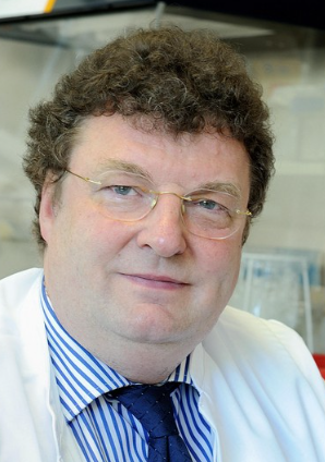 Jürgen Hescheler合作论文数Institute of Neurophysiology, University of Cologne23
 Pidder Jansen Dürr合作论文数Institute for Biomedical Ageing Research, University of Innsbruck18
 von Knebel Doeberitz Magnus合作论文数10
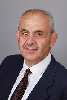 Agapios Sachinidis合作论文数Zentrum Physiologie, Medizinische Fakultät, Universität zu Köln10
 Almut Schulze合作论文数Division of Tumor Metabolism and Microenvironment, Deutsches Krebsforschungszentrum;German Cancer Research Center;Medical Faculty, Ruprecht-Karls University9
 Kurt Pfannkuche合作论文数8K Zerfass合作论文数6F. Aengeneyndt合作论文数6B Henglein合作论文数5Khalil Markus合作论文数5
 Rudolf J Wiesner合作论文数5
 Jan G. Hengstler合作论文数IfADo – Leibniz Research Centre for Working Environment and Human Factors4Konrad Brockmeier合作论文数4
 Martin Eilers合作论文数IUniversität Würzburg4Johannes Winkler合作论文数4S Joswig合作论文数3Ruediger Ridder合作论文数3
 Martin S Staege合作论文数3
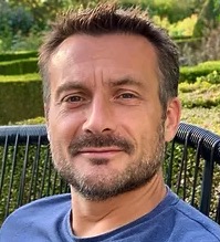 Mathieu Vinken合作论文数Laboratory of In Vitro Toxicology and Dermato-Cosmetology, Vrije Universiteit Brussel-Belgium;Department of Pathology, School of Veterinary Medicine and Animal Science, University of Sao Paulo-Brazil3Sylvia Escher合作论文数3
 Vera Rogiers合作论文数Department of Toxicology, Dermato-Cosmetology and Pharmacognosy, Faculty of Medicine and Pharmacy, Vrije Universiteit Brussel3Jiri Lukas合作论文数3Alexander Pajic合作论文数3
 Bob van de Water合作论文数Division of Toxicology;Center for Drug Research;Leiden University;Center for Drug Research, Leiden University3Axel Polack合作论文数Institut fur Virologie im Zentrum fur Hygiene, der Albert-Ludwigs-Universitet Freiburg3
 Tomo Saric合作论文数Institute for Neurophysiology, Zentrum Physiologie, Zentrum Physiologie, Universität zu Köln;Center for Physiology and Pathophysiology, Medical School, University of Cologne3Georg W. Bornkamm合作论文数3
 Jos Kleinjans合作论文数Department of Health Risk Analysis and Toxicology, Faculties of Health Sciences and Medicine, Maastricht University3P Steiner合作论文数2
 Denis Angoulvant合作论文数2Kriengchai Prasongsukarn合作论文数2
 Swapan Maiti合作论文数2A Ullmann合作论文数2Rodrigues Robim M合作论文数European Commission Joint Research Centre, Institute for Health and Consumer Protection2
 Hoffmann Simone合作论文数Department of Anatomical Sciences, Stony Brook University2Wouters Guy合作论文数2Kempkes Bettina合作论文数2
 Regina Stoeber合作论文数2U. Schäfer合作论文数2
 Edmund Neugebauer合作论文数Brandenburg Medical School;Health Services Research2
 Albert Sickmann合作论文数Rudolf-Virchow-Center for Experimental Biomedicine, Universitaet Wuerzburg2
 Massimo Tommasino合作论文数2
 Maya Simionescu合作论文数Institute of Cellular Biology and Pathology N. Simionescu2
 Hai Tian (田海)合作论文数Department of Cardiovascular Surgery,The Second Clinical College, Harbin Medical University,Harbin ,China2
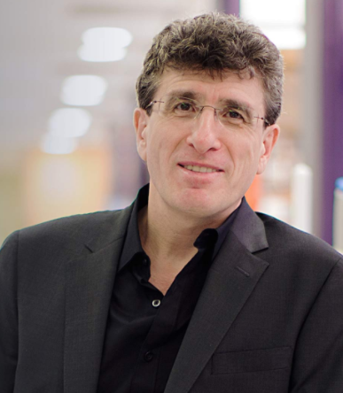 Michele Pagano合作论文数Department of Biochemistry and Molecular Pharmacology, New York University Grossman School of Medicine2
 Annette Kopp-Schneider合作论文数Division of Biostatistics, German Cancer Research Center2
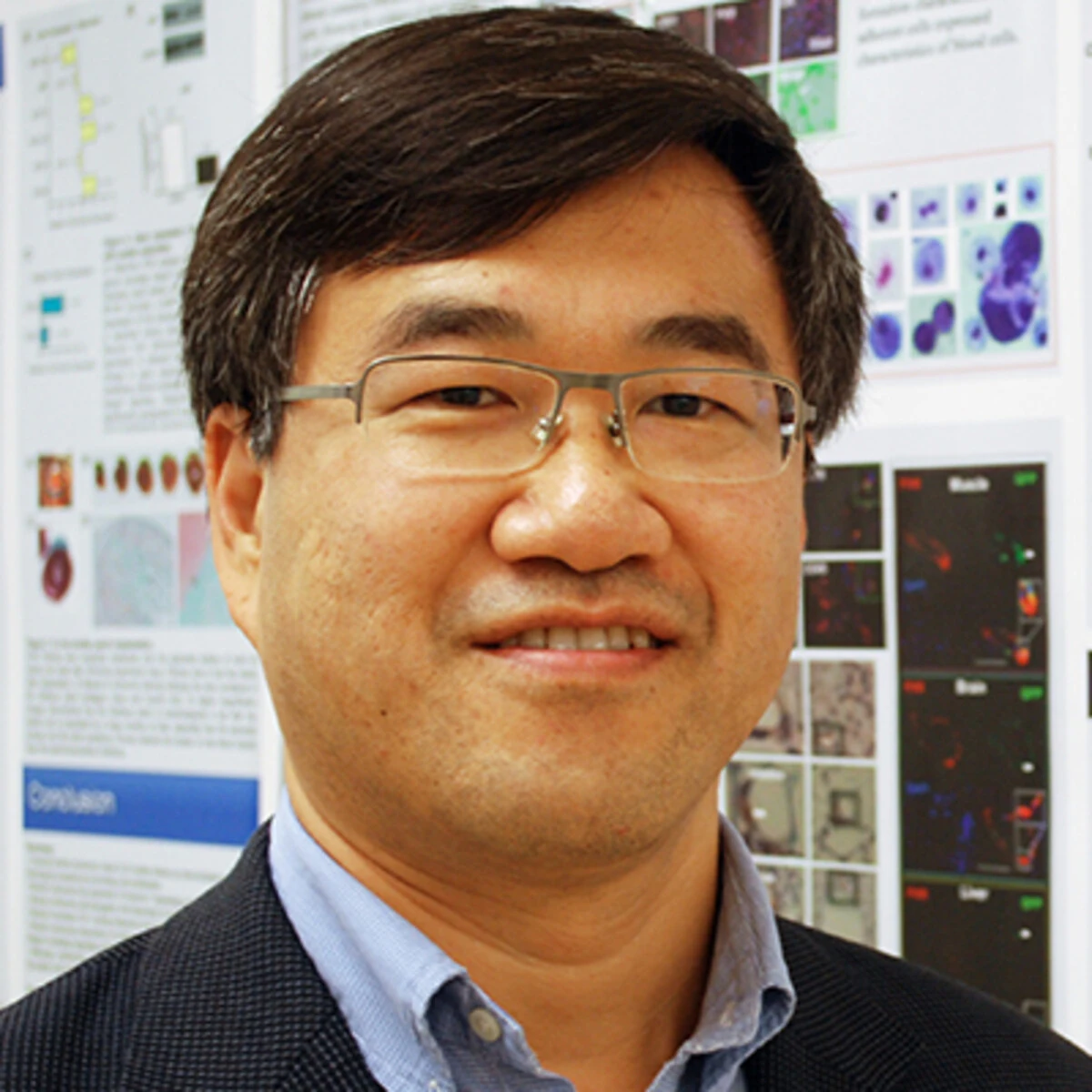 Ren-Ke Li合作论文数Department of Surgery, Division of Cardiac Surgery, Temerty Faculty of Medicine, University of Toronto;Toronto General Hospital Research Institute, University Health Network2Frank Thomas Wunderlich合作论文数2
 Markus Brielmeier合作论文数Institut fer Klinische;Molekularbiologie und Tumorgenetik;Institut fer Klinische|Molekularbiologie und Tumorgenetik2




