BACKGROUND:Cardiac fibrosis is a major cause of cardiac dysfunction and is associated with virtually all forms of heart disease. Recently, single-cell genomic approaches have revealed in unprecedented resolution the orchestrated cellular responses driving cardiac fibrosis. Yet, the fibrosis-inducing phenotypes that emerge in the heart after nonischemic cardiac stress and the transcriptional circuits that govern fibrogenic cellular phenotypes are not well understood.METHODS:Applying a single-cell paired-multiomic approach-by which both transcriptomic and epigenetic information is captured from individual cells-we reveal key transcription factors, in mouse and human hearts, associated with fibrosis development after nonischemic cardiac insults. Using high-throughput bulk transcriptomic and proteomic analyses, microscopy, and functional in vitro assays, we validate the distinct roles of new and established transcription factors in cardiac fibrosis.RESULTS:Analysis of mouse hearts undergoing reverse remodeling after angiotensin II stimulation, where cardiac fibrosis dissipates, we find these factors are reversibly activated. Further, silencing transcription factors-including those we have identified that are previously unlinked to cardiac fibrosis, such as CREB3L2 (CAMP Responsive Element Binding Protein 3 Like 2), BNC2 (Basonuclin Zinc Finger Protein 2), and NFAT5 (Nuclear Factor of Activated T Cells 5)-modulates induction of extracellular matrix gene expression by human cardiac fibroblasts. Detailed analysis of CREB3L2 showed that it regulates cardiac fibrosis by modulating extracellular matrix synthesis through a dual mechanism-involving its N-terminal transactivation domain and a paracrine-acting C-terminal fragment-which is triggered after endoplasmic reticular stress.CONCLUSIONS:This study identifies critical transcription factors regulating cardiac fibrosis and offers promising new targets to ameliorate the development of fibrosis in the context of stressors that cause cardiac dysfunction.
HIV infection is accompanied by chronic inflammation-related co-morbidities, even when viral replication is suppressed by therapy. This persistent inflammatory state suggests that long-lived immune cell lineages may acquire stable pro-inflammatory programming. Here, we investigate whether inflammatory programming can be imprinted within hematopoietic lineages, following the exposure of mice and bone marrow-derived macrophages (BMDMs) to extracellular vesicles (EVs) carrying Nef, a key inflammatory factor of HIV. Multi-omics profiling shows that hematopoietic cells exposed to Nef-EVs undergo epigenetic remodeling and reprogramming of energy and lipid metabolism characteristic of trained innate immunity. The inflammatory phenotype in BMDMs is partially reversed by inhibition of glycolysis, a key metabolic driver of trained immunity. We demonstrate that following competitive bone marrow transplantation, hematopoiesis in mice receiving bone marrow from Nef-EV-treated donors displays a sustained bias toward myelopoiesis, and BMDMs retain enhanced inflammatory potential. These findings demonstrate that Nef-EVs can imprint a lasting inflammatory memory, mechanistically similar to trained immunity, in hematopoietic cells. This memory persists beyond the initial exposure and may contribute to chronic inflammation in people with HIV.
The biological mechanisms that sustain the vast blood production required for healthy life remain incompletely understood. To search for cell intrinsic regulators of hematopoiesis, we perform a genome-wide in vivo hematopoietic stem and progenitor cell (HSPC)-based CRISPR knockout screen. We discover SAGA complex members, including Tada2b and Taf5l, as key regulators of hematopoiesis. Loss of Tada2b or Taf5l strongly inhibits hematopoiesis in vivo, causing a buildup of immature hematopoietic cells in the bone marrow. The SAGA complex deposits histone H3 lysine 9 acetylation (H3K9ac) and removes histone H2B ubiquitination (H2Bub). Loss of Tada2b leads to a reduction in H3K9ac levels and altered H2Bub enrichment in HSPCs, implicating disruption of SAGA complex activity. This is associated with upregulation of interferon pathway genes, reduced mitochondrial activity, and increased megakaryocyte progenitor cell commitment. Loss of these factors also enhances the cell outgrowth and the interferon pathway in an in vivo human myelodysplastic syndrome cell line model. In summary, this study identifies the SAGA complex as an important regulator of hematopoiesis.
These preclinical trials provide the first evidence of cluster of differentiation 14 (CD14) blockade with a murine analogue of atibuclimab, a CD14-neutralizing antibody, preventing secondary immunological exacerbation of cardiac injury in a translational mouse model of reperfused ST-segment elevation myocardial infarction (STEMI), assessed using multiple clinical modalities. Multiomic studies suggest CD14 blockade downregulated macrophage-specific proinflammatory and tissue-wide remodeling processes without suppression of monocyte-macrophage infiltration or repair. These findings support a clinically practicable targeted immunomodulatory strategy of CD14 blockade initiated at reperfusion to prevent chronic immunological progression toward ischemic heart failure, and provide new insights into the pleiotropic roles of CD14 in inflammation and myocardial injury.
Background: Aortic stiffening is a consequence of hypertension and a major contributor to end organ damage. A key driver of aortic stiffening is fibrosis involving the excess production of extracellular matrix (ECM) proteins such as collagen, fibronectin and laminin. The present study aimed to identify the cell types and signalling mechanisms that contribute to aortic fibrosis in hypertension. Methods and Results: Male C57BL/6 mice (10-12-week-old) were randomly assigned to a 28-day angiotensin II (0.7 mg/kg/day) or vehicle (saline) infusion via osmotic minipump (s.c.). At endpoint, scRNA-seq analysis of 26,196 cells recovered all major aortic cell populations. Among these, fibroblasts exhibited the greatest heterogeneity and shift in gene expression after angiotensin II compared to all other cell types. Gene ontology analyses revealed that after angiotensin II treatment, a particular subcluster of fibroblasts (Fibro-Cthrc1) - characterised by its high expression of Cthrc1 - was especially fibrogenic. Fibro-Cthrc1 cells were nearly undetectable in aortas from vehicle-infused mice. Transcripts relating to ECM remodelling (Thbs2, Cdh11 and Postn) and collagen production (specifically collagen type I, III and V) were more highly enriched in Fibro-Cthrc1 compared to other fibroblasts within hypertensive aortas. Moreover, GO terms corresponding to profibrotic signalling pathways (i.e., cell adhesion, extracellular matrix organisation and collagen fibril organisation) were significantly enriched in Fibro-Cthrc1. Spatial transcriptomics and immunohistochemistry confirmed the presence of Fibro-Cthrc1 in the adventitial layer of angiotensin II-infused but not vehicle-infused mice. Finally, analysis of plasma analytes in approximately 24,000 participants of the UK Biobank collection revealed CTHRC1 to be strongly associated with raised systolic blood pressure and pulse pressure, and a strong predictor of the risk of developing hypertension over a 15-year follow-up. Conclusion: Our study identifies a novel fibroblast subcluster, Fibro-Cthrc1, as a potential driver of aortic fibrosis and stiffening in hypertension. This cluster is absent in normotensive aortas, suggesting that targeting Fibro-Cthrc1 therapeutically could prevent aortic fibrosis and its associated hypertensive end-organ damage. Notably, such an approach may avoid compromising physiological extracellular matrix production and vessel integrity. ### Competing Interest Statement The authors have declared no competing interest.
ABSTRACT:Self-renewing multipotent hematopoietic stem cells (HSCs) are a rare but important cell population that can reconstitute the entire blood and immune system after transplantation. Due to their rarity, it has been difficult to comprehensively study the mechanisms regulating HSC activity. However, recent improvements in hematopoietic stem and progenitor cell (HSPC) culture methods using polyvinyl alcohol-based media now facilitate large-scale ex vivo HSC expansion. Here, we performed a genome-wide CRISPR knockout (KO) screen in primary mouse HSPCs to discover novel regulators of ex vivo expansion. The screen identified Runx2 as a strong negative regulator of HSC expansion, which we validated using ex vivo and in vivo assays. Loss of Runx2 increased the frequency of immunophenotypic HSCs in HSPC cultures by approximately threefold. After expansion, these Runx2-KO HSCs engrafted at approximately fivefold higher levels in transplantation assays. Noncultured Runx2-KO HSCs also displayed enhanced reconstitution potential, but loss of Runx2 did not alter blood parameters. Notably, however, T-cell reconstitution was diminished from Runx2-KO HSCs, and we further validated an additional role for Runx2 in T-cell commitment using ex vivo and in vivo assays. In summary, we have identified a multifaceted role for Runx2 in HSCs, as a negative regulator of HSC self-renewal and as a facilitator of T-cell commitment. These results contribute to our understanding of the transcriptional regulation of hematopoiesis and HSC therapies.
The hematopoietic system declines with age, and its dysfunction is associated with many diseases, ranging from infections to cancers. However, the processes that sustain lifelong hematopoiesis remain incompletely understood. To identify genetic regulators of hematopoiesis, we developed an in vivo hematopoietic stem cell (HSC)-based large-scale CRISPR knockout screening platform. Targeting ~2000 genes with this platform, we identified SAGA complex members Tada2b and Taf5l as key regulators of hematopoiesis. Genetic perturbation of Tada2b or Taf5l in murine HSCs led to significant HSC expansion ex vivo, strongly inhibited hematopoiesis in vivo, led to a buildup of immature hematopoietic cells in the bone marrow, and was associated with upregulation of interferon pathway genes. These results were validated in an ex vivo human HSC culture as well, and loss of these SAGA complex components enhanced the cell outgrowth and drives interferon pathway gene expression in an in vivo human myelodysplastic syndrome model, suggesting that loss of SAGA complex activity could contribute to hematological disease progression in humans. Altogether, this study offers a new platform to screen genetic regulators of hematopoiesis and identifies novel regulators of hematopoiesis.
Self-renewing multipotent haematopoietic stem cells (HSCs) are a rare but indispensable cell population for the life-long maintenance of the haematopoietic system. The loss or dysfunction of haematopoiesis underlies a wide range of disorders which collectively contribute to an estimated 70,000 deaths per year in the US. HSCs are also important therapeutically because they are used in stem cell transplantation and associated gene therapies for various blood diseases. A major hurdle in studying the haematopoietic system has been the availability of tractable ex vivo models of human haematopoiesis that supports deep experimental interrogation. This has at least in part been due to a lack of culture conditions that stably maintain and expand HSCs ex vivo.Building on recent advances in polymer-based expansion of functional human HSCs, we have optimised ex vivo culture conditions for human HSC expansion and the directed differentiation of expanded HSCs into 6 major blood lineages – erythroid, megakaryocytic, monocytic, granulocytic, and the B and T lymphoid lineages. To enhance reproducibility and tractability of this model, we have focused on generating serum-free, feeder-free protocols that are amenable to genetic modification. We have validated these ex vivo culture-derived cell types using flow cytometry, imaging, RNA-sequencing, and functional assays. We hope that this ex vivo human model system will support investigations into the molecular regulation of healthy and diseased haematopoiesis, and facilitate the development of new HSC-based therapies.
ABSTRACT Cardiac fibrosis is a major cause of cardiac dysfunction. Recently, single-cell genomic approaches have revealed in unprecedented resolution the orchestrated cellular responses driving cardiac fibrosis. Yet, the fibrosis-causing phenotypes that emerge in the heart following non-ischemic cardiac stress, and the transcriptional circuits that govern cell identity and drive fibrosis, are not well understood. Applying a paired multiomic approach, we reveal key transcriptional circuits, in mouse and human hearts, which are associated with fibrosis development following non-ischemic cardiac insults, independent of disease model, species or biological sex. Strikingly, we find the key regulatory events driving fibrosis are reversible at the single-cell transcriptional and epigenomic level, further pointing to key factors regulating fibrosis development and resolution. The transcriptional regulators identified in this study represent promising targets to ameliorate the development of fibrosis in the context of chronic stressors such as aging and hypertension.
Excessive adipose tissue expansion is often linked with type-2 diabetes. Despite recent efforts mapping adipose tissue changes in obesity using single-cell omics, an understanding of cellular and gene expression changes in a model of type 2 diabetes, and the transcriptional circuitry controlling it, is still lacking. Here, we use single-nucleus RNA sequencing to analyse the remodelling of gonadal white and interscapular brown adipose tissue from female and male mice with or without diabetes. Analysis of 51,877 nuclei revealed altered phenotypes in every cell population in type 2 diabetes. This included an immunoregulatory response, and changes in extracellular matrix organisation, energy generation, and hormone stimuli. Key transcription factors were inferred as cell-specific and non-specific nodes controlling diabetes-linked phenotypes. Finally, female-to-male population heterogeneity and gene expression differences were observed. Here we provide a resource detailing how adipose tissue remodelling, and the molecular mechanisms governing it, may contribute to cardiometabolic disease.### Competing Interest StatementThe authors have declared no competing interest.
Haematopoietic stem cells (HSCs) sustain life-long blood system homeostasis through their capacities for self-renewal and multilineage differentiation. HSCs can also reconstitute the entire blood and immune system following transplantation, which is used clinically in stem cell transplantation and associated gene therapies. HSC therapies can be used to treat a wide array of life-threatening diseases, including malignancies, hemoglobinopathies, metabolic diseases, bone marrow failure and immune disorders. The clinical and scientific utility of HSCs is currently constrained by the paucity of HSCs and the limited availability of suitable donors, underscoring the need for alternative sources of HSCs. Ex vivo HSC expansion represents an important approach to help improve clinical HSC therapies and to ask biological questions in molecular haematopoiesis. However, stable ex vivo HSC expansion culture conditions have long remained elusive.Drawing on recent advancements in polymer-based expansion of functional HSCs (Wilkinson et al., 2019; Sakurai et al., 2023), we have refined ex vivo culture conditions to enhance long-term expansion of human HSCs. We achieved this by optimising HSC signalling pathway stimulation and modulating oxygen availability. These optimisations have boosted human HSC expansion from ∼75-fold to ∼400-fold (in 4-week cultures). To characterise the HSCs grown in these enhanced culture conditions, we employed a multi-faceted approach including flow cytometry, RNA-sequencing, and xenotransplantation assays. Furthermore, we have validated that HSCs cultured under these conditions are amenable to genetic modification and directed differentiation. We anticipate that this optimised ex vivo human HSC model system will support efforts to understand the molecular mechanisms governing healthy and diseased haematopoiesis and lead to new clinical advances.
CRISPR-Cas9 paired with adeno-associated virus serotype 6 (AAV6) is among the most efficient tools for producing targeted gene knockins. Here, we report that this system can lead to frequent concatemeric insertions of the viral vector genome at the target site that are difficult to detect. Such errors can cause adverse and unreliable phenotypes that are antithetical to the goal of precision genome engineering. The concatemeric knockins occurred regardless of locus, vector concentration, cell line or cell type, including human pluripotent and hematopoietic stem cells. Although these highly abundant errors were found in more than half of the edited cells, they could not be readily detected by common analytical methods. We describe strategies to detect and thoroughly characterize the concatemeric viral vector insertions, and we highlight analytical pitfalls that mask their prevalence. We then describe strategies to prevent the concatemeric inserts by cutting the vector genome after transduction. This approach is compatible with established gene editing pipelines, enabling robust genetic knockins that are safer, more reliable and more reproducible. AAV vectors form difficult-to-detect concatemers at Cas9 target sites.
Cardiac fibrosis—the deposition of excess extracellular matrix in the heart—occurs during pathological cardiac remodelling and precedes cardiac dysfunction and heart failure. Experimental and clinical data show that both cardiac fibrosis and heart failure exhibit sex-specific differences, particularly in the absence of ischemic injury. However, no study to date has systematically examined this, or the hormonal mechanisms driving these differences, using high-resolution single-cell and spatial omics. To address this gap, we applied single-cell and spatial omics to map the quality and distribution of non-ischemic cardiac fibrosis in human and mouse hearts. Further, to query the impact of sex hormones on cardiac fibrosis development, we analysed hearts of castrated (CAST) and ovariectomized (OVX) mice, which were ectopically infused with angiotensin-II (AngII). Using single cell transcriptomic data of hypertensive mouse hearts, we identified a fibroblast population driving cardiac fibrosis with a unique gene signature. We confirmed the presence of these cells in multiple mouse models and aged donor human hearts. Spatial mapping of fibrosis showed a sex-specific distribution of fibrosis, specifically in the development of perivascular fibrosis in the heart. Hypertensive male mice exhibited extensive perivascular fibrosis, and coronary artery adventitial hypertrophy, which was absent in females. However, OVX resulted in the development of perivascular fibrosis in hypertensive mice suggesting a cardioprotective role of estrogen. Confirming these observations, spatial transcriptomic analyses showed distinct fibrotic signatures driving perivascular and interstitial fibrosis, and transcriptomic differences between females and males. Our findings provide fundamental new insights towards the importance of biological sex in driving cardiac fibrosis, pointing to new mechanisms that may be manipulated to ameliorate development of heart failure.
The biological mechanisms that sustain the vast blood production required for healthy life remain incompletely understood. To address this knowledge gap, we developed an in vivo hematopoietic stem cell (HSC)-based large-scale CRISPR knockout screening platform to enable the genetic interrogation of hematopoiesis and broad aspects of immune cell function in vivo. Targeting ∼7000 genes with this methodology, we discovered SAGA complex members Tada2b and Taf5l as key regulators of HSC lineage commitment. Loss of Tada2b or Taf5l inhibited hematopoiesis in vivo and was associated with upregulation of interferon response gene expression. SAGA complex member expression is significantly reduced in aged HSCs and upregulated with heterochronic parabiosis, suggesting a novel mechanism of age-associated hematopoietic decline and rejuvenation. Our study provides a rich functional genetics resource of hematopoiesis regulators accessible through a public interactive database (www.hematopoiesiscrisprscreens.com), a novel mechanism regulating age-related decline of hematopoiesis, and a new methodology with broad applications to systematically probe the development and functions of the lymphohematopoietic system.
Diabetes is associated with a significantly elevated risk of heart failure. However, despite extensive efforts to characterize the phenotype of the diabetic heart, the molecular and cellular protagonists that underpin cardiac pathological remodeling in diabetes remain unclear, with a notable paucity of data regarding the impact of diabetes on non-myocytes within the heart. Here we aimed to define key differences in cardiac non-myocytes between spontaneously type-2 diabetic (db/db) and healthy control (db/h) mouse hearts. Single-cell transcriptomic analysis revealed a concerted diabetes-induced cellular response contributing to cardiac remodeling. These included cell-specific activation of gene programs relating to fibroblast hyperplasia and cell migration, and dysregulation of pathways involving vascular homeostasis and protein folding. This work offers a new perspective for understanding the cellular mediators of diabetes-induced cardiac pathology, and pathways that may be targeted to address the cardiac complications associated with diabetes.
Hematopoietic stem cells (HSCs) are a rare type of hematopoietic cell that can entirely reconstitute the blood and immune system after transplantation. Allogeneic HSC transplantation (HSCT) is used clinically as a curative therapy for a range of hematolymphoid diseases; however, it remains a high-risk therapy because of its potential side effects, including poor graft function and graft-versus-host disease (GVHD). Ex vivo HSC expansion has been suggested as an approach to improve hematopoietic reconstitution in low-cell dose grafts. Here, we demonstrate that the selectivity of polyvinyl alcohol (PVA)-based mouse HSC cultures can be improved using physioxic culture conditions. Single-cell transcriptomic analysis helped confirm the inhibition of lineage-committed progenitor cells in physioxic cultures. Long-term physioxic expansion also afforded culture-based ex vivo HSC selection from whole bone marrow, spleen, and embryonic tissues. Furthermore, we provide evidence that HSC-selective ex vivo cultures deplete GVHD-causing T cells and that this approach can be combined with genotoxic-free antibody-based conditioning HSCT approaches. Our results offer a simple approach to improve PVA-based HSC cultures and the underlying molecular phenotype, and highlight the potential translational implications of selective HSC expansion systems for allogeneic HSCT.
 Adam Wilkinson合作论文数School of Animal and Microbial Sciences;University of Reading;School of Animal and Microbial Sciences, University of Reading16
 Crisdion Krstevski合作论文数Baker Heart and Diabetes Institute15
 Gabriella Farrugia合作论文数Baker Heart and Diabetes Institute15
 Malathi Imiyage Dona合作论文数Baker Heart and Diabetes Research Institute12
 Taylah L Gaynor合作论文数Dept Microbiol Anat Physiol & Pharmacol, La Trobe Univ12
 Alexander Pinto合作论文数Baker Department of Cardiovascular Research, Translation and Implementation, La Trobe University;Cardiac Cellular Systems Laboratory, Baker Heart and Diabetes Institute12
 Hiromitsu (Hiro) Nakauchi合作论文数Department of Genetics, Stanford University12
 Charles D Cohen合作论文数Baker Heart and Diabetes Institute10
 Joydeep Bhadury合作论文数Institute of Clinical Sciences, University of Gothenburg;Institute of Clinical Sciences, University of Gothenburg9
 Alan M Moses合作论文数Department of Computer Science, Faculty of Arts and Science, University of Toronto7Carsten T Charlesworth合作论文数Stanford Univ, Dept Pediat, Stanford, CA 94305 USA6Helen Kiriazis合作论文数6Bob Strome合作论文数5M. Dona合作论文数4
 Grant Drummond合作论文数Department of Physiology, Anatomy and Microbiology, La Trobe University;School of Agriculture, Biomedicine and Environment, La Trobe University4Hwei Minn Khoo合作论文数4Kyomi J Igarashi合作论文数4
 Archana Shankar合作论文数Inst Genom Med, Univ Calif San Diego4
 Daniel G Donner合作论文数4
 Tony Wyss-Coray合作论文数Department of Neurology and Neurological Sciences, School of Medicine, Stanford University4
 David W. Greening合作论文数Joint Proteomics Laboratory, Ludwig Institute for Cancer Research and the Walter and Eliza Hall Institute of Medical Research4Jinyu Zhang合作论文数3Maimi Higuchi合作论文数3Tze-Kai Tan合作论文数3
 Darnel Prakoso合作论文数Drug Discovery Biol, Monash Inst Pharmaceut Sci3Yavor Bozhilov合作论文数3Ron Baik合作论文数Department of Pediatrics, Stanford University3
 David M Nash合作论文数Drug Discovery Biol, Monash Univ3Minh Deo合作论文数Baker Heart & Diabet Inst, Melbourne, Vic, Australia3Chika Morita合作论文数3Mandy Li合作论文数3
 Miles J De Blasio合作论文数Baker Heart and Diabetes Institute3
 Toshiya Nishimura合作论文数3
 Fabian Suchy合作论文数Center for Eukaryotic Structural Genomics, University of Wisconsin-Madison3
 Nishimura Toshinobu合作论文数The Institute of Medical Science, The University of Tokyo3
 Ravi Majeti合作论文数Institute for Stem Cell Biology and Regenerative Medicine, Cancer Institute, Division of Hematology, Department of Medicine, Stanford University3
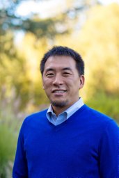 Yusuke Nakauchihiroshi合作论文数Department of Hematology, Tokyo Medical and Dental University3
 Julie R. Mcmullen合作论文数Heart and Diabetes Institute, Baker IDI3Róbert Pálovics合作论文数2Christy Munson合作论文数2
 Francesco E Emiliani合作论文数Molecular and Systems Biology, Dartmouth College2
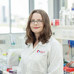 Bethany Claridge合作论文数Baker Heart and Diabetes Institute2
 Amy Fan合作论文数Stanford University2
 Joab Camarena合作论文数Stanford Univ, Dept Pediat, Stanford, CA 94305 USA2
 Sergey V. Plotnikov合作论文数Department of Cell & Systems Biology, Faculty of Arts and Science, University of Toronto2
 Sean Lal合作论文数School of Medicine Sciences, Faculty of Medicine and Health, The University of Sydney2Karigane Daiki合作论文数2
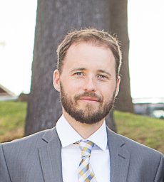 Aaron McKenna合作论文数The Broad Institute of MIT and Harvard, Cambridge, MA 02142;The Broad Institute of MIT and Harvard, Cambridge, MA 02142;2
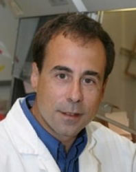 Mark A. Kay合作论文数Departments of Pediatrics and Genetics, School of Medicine, Stanford University;Division of Human Gene Therapy, Department of Pediatrics, School of Medicine, Stanford University2




