The biological mechanisms that sustain the vast blood production required for healthy life remain incompletely understood. To search for cell intrinsic regulators of hematopoiesis, we perform a genome-wide in vivo hematopoietic stem and progenitor cell (HSPC)-based CRISPR knockout screen. We discover SAGA complex members, including Tada2b and Taf5l, as key regulators of hematopoiesis. Loss of Tada2b or Taf5l strongly inhibits hematopoiesis in vivo, causing a buildup of immature hematopoietic cells in the bone marrow. The SAGA complex deposits histone H3 lysine 9 acetylation (H3K9ac) and removes histone H2B ubiquitination (H2Bub). Loss of Tada2b leads to a reduction in H3K9ac levels and altered H2Bub enrichment in HSPCs, implicating disruption of SAGA complex activity. This is associated with upregulation of interferon pathway genes, reduced mitochondrial activity, and increased megakaryocyte progenitor cell commitment. Loss of these factors also enhances the cell outgrowth and the interferon pathway in an in vivo human myelodysplastic syndrome cell line model. In summary, this study identifies the SAGA complex as an important regulator of hematopoiesis.
Organ shortage remains a major challenge in transplantation medicine. Interspecies blastocyst complementation offers a promising strategy to generate human organs in livestock. However, efficient xenogeneic donor cell engraftment remains challenging. Here, we identify an innate immune barrier wherein host macrophages selectively eliminate viable xenogeneic donor cells, a process we term xenophagocytosis. Mechanistically, xenogeneic cells display elevated phosphatidylserine, an "eat-me" signal recognized by host macrophages through phagocytic receptor Axl. We demonstrate three orthogonal strategies for xenophagocytosis blockade: genetic ablation of macrophages or the Axl receptor in the host embryo or overexpression of the "don't-eat-me" signal CD47 or the phosphatidylserine-regulating flippase ATP11C in donor cells. Xenophagocytosis blockade enhances rat and human donor chimerism in mouse embryos and improves interspecies pancreas complementation efficiency. These findings reveal a previously unrecognized innate immune barrier that safeguards species integrity during early embryogenesis and provide mechanistic insights to enhance xenogeneic chimerism for generating human organs in livestock.
Hematopoietic stem cells (HSCs) are blood-forming stem cells that can reconstitute the entire blood system after transplantation, which provides curative options for both blood and non-blood diseases. Despite their known utility, broader applications of human HSCs are hampered by the lack of a stable culture protocol. Previously, we developed a long-term mouse HSC expansion protocol using a synthetic polymer to replace serum albumin, thus mitigating inflammatory responses that trigger HSC differentiation. Although this approach, with ongoing improvements, has been applied to human HSC cultures, achieving long-term expansion of human HSCs remains challenging. To address this, we optimized a previously published cytokine-free culture for human HSCs, which utilizes butyzamide, a thrombopoietin (TPO) receptor agonist (TPO-RA), and 740 Y-P, a PI3K activator, as replacements for TPO and stem cell factor (SCF), respectively. In a short-term (7-day) culture, we found that supplementing SCF in addition to 740 Y-P, or substituting butyzamide with lusutrombopag, an FDA-approved TPO-RA, enhanced the ex vivo expansion of phenotypic hematopoietic stem and progenitor cells (lineage⁻CD41⁻CD34⁺) from CD34+ umbilical cord blood (UCB) cells. In xenograft transplantation model using immunodeficient NSG mice, a mid-term (14-day) physioxic (5% O₂) culture supplemented with lusutrombopag and SCF, named “5LS culture”, resulted in robust expansion of engraftable human HSCs (mean human chimerism: 22.9% [14-day expanded] vs. 0.96% [unmanipulated], with multilineage reconstitution of CD33⁺ myeloid and CD19⁺ B cells; N = 7 mice, 16 weeks post-transplantation). Molecularly, SCF consistently upregulated phosphorylation of AKT, STAT5, and p38 in phenotypic multipotent progenitors/HSCs (pMPPs/pHSCs: lineage⁻CD41⁻CD34⁺EPCR⁺) across different samples, whereas 740 Y-P did not, suggesting a mechanistic insight why SCF addition improves human HSC expansion. We next extended the culture duration to 28 days. Interestingly, despite successful 14-day expansion in the 5LS culture, CD34⁺ UCB cells cultured for 28 days predominantly reconstituted CD33⁺ myeloid cells but lost their lymphoid reconstitution potential in NSG mice. Further optimization identified that FLT3 ligand (FLT3-L) supplementation in a 5LS culture (resulting in the “5LSF culture”) improved both the expansion and the maintenance of phenotypic HSCs (pHSCs: lineage⁻CD41⁻CD34⁺EPCR⁺CD90⁺ITGA3⁺) over 28-day culture of CD34+ UCB cells. Similar results were observed using CD34⁺ mobilized peripheral blood cells. Single-cell RNA sequencing with UMAP visualization and clustering analyses revealed that while HSC/MPP clusters from 5LS cultures were skewed toward granulocyte-monocyte progenitors (GMPs), this skewing was absent in the 5LSF condition. Notably, xenotransplantation demonstrated partial restoration of CD19⁺ B cell reconstitution from 28-day expanded CD34⁺ UCB cells in the 5LSF culture (mean B cell contribution among human cells: 0% [5LS culture] vs. 32.1% [5LSF culture], N = 7 mice, 16 weeks post-transplantation). These results highlight that FLT3-L is critical for maintaining multipotency of HSCs in long-term culture.To assess whether the 5LSF culture supports genetic modification, we performed lentiviral transduction and CRISPR-Cas9/AAV6-mediated targeted gene editing in CD34⁺ UCB cells. Lentiviral vectors successfully transduced the pHSC population. Targeted editing of a reporter cassette in a HLF (a transcription factor specifically expressed in HSCs) locus confirmed efficient editing in HLF⁺ HSCs, underscoring the utility of 5LSF culture for HSC genetic engineering. Finally, a limiting dilution assay using unmanipulated, 14-day-expanded, and 28-day-expanded CD34+ UCB cells enabled estimation of functional HSC frequencies and fold expansion over a 28-day culture. The 5LSF culture exhibited the highest fold expansion of functional HSCs (109.0-fold expansion) after 28 days. In contrast, the 5LS culture demonstrated greater fold expansion of functional HSCs after 14 days (60.6-fold expansion) compared to the 5LSF culture (23.1-fold expansion), which is consistent with the results from the phenotypic characterization. These findings provide evidence that human functional HSC can stably expands long-term in the 5LSF culture. In conclusion, we have developed a long-term expansion protocol for human HSCs that facilitates both clinical and research applications of these functional stem cells.
Intercellular transmission of messenger RNA (mRNA) is being explored in mammalian species using immortal cell lines. Here, we uncover an intercellular mRNA transfer phenomenon that allows for the adaptation and reprogramming of human primed pluripotent stem cells (hPSCs). This process is induced by the direct cell contact-mediated coculture with mouse embryonic stem cells under the condition impermissible for primed hPSC culture. Mouse-derived mRNA contents are transmitted into adapted hPSCs only in the coculture. Transfer-specific mRNA analysis shows the enrichment for divergent biological pathways involving transcription/translational machinery and stress-coping mechanisms, wherein such transfer is diminished when direct cell contacts are lost. After 5 d of coculture with mouse embryonic stem cells, surface marker analysis and global gene profiling confirmed that mRNA transfer-prone hPSC efficiently gains a naïve-like state. Furthermore, transfer-specific knockdown experiments targeting mouse-specific transcription factor-coding mRNAs in hPSC show that mouse-derived Tfcp2l1, Tfap2c, and Klf4 are indispensable for human naïve-like conversion. Thus, interspecies mRNA transfer triggers cellular reprogramming in mammalian cells. Our results support that episodic mRNA transfer can occur in cell cooperative and competitive processes, which provides a fresh perspective on understanding the roles of mRNA mobility for intra- and interspecies cellular communications.
ABSTRACT:Self-renewing multipotent hematopoietic stem cells (HSCs) are a rare but important cell population that can reconstitute the entire blood and immune system after transplantation. Due to their rarity, it has been difficult to comprehensively study the mechanisms regulating HSC activity. However, recent improvements in hematopoietic stem and progenitor cell (HSPC) culture methods using polyvinyl alcohol-based media now facilitate large-scale ex vivo HSC expansion. Here, we performed a genome-wide CRISPR knockout (KO) screen in primary mouse HSPCs to discover novel regulators of ex vivo expansion. The screen identified Runx2 as a strong negative regulator of HSC expansion, which we validated using ex vivo and in vivo assays. Loss of Runx2 increased the frequency of immunophenotypic HSCs in HSPC cultures by approximately threefold. After expansion, these Runx2-KO HSCs engrafted at approximately fivefold higher levels in transplantation assays. Noncultured Runx2-KO HSCs also displayed enhanced reconstitution potential, but loss of Runx2 did not alter blood parameters. Notably, however, T-cell reconstitution was diminished from Runx2-KO HSCs, and we further validated an additional role for Runx2 in T-cell commitment using ex vivo and in vivo assays. In summary, we have identified a multifaceted role for Runx2 in HSCs, as a negative regulator of HSC self-renewal and as a facilitator of T-cell commitment. These results contribute to our understanding of the transcriptional regulation of hematopoiesis and HSC therapies.
Hematopoietic stem cells (HSCs) are somatic stem cells that continuously generate lifelong supply of blood cells through a balance of symmetric and asymmetric divisions. It is well established that the HSC pool increases with age. However, not much is known about the underlying cause for these observed changes. Here, using a novel method combining single-cell ex vivo HSC expansion with mathematical modeling, we quantify HSC division types (stem cell-stem cell (S-S) division, stem cell-progenitor cell (S-P) division, and progenitor cell-progenitor cell (P-P) division) as a function of the aging process. Our time-series experiments reveal how changes in these three modes of division can explain the increase in HSC numbers with age. Contrary to the popular notion that HSCs divide predominantly through S-P divisions, we show that S-S divisions are predominant throughout the lifespan of the animal, thereby expanding the HSC pool. We, therefore, provide a novel mathematical model-based experimental validation for reflecting HSC dynamics in vivo.
The hematopoietic system declines with age, and its dysfunction is associated with many diseases, ranging from infections to cancers. However, the processes that sustain lifelong hematopoiesis remain incompletely understood. To identify genetic regulators of hematopoiesis, we developed an in vivo hematopoietic stem cell (HSC)-based large-scale CRISPR knockout screening platform. Targeting ~2000 genes with this platform, we identified SAGA complex members Tada2b and Taf5l as key regulators of hematopoiesis. Genetic perturbation of Tada2b or Taf5l in murine HSCs led to significant HSC expansion ex vivo, strongly inhibited hematopoiesis in vivo, led to a buildup of immature hematopoietic cells in the bone marrow, and was associated with upregulation of interferon pathway genes. These results were validated in an ex vivo human HSC culture as well, and loss of these SAGA complex components enhanced the cell outgrowth and drives interferon pathway gene expression in an in vivo human myelodysplastic syndrome model, suggesting that loss of SAGA complex activity could contribute to hematological disease progression in humans. Altogether, this study offers a new platform to screen genetic regulators of hematopoiesis and identifies novel regulators of hematopoiesis.
ABSTRACTA multitude of tools now exist that allow us to precisely manipulate the human genome in a myriad of different ways. However, successful delivery of these tools to the cells of human patients remains a major barrier to their clinical implementation. Here we introduce a new cellular approach forin vivogenetic engineering,SecretedParticleInformationTransfer (SPIT) that utilizes human cells as delivery vectors forin vivogenetic engineering. We demonstrate the application of SPIT for cell-cell delivery of Cre recombinase and CRISPR-Cas9 enzymes, we show that genetic logic can be incorporated into SPIT and present the first demonstration of human cells as a delivery platform forin vivogenetic engineering in immunocompetent mice. We successfully applied SPIT to genetically modify multiple organs and tissue stem cellsin vivoincluding the liver, spleen, intestines, peripheral blood, and bone marrow. We anticipate that by harnessing the large packaging capacity of a human cell’s nucleus, the ability of human cells to engraft into patients’ long term and the capacity of human cells for complex genetic programming, that SPIT will become a paradigm shifting approach forin vivogenetic engineering.
A multitude of tools now exist that allow us to precisely manipulate the human genome in a myriad of different ways. However, successful delivery of these tools to the cells of human patients remains a major barrier to their clinical implementation. Here we introduce a new cellular approach for in vivo genetic engineering, Secreted Particle Information Transfer (SPIT) that utilizes human cells as delivery vectors for in vivo genetic engineering. We demonstrate the application of SPIT for cell-cell delivery of Cre recombinase and CRISPR-Cas9 enzymes, we show that genetic logic can be incorporated into SPIT and present the first demonstration of human cells as a delivery platform for in vivo genetic engineering in immunocompetent mice. We successfully applied SPIT to genetically modify multiple organs and tissue stem cells in vivo including the liver, spleen, intestines, peripheral blood, and bone marrow. We anticipate that by harnessing the large packaging capacity of a human cell's nucleus, the ability of human cells to engraft into patients' long term and the capacity of human cells for complex genetic programming, that SPIT will become a paradigm shifting approach for in vivo genetic engineering.
CRISPR-Cas9 paired with adeno-associated virus serotype 6 (AAV6) is among the most efficient tools for producing targeted gene knockins. Here, we report that this system can lead to frequent concatemeric insertions of the viral vector genome at the target site that are difficult to detect. Such errors can cause adverse and unreliable phenotypes that are antithetical to the goal of precision genome engineering. The concatemeric knockins occurred regardless of locus, vector concentration, cell line or cell type, including human pluripotent and hematopoietic stem cells. Although these highly abundant errors were found in more than half of the edited cells, they could not be readily detected by common analytical methods. We describe strategies to detect and thoroughly characterize the concatemeric viral vector insertions, and we highlight analytical pitfalls that mask their prevalence. We then describe strategies to prevent the concatemeric inserts by cutting the vector genome after transduction. This approach is compatible with established gene editing pipelines, enabling robust genetic knockins that are safer, more reliable and more reproducible. AAV vectors form difficult-to-detect concatemers at Cas9 target sites.
Figure S1: Molecular modelling and structure determination by X ray crystallography. Expanded data related to Figure 1B. Figure S2-S3: Ingenuity Pathway analyses of microarray data. Expanded analyses related to Figure 4 and Supplemental dataset 1. Figure S4: Gene-set enrichment analyses of microarray data. Expanded analyses related to Figure 4 and Supplemental dataset 1.
The biological mechanisms that sustain the vast blood production required for healthy life remain incompletely understood. To address this knowledge gap, we developed an in vivo hematopoietic stem cell (HSC)-based large-scale CRISPR knockout screening platform to enable the genetic interrogation of hematopoiesis and broad aspects of immune cell function in vivo. Targeting ∼7000 genes with this methodology, we discovered SAGA complex members Tada2b and Taf5l as key regulators of HSC lineage commitment. Loss of Tada2b or Taf5l inhibited hematopoiesis in vivo and was associated with upregulation of interferon response gene expression. SAGA complex member expression is significantly reduced in aged HSCs and upregulated with heterochronic parabiosis, suggesting a novel mechanism of age-associated hematopoietic decline and rejuvenation. Our study provides a rich functional genetics resource of hematopoiesis regulators accessible through a public interactive database (www.hematopoiesiscrisprscreens.com), a novel mechanism regulating age-related decline of hematopoiesis, and a new methodology with broad applications to systematically probe the development and functions of the lymphohematopoietic system.
Hematopoietic stem cells (HSCs) are a rare type of hematopoietic cell that can entirely reconstitute the blood and immune system after transplantation. Allogeneic HSC transplantation (HSCT) is used clinically as a curative therapy for a range of hematolymphoid diseases; however, it remains a high-risk therapy because of its potential side effects, including poor graft function and graft-versus-host disease (GVHD). Ex vivo HSC expansion has been suggested as an approach to improve hematopoietic reconstitution in low-cell dose grafts. Here, we demonstrate that the selectivity of polyvinyl alcohol (PVA)-based mouse HSC cultures can be improved using physioxic culture conditions. Single-cell transcriptomic analysis helped confirm the inhibition of lineage-committed progenitor cells in physioxic cultures. Long-term physioxic expansion also afforded culture-based ex vivo HSC selection from whole bone marrow, spleen, and embryonic tissues. Furthermore, we provide evidence that HSC-selective ex vivo cultures deplete GVHD-causing T cells and that this approach can be combined with genotoxic-free antibody-based conditioning HSCT approaches. Our results offer a simple approach to improve PVA-based HSC cultures and the underlying molecular phenotype, and highlight the potential translational implications of selective HSC expansion systems for allogeneic HSCT.
Animal chimeras are widely used for biomedical discoveries, from developmental biology to cancer research. However, the accurate quantitation of mixed cell types in chimeric and mosaic tissues is complicated by sample preparation bias, transgenic silencing, phenotypic similarity, and low-throughput analytical pipelines. Here, we have developed and characterized a droplet digital PCR single-nucleotide discrimination assay to detect chimerism among common albino and non-albino mouse strains. In addition, we validated that this assay is compatible with crude lysate from all solid organs, drastically streamlining sample preparation. This chimerism detection assay has many additional advantages over existing methods including its robust nature, minimal technical bias, and ability to report the total number of cells in a prepared sample. Moreover, the concepts discussed here are readily adapted to other genomic loci to accurately measure mixed cell populations in any tissue.
As our closest living relatives, non-human primates uniquely enable explorations of human health, disease, development, and evolution. Considerable effort has thus been devoted to generating induced pluripotent stem cells (iPSCs) from multiple non-human primate species. Here, we establish improved culture methods for chimpanzee (Pan troglodytes) and pig-tailed macaque (Macaca nemestrina) iPSCs. Such iPSCs spontaneously differentiate in conventional culture conditions, but can be readily propagated by inhibiting endogenous WNT signaling. As a unique functional test of these iPSCs, we injected them into the pre-implantation embryos of another non-human species, rhesus macaques (Macaca mulatta). Ectopic expression of gene BCL2 enhances the survival and proliferation of chimpanzee and pig-tailed macaque iPSCs within the pre-implantation embryo, although the identity and long-term contribution of the transplanted cells warrants further investigation. In summary, we disclose transcriptomic and proteomic data, cell lines, and cell culture resources that may be broadly enabling for non-human primate iPSCs research.
The biological mechanisms that sustain the vast blood production required for healthy life remain incompletely understood. To address this knowledge gap, we developed an in vivo hematopoietic stem cell (HSC)-based large-scale CRISPR knockout screening platform to enable the genetic interrogation of hematopoiesis and broad aspects of immune cell function in vivo. Targeting ∼7000 genes with this methodology, we discovered SAGA complex members Tada2b and Taf5l as key regulators of HSC lineage commitment. Loss of Tada2b or Taf5l inhibited hematopoiesis in vivo and was associated with upregulation of interferon response gene expression. SAGA complex member expression is significantly reduced in aged HSCs and upregulated with heterochronic parabiosis, suggesting a novel mechanism of age-associated hematopoietic decline and rejuvenation. Our study provides a rich functional genetics resource of hematopoiesis regulators accessible through a public interactive database ([www.hematopoiesiscrisprscreens.com][1]), a novel mechanism regulating age-related decline of hematopoiesis, and a new methodology with broad applications to systematically probe the development and functions of the lymphohematopoietic system. ### Competing Interest Statement H.N. is a co-founder and shareholder in ReproCELL, Megakaryon, and Century Therapeutics. [1]: http://www.hematopoiesiscrisprscreens.com
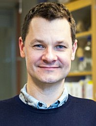 Jonas A Nilsson合作论文数University of Gothenburg15
 Hiromitsu (Hiro) Nakauchi合作论文数Department of Genetics, Stanford University15
 Lisa M Nilsson合作论文数Univ Gothenburg, Gothenburg, Sweden11
 Somsundar Veppil Muralidharan合作论文数Institute of Clinical Sciences, University of Gothenburg;Institute of Clinical Sciences, University of Gothenburg10
 Ian Hsu合作论文数Institute for Stem Cell Biology and Regenerative Medicine, Stanford University School of Medicine, Stanford, CA9
 Fabian Suchy合作论文数Center for Eukaryotic Structural Genomics, University of Wisconsin-Madison9
 Adam Wilkinson合作论文数School of Animal and Microbial Sciences;University of Reading;School of Animal and Microbial Sciences, University of Reading8Jinyu Zhang合作论文数7Lydia C. Green合作论文数7Tze-Kai Tan合作论文数5Kyomi J Igarashi合作论文数5Carsten T Charlesworth合作论文数Stanford Univ, Dept Pediat, Stanford, CA 94305 USA5
 Marcela Davila Lopez合作论文数5
 Archana Shankar合作论文数Inst Genom Med, Univ Calif San Diego4
 Toshiya Nishimura合作论文数4
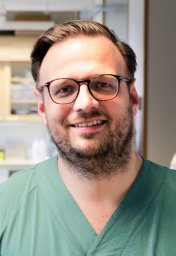 Roger Olofsson Bagge合作论文数Department of Surgery, Institute of Clinical Sciences;Sahlgrenska University Hospital, the University of Gothenburg;roger.olofsson@surgery.gu.se.;Sahlgrenska University Hospital, the University of Gothenburg4Lars Ny合作论文数4
 Berglind Einarsdottir合作论文数Sahlgrenska University Hospital, Sahlgrenska Academy at the University of Gothenburg4McLure Kevin G合作论文数4Miyauchi Masashi合作论文数4
 Tony Wyss-Coray合作论文数Department of Neurology and Neurological Sciences, School of Medicine, Stanford University4Ulrika Stierner合作论文数4Dagsu Demir合作论文数3Maimi Higuchi合作论文数3Hwei Minn Khoo合作论文数3Karigane Daiki合作论文数3
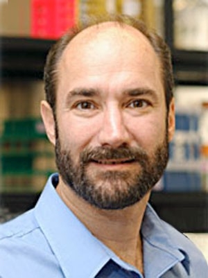 Michael Snyder合作论文数Department of Genetics, School of Medicine, Stanford University;Center for Genomics and Personalized Medicine;Karolinska Institute3Satoshi Yamazaki合作论文数3Martin Welin合作论文数3
 Bjorn Walse合作论文数3
 Derek T Logan合作论文数Department of Molecular Biophysics, Lund University3Róbert Pálovics合作论文数2Tomoya Kakegawa合作论文数2Shota Homma合作论文数2Hans Jiro Becker合作论文数2Christy Munson合作论文数2
 Amy Fan合作论文数Stanford University2
 Anais K Amaya合作论文数Children's Medical Research Institute and Sydney Children's Hospitals Network, The University of Sydney2
 Joab Camarena合作论文数Stanford Univ, Dept Pediat, Stanford, CA 94305 USA2
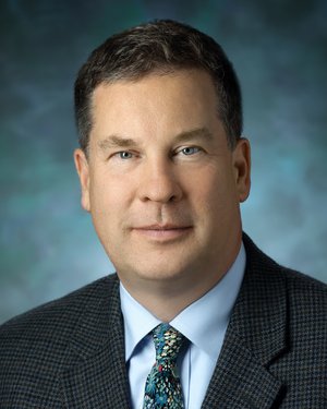 Joseph L. Mankowski合作论文数School of Medicine, Johns Hopkins University2
 Bajpai Vivek K合作论文数School of Biotechnology, Yeungnam University2Emily M. Gesner合作论文数2
 Zhoulei Li合作论文数2
 Lena Maria Nilsson合作论文数Dept Publ Hlth & Clin Med, Umea Univ2Jan Mattsson合作论文数2Masaki Hideki合作论文数Institute of Medical Science, University of Tokyo2
 Mark A. Kay合作论文数Departments of Pediatrics and Genetics, School of Medicine, Stanford University;Division of Human Gene Therapy, Department of Pediatrics, School of Medicine, Stanford University2
 Jespersen Henrik合作论文数Sahlgrenska University Hospital, Sahlgrenska Academy at the University of Gothenburg2Kouta Niizuma合作论文数2




