A 70-year-old man presented to our hospital with non-ST elevation myocardial infarction and severely depressed left ventricular systolic function. Coronary angiogram revealed a giant fusiform aneurysm of the proximal left anterior descending artery with significant stenoses immediately proximal and distal to it and a left circumflex chronic total occlusion. The patient was treated surgically, with ligation and bypass of the aneurysm using a radial artery graft and a vein graft to the first obtuse marginal branch.
Objective: Accelerated atherosclerosis in diabetes constitutes an ongoing challenge despite optimal medical therapies. This study aimed to identify evolutionarily conserved lesion-based regulatory signaling networks in diabetic versus nondiabetic conditions during the development of atherosclerosis in an initial translational effort to provide insights for targets. Approach and Results: Serial 3-mm coronary artery segments of hypercholesterolemic Yorkshire swine and diabetic-hypercholesterolemic swine were characterized as mild, moderate, or severe phenotypic manifestations of coronary atherosclerosis based on histopathologic examination. Lesional RNA sequencing was performed (n=3–8 lesions per group) corresponding to increasing phenotypic severity. Differentially expressed genes, transcription factors, upstream regulators, and hubs were validated using the NanoString technology and a human atherosclerotic specimen cohort. Despite similar stage histopathologic characterization of lesions, genome-wide transcriptomics revealed gene sets and nodal signaling pathways uniquely expressed in diabetic lesions including signaling pathways for Th17, IL (interleukin)-17F, TWEAK (TNF [tumor necrosis factor]-related weak inducer of apoptosis), CD27, and PI3K/Akt. In contrast, pathways of nondiabetic lesions involved TREM-1 and Th1 and Th2 responses during the initiation stage, whereas networks for mitochondrial dysfunction, oxidative phosphorylation, and lipid metabolism emerged with progression. RNA sequencing data were validated in a human atherosclerosis specimen cohort using machine learning algorithms. F8 , MAPKAPK3 , and ITGB1 emerged as powerful genes for clustering diabetic versus nondiabetic lesions and for separating different degrees of atherosclerosis progression. Conclusions: This study identifies evolutionarily conserved gene signatures and signaling pathways in a stage-specific manner that successfully distinguishes diabetes- and non–diabetes-associated atherosclerosis. These findings establish new molecular insights and therapeutic opportunities to address accelerated atherosclerotic lesion formation in diabetes.
Stents are an indispensable tool in the percutaneous treatment of symptomatic coronary artery disease. Yet, stent failure due to restenosis or thrombosis may compromise their clinical benefit, carrying substantial morbidity and mortality. Despite improvements in device design and adjunctive medical treatment, stent failure still occurs during long-term follow-up, suggesting that this may be an issue that persists for many years, perhaps indefinitely. Numerous studies during the last decade have highlighted the previously underappreciated pivotal role of atherosclerosis in stent failure. We review evolving evidence on the role of atherosclerosis in stent restenosis and thrombosis, differentiating between de novo in-stent atherosclerosis development (i.e., neoatherosclerosis) and progression of pre-existing underlying atherosclerosis (i.e., paleoatherosclerosis), a distinction with potentially important clinical implications. We conclude with a concept that provides a unifying pathophysiology for these significant problems in the field of interventional cardiology based on the progression and destabilization of atherosclerosis.
Dual antiplatelet therapy (DAPT) duration in patients undergoing percutaneous coronary intervention (PCI) has long been considered a matter of controversy. Complex-PCI (C-PCI) is considered to be associated with an increased ischemic risk that tends to be greater with progressively higher procedural complexity. Thus, with a view to balance ischemic versus bleeding risks, high complexity of PCI intuitively represents an advocate of prolonged DAPT duration. However, the optimal DAPT strategy in this high ischemic risk subset of patients remains unclear, a fact that is exacerbated by the absence of a universal definition of C-PCI, resulting in a significant between-study heterogeneity. The aim of this review is to highlight the increased risks associated with C-PCI, compare long- versus short-term DAPT regimens regarding safety and efficacy endpoints as well as investigate outcomes in special C-PCI cohorts, such as patients with bifurcation, left main or chronic total occlusion lesions. Furthermore, controversial issues, such as antithrombotic regimens in C-PCI patients with atrial fibrillation, and future perspectives are addressed.
BACKGROUND AND PURPOSE:Cardioembolism is a postulated mechanism of embolic stroke of undetermined source (ESUS). We investigated endothelial glycocalyx, aortic elastic properties, oxidative stress, and their association with left atrial (LA) function in ESUS and healthy individuals. METHODS:In 90 ESUS patients (age 50.4 ± 13.2) and 90 controls with similar risk factors, we measured: (1) perfused boundary region (PBR) of the sublingual arterial microvessels (range 5-25 µm), a marker inversely related with glycocalyx thickness, (2) pulse wave velocity (PWV), central systolic blood pressure (cSBP), and augmentation index (AIx), (3) LA volume and strain using speckle-tracking imaging, and (4) malondialdehyde (MDA) and protein carbonyls (PCs), as oxidative stress markers. RESULTS:Compared with controls, ESUS had higher PWV, PBR, MDA, and PC levels as well as higher LA volume and reduced reservoir LA strain (p < 0.05). PBR > 1.2 μm of microvessel ranging from 5 to 9 μm and PWV > 10.2 m/s were associated with ESUS on multivariable analysis (odds ratio: 2.374 and 5.429, p < 0.05, respectively) and increased the c-statistic of the initial model from 0.54 to 0.71. In ESUS, glycocalyx damage (increased PBR) was related with increased PWV (p < 0.01) which was linked with LA reservoir strain after controlling for age, sex, and risk factors (p = 0.03). Increased MDA and PC were related with glycocalyx damage, increased PWV (r = 0.67 and r = 0.52), AIx, cSBP, and aortic atheroma (p < 0.01). CONCLUSION:Arterial function and endothelial glycocalyx are severely impaired in ESUS and are linked to LA dysfunction suggesting their contribution to ESUS pathogenesis. CLINICAL TRIAL REGISTRATION:URL-http://www.clinicaltrials.gov. Unique identifier: NCT03609437.
PURPOSE:Clopidogrel is the standard P2Y12 receptor inhibitor used in patients requiring both antiplatelet therapy and oral anticoagulation (OAC). We investigated the safety and efficacy of ticagrelor as an alternative to clopidogrel in patients on OAC.METHODS:A systematic electronic literature search was performed in MEDLINE, EMBASE, and the Cochrane Library for randomised controlled studies that examined the relative safety and efficacy of clopidogrel versus ticagrelor among patients requiring therapy with antiplatelet agents plus OAC.RESULTS:Three randomised controlled trials were identified with a total of 5659 patients. The risk of clinically significant bleeding was significantly increased among patients on dual or triple antithrombotic therapy who received ticagrelor compared with patients on clopidogrel (OR 1.52, 95% CI 1.12 to 2.06, and OR 1.7, 95% CI 1.24 to 2.33, respectively). Among those on triple therapy, ticagrelor was associated with a significantly higher risk of major adverse cardiovascular events (MACE) compared to clopidogrel (OR 1.88, 95% CI 1.26 to 2.80). Patients who received dual therapy exhibited similar risk of MACE and stroke with ticagrelor versus clopidogrel (OR 1.14, 95% CI 0.83 to 1.56, and OR 0.42, 95% CI 0.10 to 1.74, respectively).CONCLUSION:The use of ticagrelor as part of dual or triple antithrombotic therapy is associated with significantly higher rates of clinically relevant haemorrhagic complications compared with clopidogrel. Among triple therapy-treated patients, the use of ticagrelor might increase thromboembolic and ischaemic cardiac events.
Chronic kidney disease (CKD) is a major health burden affecting roughly 11% of the population with a disproportionally increased propensity to develop cardiovascular disease and events.1 CKD perpetuates systemic inflammation and oxidative stress, well-known instigators in cardiovascular disease. Proposed mechanisms by which renal dysfunction causes cardiac dysfunction include uraemic toxins, altered levels of renal endocrine factors, and haemodynamic overload. There is mounting evidence to support that CKD patients have an accelerated disease process with premature atherosclerosis, calcification, and cardiac remodelling resulting in left ventricular (LV) hypertrophy and diastolic dysfunction in early stages of the disease. LV systolic function when measured by LV ejection fraction (LVEF) is often preserved. We have a limited understanding of how to use echocardiographic parameters to risk stratify these patients. Although the relative risk of adverse events and death increases with decreasing estimated glomerular filtration rate and increasing proteinuria, patients within the same CKD classification may have very different absolute risks, while there is substantial overlap between the categories. In patients with heart failure and preserved LVEF, CKD is independently associated with increased cardiac remodelling (high LV mass, impaired diastolic relaxation), significantly worse cardiac mechanics (impaired LV longitudinal, left atrial, and right ventricular free wall strain), and worse outcome.2,3 However, the role of CKD in patients with heart failure and preserved LVEF remains controversial as it was recently reported that although CKD is more common among these patients, it is less important with a weaker association and a less discriminatory power for mortality compared to heart failure with reduced or mildly reduced LVEF.4
OBJECTIVES This study sought to determine whether low endothelial shear stress (ESS) adds independent prognostication for future major adverse cardiac events (MACE) in coronary lesions in patients with high-risk acute coronary syndrome (ACS) from the United States and Europe. BACKGROUND Low ESS is a proinflammatory, proatherogenic stimulus associated with coronary plaque development, progression, and destabilization in human-like animal models and in humans. Previous natural history studies including baseline ESS characterization investigated low-risk patients. METHODS In the PROSPECT (Providing Regional Observations to Study Predictors of Events in the Coronary Tree) study, 697 patients with ACS underwent 3-vessel intracoronary imaging. Independent predictors of MACE attributable to untreated nonculprit (nc) coronary lesions during 3.4-year follow-up were large plaque burden (PB), small minimum lumen area (MLA), and thin-cap fibroatheroma (TCFA) morphology. In this analysis, baseline ESS of nc lesions leading to new MACE (nc-MACE lesions) and randomly selected control nc lesions without MACE (nc-non-MACE lesions) were calculated. A propensity score for ESS was constructed for each lesion, and the relationship between ESS and subsequent nc-MACE was examined. RESULTS A total of 145 lesions were analyzed in 97 patients: 23 nc-MACE lesions (13 TCFAs, 10 thick-cap fibroatheromas [ThCFAs]), and 122 nc-non-MACE lesions (63 TCFAs, 59 ThCFAs). Low local ESS (<1.3 Pa) was strongly associated with subsequent nc-MACE compared with physiological/high ESS (>= 1.3 Pa) (23 of 101 [22.8%]) versus (0 of 44 [0%]). In propensity-adjusted Cox regression, low ESS was strongly associated with MACE (hazard ratio: 4.34; 95% confidence interval: 1.89 to 10.00; p < 0.001). Categorizing plaques by anatomic risk (high risk: >= 2 high-risk characteristics PB >= 70%, MLA <= 4m m(2), or TCFA), high anatomic risk, and low ESS were prognostically synergistic: 3-year nc-MACE rates were 52.1% versus 14.4% versus 0.0% in high-anatomic risk/low-ESS, low-anatomic risk/low-ESS, and physiological/high-ESS lesions, respectively (p < 0.0001). No lesion without low ESS led to nc-MACE during follow-up, regardless of PB, MLA, or lesion phenotype at baseline. CONCLUSIONS Local low ESS provides incremental risk stratification of untreated coronary lesions in high-risk patients, beyond measures of PB, MLA, and morphology. (C) 2018 by the American College of Cardiology Foundation.
Low endothelial shear stress (ESS) is pro-inflammatory, and was an independent predictor of MACE in the PROSPECT study. The site of low ESS along the lesion course is unknown and may have important implications for evaluation of high-risk lesions. Baseline untreated non-culprit (nc) lesions in
Purpose of Review Despite the important progress in identifying high-risk atherosclerotic plaques, many key elements are elusive. Advanced imaging modalities provide valuable information about the anatomic and functional plaque characteristics and underscore the presence of multiple plaque morphologies. However, how the heterogeneity of atherosclerotic plaque can alter our current understanding of coronary artery disease is not fully understood. Recent Findings Along the length of an individual plaque, the morphology patterns display marked heterogeneity. Contrary to previous beliefs, plaque morphology is also highly dynamic over time, with the vast majority of high-risk plaques becoming quiescent and mild plaques becoming severely obstructive in a short period of time. Endothelial shear stress, a local hemodynamic factor known for its critical effects in plaque initiation and progression, also displays longitudinal heterogeneity contributing to the arterial wall response in all time points. Summary Risk stratification of plaques based on the morphological characteristics at one region of the plaque, usually the minimal lumen diameter, and at one point in time may be misleading. The evaluation of both morphological and hemodynamic characteristics along the length of a plaque will improve the risk assessment of individual plaques.
Background In‐stent hyperplasia ( ISH ) may develop in regions of low endothelial shear stress ( ESS ), but the relationship between the magnitude of low ESS , the extent of ISH, and subsequent clinical events has not been investigated. Methods and Results We assessed the association of poststent ESS with neointimal ISH and clinical outcomes in patients treated with percutaneous coronary interventions ( PCI ). Three‐dimensional coronary reconstruction was performed in 374 post‐ PCI patients at baseline and 6 to 10 months follow‐up as part of the PREDICTION Study. Each vessel was divided into 1.5‐mm‐long segments, and we calculated the local ESS within each stented segment at baseline. At follow‐up, we assessed ISH and the occurrence of a clinically indicated repeat PCI for in‐stent restenosis. In 246 total stents (54 overlapping), 100 (40.7%) were bare‐metal stents ( BMS ), 104 (42.3%) sirolimus‐eluting stents, and 42 (17.1%) paclitaxel‐eluting stents. In BMS , low ESS post‐ PCI at baseline was independently associated with ISH (β=1.47 mm 2 per 1‐Pa decrease; 95% CI , 0.38–2.56; P <0.01). ISH was minimal in drug‐eluting stents. During follow‐up, repeat PCI in BMS was performed in 21 stents (8.5%). There was no significant association between post‐ PCI ESS and in‐stent restenosis requiring PCI . Conclusions Low ESS after BMS implantation is associated with subsequent ISH . ISH is strongly inhibited by drug‐eluting stents. Post‐ PCI ESS is not associated with in‐stent restenosis requiring repeat PCI . ESS is an important determinant of ISH in BMS , but ISH of large magnitude to require PCI for in‐stent restenosis is likely attributed to factors other than ESS within the stent.
Ioannis Andreou, MD, PhD Peter H. Stone, MD A ccumulating histopathologic and intravascular imaging studies indicate that atherosclerosis development inside a coronary stent is a complication that may be identified as the substrate in a considerable proportion of late/very late stent thrombosis and restenosis cases.1 The frequency of in-stent atherosclerosis-related very late stent thrombosis is similar between early and new-generation drug-eluting stents but with considerably shorter implantation-to-thrombosis interval in the latter.2 Although in-stent atherosclerosis has recently been the subject of continued concern and investigation, the mechanisms of its development remain unknown. The prevailing hypothesis is that it occurs due to the formation of de novo atherosclerosis within the neointima, which has led to the widespread adoption of the term “neoatherosclerosis.” However, the underlying native atherosclerotic plaque might as well contribute to the pathogenesis of this disease entity, a hypothesis largely overlooked in recent reports. Our current understanding of in-stent atherosclerosis derives solely from pathological and retrospective observational clinical studies, which only provide a single snapshot of atherosclerotic lesion evolution. The term “neoatherosclerosis” has been adopted on the assumption that the atherosclerotic tissue within the stent does not communicate with the underlying native atherosclerotic plaque (ie, de novo atherosclerosis). However, whereas in autopsy studies it may be feasible to identify lesions without evidence of direct communication between the underlying plaque and overlying neointima, likely signifying neoatherosclerosis development, clearly the currently used intravascular imaging modalities, mainly optical coherence tomography, cannot discriminate between the 2 hypotheses concerning the preceding pathobiologic mechanism. This fact may account, at least partly, for the substantially conflicting data on the prevalence and complications of in-stent atherosclerosis between autopsy and clinical optical coherence tomography studies, both reported much higher in the latter studies.1 What is the evidence for de novo in-stent atherosclerosis development? A suggested explanation for the neoatherosclerosis hypothesis has been that neointima transforms into atherosclerotic tissue primarily due to delayed or impaired reendothelialization after poststent endothelial denudation.1 Indeed, drug-eluting stents not only target vascular smooth muscle cells but also endothelial cells, resulting in incompetent endothelial coverage of the stented segment with poorly formed intercellular junctions favoring a greater lipid diffusion and inflammatory cell migration into neointima. However, it needs to be noted that this mechanism would not apply to bare metal stents that may develop atherosclerosis very late after their placement despite complete endothelial coverage within 3 to 4 months. Neoatherosclerosis formation could also be promoted by the development of small regions with flow reversal and disturbed shear stress between nonstreamlined stent struts, contributing to the continued activation of the regenerating endothelial cells toward a proinflammatory phenotype. Again, one could speculate that disturbed shear stress might also contribute to the progression of the underlyIn-Stent Atherosclerosis at a Crossroads
HomeCirculationVol. 134, No. 19In-Stent Atherosclerosis at a Crossroads Free AccessResearch ArticlePDF/EPUBAboutView PDFView EPUBSections ToolsAdd to favoritesDownload citationsTrack citationsPermissions ShareShare onFacebookTwitterLinked InMendeleyReddit Jump toFree AccessResearch ArticlePDF/EPUBIn-Stent Atherosclerosis at a CrossroadsNeoatherosclerosis … or Paleoatherosclerosis? Ioannis Andreou, MD, PhD and Peter H. Stone, MD Ioannis AndreouIoannis Andreou From Cardiovascular Division, Brigham and Women's Hospital, Harvard Medical School, Boston, MA (I.A., P.H.S.); and Institute for Medical Engineering and Science, Massachusetts Institute of Technology, Cambridge (I.A.). and Peter H. StonePeter H. Stone From Cardiovascular Division, Brigham and Women's Hospital, Harvard Medical School, Boston, MA (I.A., P.H.S.); and Institute for Medical Engineering and Science, Massachusetts Institute of Technology, Cambridge (I.A.). Originally published8 Nov 2016https://doi.org/10.1161/CIRCULATIONAHA.116.025129Circulation. 2016;134:1413–1415IntroductionAccumulating histopathologic and intravascular imaging studies indicate that atherosclerosis development inside a coronary stent is a complication that may be identified as the substrate in a considerable proportion of late/very late stent thrombosis and restenosis cases.1 The frequency of in-stent atherosclerosis-related very late stent thrombosis is similar between early and new-generation drug-eluting stents but with considerably shorter implantation-to-thrombosis interval in the latter.2 Although in-stent atherosclerosis has recently been the subject of continued concern and investigation, the mechanisms of its development remain unknown. The prevailing hypothesis is that it occurs due to the formation of de novo atherosclerosis within the neointima, which has led to the widespread adoption of the term "neoatherosclerosis." However, the underlying native atherosclerotic plaque might as well contribute to the pathogenesis of this disease entity, a hypothesis largely overlooked in recent reports.Our current understanding of in-stent atherosclerosis derives solely from pathological and retrospective observational clinical studies, which only provide a single snapshot of atherosclerotic lesion evolution. The term "neoatherosclerosis" has been adopted on the assumption that the atherosclerotic tissue within the stent does not communicate with the underlying native atherosclerotic plaque (ie, de novo atherosclerosis). However, whereas in autopsy studies it may be feasible to identify lesions without evidence of direct communication between the underlying plaque and overlying neointima, likely signifying neoatherosclerosis development, clearly the currently used intravascular imaging modalities, mainly optical coherence tomography, cannot discriminate between the 2 hypotheses concerning the preceding pathobiologic mechanism. This fact may account, at least partly, for the substantially conflicting data on the prevalence and complications of in-stent atherosclerosis between autopsy and clinical optical coherence tomography studies, both reported much higher in the latter studies.1What is the evidence for de novo in-stent atherosclerosis development? A suggested explanation for the neoatherosclerosis hypothesis has been that neointima transforms into atherosclerotic tissue primarily due to delayed or impaired reendothelialization after poststent endothelial denudation.1 Indeed, drug-eluting stents not only target vascular smooth muscle cells but also endothelial cells, resulting in incompetent endothelial coverage of the stented segment with poorly formed intercellular junctions favoring a greater lipid diffusion and inflammatory cell migration into neointima. However, it needs to be noted that this mechanism would not apply to bare metal stents that may develop atherosclerosis very late after their placement despite complete endothelial coverage within 3 to 4 months. Neoatherosclerosis formation could also be promoted by the development of small regions with flow reversal and disturbed shear stress between nonstreamlined stent struts, contributing to the continued activation of the regenerating endothelial cells toward a proinflammatory phenotype. Again, one could speculate that disturbed shear stress might also contribute to the progression of the underlying native plaque. Moreover, persistent stent metal/polymer-evoked foreign body inflammatory response and subsequent neovessel formation leading to macrophage recruitment might enhance neointimal atherosclerotic changes. Nevertheless, atherosclerosis development within the neointima has been reported even after bioresorbable vascular scaffolds or drug-coated balloons, which avoid the proinflammatory effects of stent struts.3 Collectively, although the neoatherosclerosis hypothesis has a sound theoretical basis, it remains controversial and unproven.What happens with the native plaque after stent implantation? An alternative and potential complement to the previous hypothesis is that the underlying native plaque plays an important role in the development of in-stent atherosclerosis. During stent deployment, the vascular wall undergoes significant expansion triggering an inflammatory response and leading, among others, to the compression of the plaque. In the absence of a potent antiatherosclerotic-eluted drug, this plaque might evolve over time or be a source for cells, growth factors, and chemoattractant chemokines and cytokines, contributing to in-stent atherosclerotic process. In support of this concept, we have shown in a serial intravascular ultrasound study that the decrease in atherosclerotic plaque area located behind the stent over time is significantly associated with the magnitude of neointimal development at follow-up, raising the possibility of a communication between the lesion within the stent and the underlying native atherosclerotic plaque with potential tissue shifts across the stent struts.4 Constrictive arterial remodeling that often occurs after stenting might significantly contribute to this process.4 Furthermore, stent implantation is almost invariably associated with varying degrees of tissue protrusion.5 Accordingly, because plaque may be squeezed through the stent strut interstices, atherosclerotic foci could remain and progress within the intima overlying the struts immediately poststenting that might erroneously be presumed as neoatherosclerotic at autopsy or intravascular imaging at follow-up. In this context, the presence of unstable underlying lesion morphology has been reported to be a significant predictor for the development of in-stent atherosclerosis in autopsy studies.1 Moreover, a recent optical coherence tomography study reported that significant poststenting tissue protrusion is an independent predictor of target lesion revascularization.5 Intriguingly, an example case of this study demonstrated that the location of intrastent tissue protrusion at postintervention well matched that of neointima at follow-up.5 Taken together, several lines of indirect evidence implicate the underlying native plaque in the pathogenesis of in-stent atherosclerosis.The identification of in-stent atherosclerosis pathomechanisms remains highly challenging (Figure). It is plausible that both hypotheses hold true, and the prevalence of underlying mechanisms differs according to stent type (eg, drug-eluting vs bare metal stents) or atherosclerotic foci location (eg, adjacent to the luminal surface vs adjacent to the struts). The discrimination between neoatherosclerosis and paleoatherosclerosis (ie, progression of preexisting disease) could be of clinical significance assuming their potential different outcomes and treatment strategies. A better understanding of the underlying processes could lead us to refinements in stent design and targeted antiatherosclerotic therapies of this emerging indicator of high-risk stents. Prospective cohort studies and increasing sophistication in coronary imaging to improve tissue characterization around the implanted stent will be fundamental to advancing the understanding of the mechanisms and natural history of in-stent atherosclerosis in the future.Download figureDownload PowerPointFigure. Potential mechanisms of in-stent atherosclerosis development.A, Coronary atherosclerotic plaque development before stenting. B, Immediate result of stent deployment with endothelial denudation, disturbed local blood flow patterns, and underlying native plaque compression. C, Mechanisms of atherosclerosis development within the neointima (identified with circled numbers) months to years after stenting. IEL indicates internal elastic lamina; LDL, low-density lipoprotein; and SMC, smooth muscle cell.DisclosuresNone.FootnotesThe opinions expressed in this article are not necessarily those of the editors or of the American Heart Association.Circulation is available at http://circ.ahajournals.org.Correspondence to: Ioannis Andreou, MD, PhD, 75 Francis St, Brigham and Women's Hospital, Harvard Medical School, Cardiovascular Division, Boston, MA 02115. E-mail [email protected]References1. Otsuka F, Byrne RA, Yahagi K, Mori H, Ladich E, Fowler DR, Kutys R, Xhepa E, Kastrati A, Virmani R, Joner M. Neoatherosclerosis: overview of histopathologic findings and implications for intravascular imaging assessment.Eur Heart J. 2015; 36:2147–2159. doi: 10.1093/eurheartj/ehv205.CrossrefMedlineGoogle Scholar2. Taniwaki M, Radu MD, Zaugg S, Amabile N, Garcia-Garcia HM, Yamaji K, Jørgensen E, Kelbæk H, Pilgrim T, Caussin C, Zanchin T, Veugeois A, Abildgaard U, Jüni P, Cook S, Koskinas KC, Windecker S, Räber L. Mechanisms of very late drug-eluting stent thrombosis assessed by optical coherence tomography.Circulation. 2016; 133:650–660. doi: 10.1161/CIRCULATIONAHA.115.019071.LinkGoogle Scholar3. Alfonso F, Jimenez-Quevedo P, Gonzalo N, Medina M, Bañuelos C. Neoatherosclerosis after paclitaxel-coated balloon angioplasty for in-stent restenosis.Circulation. 2014; 129:923–925. doi: 10.1161/CIRCULATIONAHA.112.000800.LinkGoogle Scholar4. Andreou I, Takahashi S, Tsuda M, Shishido K, Antoniadis AP, Papafaklis MI, Mizuno S, Coskun AU, Saito S, Feldman CL, Edelman ER, Stone PH. Atherosclerotic plaque behind the stent changes after bare-metal and drug-eluting stent implantation in humans: Implications for late stent failure?Atherosclerosis. 2016; 252:9–14. doi: 10.1016/j.atherosclerosis.2016.07.914.CrossrefMedlineGoogle Scholar5. Soeda T, Uemura S, Park SJ, Jang Y, Lee S, Cho JM, Kim SJ, Vergallo R, Minami Y, Ong DS, Gao L, Lee H, Zhang S, Yu B, Saito Y, Jang IK. Incidence and clinical significance of poststent optical coherence tomography findings: one-year follow-up study from a multicenter registry.Circulation. 2015; 132:1020–1029. doi: 10.1161/CIRCULATIONAHA.114.014704.LinkGoogle Scholar Previous Back to top Next FiguresReferencesRelatedDetailsCited By Wang J, Qian H, Chen S, Huang W, Huang D, Hao H, Ren K, Wang Y, Fu G and Ji J (2021) miR-22 eluting cardiovascular stent based on a self-healable spongy coating inhibits in-stent restenosis, Bioactive Materials, 10.1016/j.bioactmat.2021.04.037, 6:12, (4686-4696), Online publication date: 1-Dec-2021. Ravindran D, Karimi Galougahi K, Tan J, Kavurma M and Bursill C (2020) The multiple roles of chemokines in the mechanisms of stent biocompatibility, Cardiovascular Research, 10.1093/cvr/cvaa072, 117:11, (2299-2308), Online publication date: 28-Sep-2021. Andreou I, Stone P, Ikonomidis I, Alexopoulos D and Sabaté M (2020) Recurrent atherosclerosis complications as a mechanism for stent failure, Hellenic Journal of Cardiology, 10.1016/j.hjc.2019.04.007, 61:1, (9-14), Online publication date: 1-Jan-2020. Wang J, Xue Y, Liu J, Hu M, Zhang H, Ren K, Wang Y and Ji J (2020) Hierarchical Capillary Coating to Biofunctionlize Drug-Eluting Stent for Improving Endothelium Regeneration, Research, 10.34133/2020/1458090, 2020, (1-14), Online publication date: 20-Aug-2020. Borovac J, D'Amario D, Vergallo R, Porto I, Bisignani A, Galli M, Annibali G, Montone R, Leone A, Niccoli G and Crea F (2018) Neoatherosclerosis after drug-eluting stent implantation: a novel clinical and therapeutic challenge, European Heart Journal - Cardiovascular Pharmacotherapy, 10.1093/ehjcvp/pvy036, 5:2, (105-116), Online publication date: 1-Apr-2019. Mazin I, Paul G and Asher E (2019) Neoatherosclerosis – From basic concept to clinical implication, Thrombosis Research, 10.1016/j.thromres.2019.03.016, 178, (12-16), Online publication date: 1-Jun-2019. Moriyama N, Shishido K, Tanaka Y, Laine M and Saito S (2018) Reply, Journal of the American College of Cardiology, 10.1016/j.jacc.2018.06.039, 72:9, (1064-1065), Online publication date: 1-Aug-2018. Andreou I and Alexopoulos D (2018) In-Scaffold Neoatherosclerosis, Journal of the American College of Cardiology, 10.1016/j.jacc.2018.05.069, 72:9, (1063-1064), Online publication date: 1-Aug-2018. Jalal Z, Piechaud J, Villemain O, Sitenfane F, Malekzadeh-Milani S and Boudjemline Y (2018) Percutaneous coronary artery interventions in the paediatric population: Periprocedural and late outcome, Archives of Cardiovascular Diseases, 10.1016/j.acvd.2017.08.007, 111:11, (644-655), Online publication date: 1-Nov-2018. Kijani S, Vázquez A, Levin M, Borén J and Fogelstrand P (2017) Intimal hyperplasia induced by vascular intervention causes lipoprotein retention and accelerated atherosclerosis, Physiological Reports, 10.14814/phy2.13334, 5:14, (e13334), Online publication date: 1-Jul-2017. Sung J and Chang J (2021) Mechanically Rotating Intravascular Ultrasound (IVUS) Transducer: A Review, Sensors, 10.3390/s21113907, 21:11, (3907) Sella G, Gandelman G, Teodorovich N, Tuvali O, Ayyad O, Abu Khadija H, Haberman D, Poles L, Jonas M, Volodarsky I, George J and Blatt A (2022) Mid-Term Clinical Outcomes Following Drug-Coated Balloons in Coronary Artery Disease, Journal of Clinical Medicine, 10.3390/jcm11071859, 11:7, (1859) Tang G, Zheng N, Yang G, Li H, Ai H, Zhao Y, Sun F and Zhang H (2021) Procedural Results and Long-Term Outcomes of Percutaneous Coronary Intervention for in-Stent Restenosis Chronic Total Occlusion Compared with de novo Chronic Total Occlusion, International Journal of General Medicine, 10.2147/IJGM.S328332, Volume 14, (5749-5758) November 8, 2016Vol 134, Issue 19 Advertisement Article InformationMetrics © 2016 American Heart Association, Inc.https://doi.org/10.1161/CIRCULATIONAHA.116.025129PMID: 27821417 Originally publishedNovember 8, 2016 Keywordsneoatherosclerosisstent thrombosisin-stent restenosismechanisms atherosclerosisatherosclerosisatherosclerotic plaquePDF download Advertisement SubjectsMechanismsOptical Coherence Tomography (OCT)Percutaneous Coronary InterventionStentVascular Biology
Intracoronary hemodynamics play a pivotal role in the initiation and progression of the atherosclerotic process. Low pro-inflammatory endothelial shear stress impacts vascular physiology and leads to the occurrence of coronary artery disease and its implications.
Atherosclerosis is determined by both systemic risk factors and local vascular mechanisms. The arterial remodeling in response to plaque development plays a key role in atherosclerosis. Compensatory expansive remodeling is an adaptive mechanism that maintains lumen patency as a plaque develops. In
 Peter Howard Stone合作论文数Vascular Profiling Research Group, Brigham and Women's Hospital;Harvard Medical School20
 Michail Papafaklis合作论文数Department of Internal Medicine, School of Medicine, University of Patras;Division of Internal Medicine Ι, University of Patras16
 Charles Feldman合作论文数Department of Internal Medicine, University of Witwatersrand15
 Christodoulos Stefanadis合作论文数University of Athens15
 Gerasimos I. Siasos合作论文数14
 A. Umit Coskun合作论文数Brigham and Women’s Hospital, Harvard Medical School14
 Dimitris Tousoulis合作论文数1st Department of Cardiology, “Hippokration” General Hospital of Athens, National and Kapodistrian University of Athens Medical School13
 Antonios P Antoniadis合作论文数12
 Costas Tentolouris合作论文数10
 Charalambos Antoniades合作论文数Division of Cardiovascular Medicine, Radcliffe Department of Medicine, Medical Sciences Division, University of Oxford;Acute Multidisciplinary Imaging and Interventional Centre, Radcliffe Department of Medicine, Medical Sciences Division, University of Oxford;Oxford University Hospitals;Caristo Diagnostics8Masaya Tsuda合作论文数7Shishido Koki合作论文数7Saeko Takahashi合作论文数Cardiology and Catheterization Laboratories, Shonan Kamakura General Hospital7Marina Zaromytidou合作论文数6
 Elazer Edelman合作论文数Edelman Lab;Department of Biology, Norwegian University of Science and Technology6
 Lampros K Michalis合作论文数Medical School, University of Ioannina6
 Shigeru Saito合作论文数5
 Dimitrios Alexopoulos合作论文数National and Kapodistrian University of Athens Medical School, Attikon University Hospital4Antigoni Miliou合作论文数4
 Meletios A. Dimopoulos合作论文数Department of Clinical Therapeutics, School of Medicine, National and Kapodistrian University of Athens4XingHui Sun (孙兴会)合作论文数3
 Alexandra Lansky合作论文数Section of Cardiovascular Medicine, Departmenr of Internal Medicine, Yale School of Medicine, Yale University;Barts Heart Centre, St Bartholomew's Hospital;The William Harvey Research Institute, Faculty of Medicine and Dentistry, Queen Mary University of London3
 Mark E. Feinberg合作论文数Department of Human Development and Family Studies, The Pennsylvania State University;Edna Bennett Pierce Prevention Research Center, College of Health and Human Development, The Pennsylvania State University3Makita Yasuhiro合作论文数3
 Christina Chrysohoou合作论文数Hippocratio Hospital3
 Gregg Stone合作论文数The Zena and Michael A. Wiener Cardiovascular Institute, Icahn School of Medicine at Mount Sinai3
 Patrick Serruys合作论文数CORRIB Research Centre for Advanced Imaging and Core Laboratory, University of Galway;National University of Ireland3
 Gary Mintz合作论文数Cardiovascular Research Foundation3
 John Parissis合作论文数Second Department of Cardiology, Attikon University Hospital;Emergency Department, Attikon University Hospital;National and Kapodistrian University of Athens3
 Akiko Maehara合作论文数Seymour, Paul and Gloria Milstein Division of Cardiology, Department of Medicine, Columbia University;Intravascular Imaging Core Laboratory, CRF Clinical Trials Center;MRI Core Laboratory, CRF Clinical Trials Center3
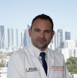 Yiannis S. Chatzizisis合作论文数AHEPA University Hospital, Aristotle University Medical School3
 Shigeru Nakamura合作论文数Department of Cardiology, Kyoto Katsura Hospital;Teikyo University School of Medicine3Shingo Mizuno合作论文数3Kostas A. Stefanou合作论文数3Charles Maynard合作论文数3Panagiota Gounari合作论文数3
 Nikos Papageorgiou合作论文数3
 Ioannis Papadakis合作论文数International Quality Certification Megistis3C. Bourantas合作论文数2
 Li-Ming Gan合作论文数2
 Rocco Vergallo合作论文数2Michelle A Cormier合作论文数2
 Johannes Wikstrom合作论文数2Michelle Lucier合作论文数2
 Ignatios Ikonomidis合作论文数2
 Sofia Stamou合作论文数2
 Koskinas Konstantinos C合作论文数Dept Cardiol, Bern Univ Hosp2
 Hang Lee合作论文数Massachusetts General Hospital Biostatistics;Harvard Medical School2




