Human pluripotent stem cells (hPSCs), either embryonic or induced, offer a plentiful platform for derivation of multiple cell types. Pericytes, generated from hPSCs, are multipotent precursors with vasculogenic features that exhibit high proliferation capability in long-term cultures. Administration of hPSC-pericytes into ischemic murine hind limb is associated with therapeutic angiogenesis and attenuation of muscle wasting. Here, we describe the protocol for derivation of large numbers of pericytes from spontaneously differentiating hPSC-embryoid bodies.
Background Cell therapy of diabetes aims at restoring the physiological control of blood glucose by transplantation of functional pancreatic islet cells. A potentially unlimited source of cells for such transplantations would be islet cells derived from an in vitro differentiation of human pluripotent stem cells (hESC/hiPSC). The islet-like clusters (ILC) produced by the known differentiation protocols contain various cell populations. Among these, the β-cells that express both insulin and the transcription factor Nkx6.1 seem to be the most efficient to restore normoglycemia in diabetes animal models. Our aim was to find markers allowing selection of these efficient cells. Methods Functional Cell-Capture Screening (FCCS) was used to identify markers that preferentially capture the cells expressing both insulin and Nkx6.1, from hESC-derived ILC cells. In order to test whether selection for such markers could improve cell therapy in diabetic mouse models, we used ILC produced from a clinical-grade line of hESC by a refined differentiation protocol adapted to up-scalable bioreactors. Re-aggregated MACS sorted cells were encapsulated in microspheres made of alginate modified to reduce foreign body reaction. Implantation was done intraperitoneally in STZ-treated C57BL/6 immuno-competent mice. Results CD49A (integrin alpha1) was identified by FCCS as a marker for cells that express insulin (or C-peptide) as well as Nkx6.1 in ILC derived by hESC differentiation. The ILC fraction enriched in CD49A + cells rapidly reduced glycemia when implanted in diabetic mice, whereas mice receiving the CD49A depleted population remained highly diabetic. CD49A-enriched ILC cells also produced higher levels of human C-peptide in the blood of transplanted mice. However, the difference between CD49A-enriched and total ILC cells remained small. Another marker, CD26 (DPP4), was identified by FCCS as binding insulin-expressing cells which are Nkx6.1 negative. Depletion of CD26 + cells followed by enrichment for CD49A + cells increased insulin + /Nkx6.1 + cells fraction to ~70%. The CD26 - /CD49A + enriched ILC exhibited improved function over non-sorted ILC or CD49A + cells in diabetic mice and maintain prolonged blood C-peptide levels. Conclusions Refining the composition of ILC differentiated from hPSC by negative selection to remove cells expressing CD26 and positive selection for CD49A expressing cells could enable more effective cell therapy of diabetes.
The limited regenerative capacity of the injured myocardium leads to remodeling and often heart failure. Novel therapeutic approaches are essential. Induced pluripotent stem cells (iPSC) differentiated into cardiomyocytes are a potential future therapeutics. We hypothesized that organ-specific reprogramed fibroblasts may serve an advantageous source for future cardiomyocytes. Moreover, exosomes secreted from those cells may have a beneficial effect on cardiac differentiation and/or function. We compared RNA from different sources of human iPSC using chip gene expression. Protein expression was evaluated as well as exosome micro-RNA levels and their impact on embryoid bodies (EBs) differentiation. Statistical analysis identified 51 genes that were altered (p ≤ 0.05), and confirmed in the protein level, cardiac fibroblasts-iPSCs (CF-iPSCs) vs. dermal fibroblasts-iPSCs (DF-iPSCs). Several miRs were altered especially miR22, a key regulator of cardiac hypertrophy and remodeling. Lower expression of miR22 in CF-iPSCs vs. DF-iPSCs was observed. EBs treated with these exosomes exhibited more beating EBs p = 0.05. vs. control. We identify CF-iPSC and its exosomes as a potential source for cardiac recovery induction. The decrease in miR22 level points out that our CF-iPSC-exosomes are naïve of congestive heart cell memory, making them a potential biological source for future therapy for the injured heart.
After becoming chief pathologist at the University of Wisconsin-Madison Wisconsin Regional Primate Center in 1995, James A. Thomson [5] began his pioneering work in deriving embryonic stem cells [6] from isolated embryos. That same year, Thomson [5] published his first paper, “Isolation of a Primate Embryonic Stem Cell Line” in Proceedings of the National Academy of Sciences [7] of the United States of America, detailing the first derivation of primate [8] embryonic stem cells [6]. In the following years, Thomson [5] and his team of scientists—Joseph Itskovitz-Eldor, Sander S. Shapiro, Michelle A. Waknitz, Jennifer J. Swiergiel, Vivienne S. Marshall, and Jeffry M. Jones—advanced their work with embryonic stem cells [6], eventually isolating and culturing human embryonic stem cells [6]. Their work with human embryos was reported in the 1998 Nature article “Embryonic Stem Cell Lines Derived from Human Blastocysts.”
Background: Amyotrophic lateral sclerosis (ALS) is a motor neuron (MN) disease characterized by the loss of MNs in the central nervous system. As MNs die, patients progressively lose their ability to control voluntary movements, become paralyzed and eventually die from respiratory/deglutition failure. Despite the selective MN death in ALS, there is growing evidence that malfunctional astrocytes play a crucial role in disease progression. Thus, transplantation of healthy astrocytes may compensate for the diseased astrocytes. Methods: We developed a good manufacturing practice-grade protocol for generation of astrocytes from human embryonic stem cells (hESCs). The first stage of our protocol is derivation of astrocyte progenitor cells (APCs) from hESCs. These APCs can be expanded in large quantities and stored frozen as cell banks. Further differentiation of the APCs yields an enriched population of astrocytes with more than 90% GFAP expression (hES-AS). hES-AS were injected intrathecally into hSOD1(G93A) transgenic mice and rats to evaluate their therapeutic potential. The safety and biodistribution of hES-AS were evaluated in a 9-month study conducted in immunodeficient NSG mice under good laboratory practice conditions. Results: In vitro, hES-AS possess the activities of functional healthy astrocytes, including glutamate uptake, promotion of axon outgrowth and protection of MNs from oxidative stress. A secretome analysis shows that these hES-AS also secrete several inhibitors of metalloproteases as well as a variety of neuroprotective factors (e.g. TIMP-1, TIMP-2, OPN, MIF and Midkine). Intrathecal injections of the hES-AS into transgenic hSOD1(G93A) mice and rats significantly delayed disease onset and improved motor performance compared to sham-injected animals. A safety study in immunodeficient mice showed that intrathecal transplantation of hES-AS is safe. Transplanted hES-AS attached to the meninges along the neuroaxis and survived for the entire duration of the study without formation of tumors or teratomas. Cell-injected mice gained similar body weight to the sham-injected group and did not exhibit clinical signs that could be related to the treatment. No differences from the vehicle control were observed in hematological parameters or blood chemistry. Conclusion: Our findings demonstrate the safety and potential therapeutic benefits of intrathecal injection of hES-AS for the treatment of ALS.
Large-scale expansion of pluripotent stem cells (PSC) in a robust, well-defined, and monitored process is essential for production of cell-based therapeutic products. The transition from laboratory-scale protocols to industrial-scale production is one of the first milestones to be achieved in order to use both human embryonic stem cells (ESC) and induced pluripotent stem cells (iPSC) as the starting material for cellular products. The methods to be developed require adjustment of the culture platforms, optimization of culture parameters, and adaptation of downstream procedures. Optimization of expansion protocols and their scalability has become much easier with the design of bioreactor systems that enable continuous monitoring of culture parameters, continuous media change, and support software for automated control. This chapter highlights the common properties that are required for production of scalable, reproducible, homogeneous, and clinically suitable cell therapy products. We describe the available platforms for large-scale expansion of PSCs and parameters that should be considered when optimizing the expansion protocols in a scalable bioreactor. All the above are detailed in the light of the requirements and challenges of bringing a cell-based therapeutic product to the clinic and ultimately to the market. We discuss some considerations that should be taken into account, such as cost-effectiveness, good manufacturing practice, and regulatory guidelines. [GRAPHICS] .
Astrocytes are the major cell population in the central nervous system (CNS) and play piv- otal role in CNS homeostasis and functionality. Malfunction of astrocytes were implicated in multiple neurodegenerative diseases and disorders, including amyotrophic lateral scle- rosis (ALS), spinal cord injury (SCI), brain stroke, Parkinson’s disease (PD), and Alzheimer disease (AD). These new insights led to the rationale that transplantation of healthy and functional human astrocytes could support survival of neurons and be of therapeutic value in treating neurodegenerative diseases. Here, we will mainly focus on the role of astrocytes in ALS disease, the major cell sources for generation of human astrocytes, or astrocyte like cells and show how multiple preclinical studies demonstrate the efficacy of these cells in animal models. In addition, we will cover immerging early stage clinical trials that are cur-rently being conducted using human astrocytes or human astrocyte like cell population. neuron transplantation of cerebrospinal (CSF) of neurotrophic factors. The combined mechanism of action provided by cell transplantation is postulated to better cope with the multifactorial nature of the disease compared to a single pathway-based drug. Preclinical studies in ALS animal models showed the high safety profile of cell-based treatments in addition to their benefits in delaying disease onset, slowing down clinical symptoms and in many cases also to extend survival. Besides behavioral measure -ments, many of the studies also demonstrated graft survival, decline in inflammation, and improvement in histopathological attributes of the disease. Translation of the preclinical stud ies into clinical trials confirmed the safety of the procedures. Efficacy in most of these trials was a secondary endpoint, and some studies showed moderate and/or transient beneficial effects. Yet, since most of the clinical trials were at early stage, with small-size groups without a control arm, it is difficult to evaluate the efficacy of the treatments. Late-stage, placebo-controlled clinical studies with greater number of patients will prove whether any of the cell-based therapies indeed change the course of the disease.
Following Jamie Thomson's lecture on primate embryonic stem cells (ESCs) at a meeting I had organized in March 1997, in Israel, to celebrate receipt of the Wolf Prize in Agriculture to my colleague and friend Neal First, frozen human embryos donated for research in Israel were shipped to Wisconsin. The five hESC lines (H1, H7, H9, H13, and H14) were established by early 1998 and transferred to my laboratory just before publication of their existence in Science, on November 6, 1998. The distribution of the cells from my institute to several laboratories, as early as 1999, enhanced the development of hESC research worldwide. My personal perspective regarding the scientific and political events surrounding this story are presented.
The liver is the main organ responsible for the modification, clearance, and transformational toxicity of most xenobiotics owing to its abundance in cytochrome P450 (CYP450) enzymes. However, the scarcity and variability of primary hepatocytes currently limits their utility. Human pluripotent stem cells (hPSCs) represent an excellent source of differentiated hepatocytes; however, current protocols still produce fetal‐like hepatocytes with limited mature function. Interestingly, fetal hepatocytes acquire mature CYP450 expression only postpartum, suggesting that nutritional cues may drive hepatic maturation. We show that vitamin K 2 and lithocholic acid, a by‐product of intestinal flora, activate pregnane X receptor (PXR) and subsequent CYP3A4 and CYP2C9 expression in hPSC‐derived and isolated fetal hepatocytes. Differentiated cells produce albumin and apolipoprotein B100 at levels equivalent to primary human hepatocytes, while demonstrating an 8‐fold induction of CYP450 activity in response to aryl hydrocarbon receptor (AhR) agonist omeprazole and a 10‐fold induction in response to PXR agonist rifampicin. Flow cytometry showed that over 83% of cells were albumin and hepatocyte nuclear factor 4 alpha (HNF4α) positive, permitting high‐content screening in a 96‐well plate format. Analysis of 12 compounds showed an R 2 correlation of 0.94 between TC50 values obtained in stem cell–derived hepatocytes and primary cells, compared to 0.62 for HepG2 cells. Finally, stem cell–derived hepatocytes demonstrate all toxicological endpoints examined, including steatosis, apoptosis, and cholestasis, when exposed to nine known hepatotoxins. Conclusion : Our work provides fresh insights into liver development, suggesting that microbial‐derived cues may drive the maturation of CYP450 enzymes postpartum. Addition of these cues results in the first functional, inducible, hPSC‐derived hepatocyte for predictive toxicology. (H epatology 2015;62:265‐278)
Catecholaminergic polymorphic ventricular tachycardia (CPVT) is an inherited arrhythmia characterized by syncope and sudden death occurring during exercise or acute emotion. CPVT is caused by abnormal intracellular Ca(2+) handling resulting from mutations in the RyR2 or CASQ2 genes. Because CASQ2 and RyR2 are involved in different aspects of the excitation-contraction coupling process, we hypothesized that these mutations are associated with different functional and intracellular Ca(²+) abnormalities. To test the hypothesis we generated induced Pluripotent Stem Cell-derived cardiomyocytes (iPSC-CM) from CPVT1 and CPVT2 patients carrying the RyR2(R420Q) and CASQ2(D307H) mutations, respectively, and investigated in CPVT1 and CPVT2 iPSC-CM (compared to control): (i) The ultrastructural features; (ii) the effects of isoproterenol, caffeine and ryanodine on the [Ca(2+) ]i transient characteristics. Our major findings were: (i) Ultrastructurally, CASQ2 and RyR2 mutated cardiomyocytes were less developed than control cardiomyocytes. (ii) While in control iPSC-CM isoproterenol caused positive inotropic and lusitropic effects, in the mutated cardiomyocytes isoproterenol was either ineffective, caused arrhythmias, or markedly increased diastolic [Ca(2+) ]i . Importantly, positive inotropic and lusitropic effects were not induced in mutated cardiomyocytes. (iii) The effects of caffeine and ryanodine in mutated cardiomyocytes differed from control cardiomyocytes. Our results show that iPSC-CM are useful for investigating the similarities/differences in the pathophysiological consequences of RyR2 versus CASQ2 mutations underlying CPVT1 and CPVT2 syndromes.
Loss of pluripotency is a gradual event whose initiating factors are largely unknown. Here we report the earliest metabolic changes induced during the first hours of differentiation. High-resolution NMR identified 44 metabolites and a distinct metabolic transition occurring during early differentiation. Metabolic and transcriptional analyses showed that pluripotent cells produced acetyl-CoA through glycolysis and rapidly lost this function during differentiation. Importantly, modulation of glycolysis blocked histone deacetylation and differentiation in human and mouse embryonic stem cells. Acetate, a precursor of acetyl-CoA, delayed differentiation and blocked early histone deacetylation in a dose-dependent manner. Inhibitors upstream of acetyl-CoA caused differentiation of pluripotent cells, while those downstream delayed differentiation. Our results show a metabolic switch causing a loss of histone acetylation and pluripotent state during the first hours of differentiation. Our data highlight the important role metabolism plays in pluripotency and suggest that a glycolytic switch controlling histone acetylation can release stem cells from pluripotency.
Isolated microvessel-residing pericytes and pericytes from human pluripotent stem cells (hPSCs) exhibit mesenchymal stem cell-like characteristics and therapeutic properties. Despite growing interest in pericyte-based stem cell therapy, their immunogenicity and immunomodulatory effects on nonactivated T cells are still poorly defined, in particular those of vasculogenic hPSC pericytes. We found that tissue-embedded and unstimulated cultured hPSC- or tissue-derived pericytes constitutively expressed major histocompatibility complex (MHC) class I and the inhibitory programmed cell death-ligand 1/2 (PD-L1/2) molecules but not MHC class II or CD80/CD86 costimulatory molecules. Pretreatment with inflammatory mediators failed to induce an antigen-presenting cell-like phenotype in stimulated perk cytes. CD146(+) pericytes from hPSCs did not induce activation and proliferation of allogeneic resting T cells independent of interferon (IFN)-gamma prestimulation, similarly to pericytes from human brain or placenta. Instead, pericytes mediated a significant increase in the frequency of allogeneic CD25(high)FoxP3(+) regulatory T cells when cocultured with nonactivated peripheral blood T cells. Furthermore, when peripheral blood CD25(high) regulatory T cells (Tregs) were depleted from isolated CD3(+) T cells, pericytes preferentially induced de novo formation of CD4(+)CD25(high)FoxP3(+)CD127(-), suppressive regulatory T cells. Constitutive expression of PD-L1/2 and secretion of transforming growth factor-beta by hPSC pericytes directly regulated generation of pericyte-induced Tregs. Pericytes cotransplanted into immunodeficient mice with allogeneic CD25(-) T cells maintained a nonimmunogenic phenotype and mediated the development of functional regulatory T cells. Together, these findings reveal a novel feature of pericyte-mediated immunomodulation distinguished from immunosuppression, shared by native tissue pericytes and hPSC pericytes, and support the notion that pericytes can be applied for allogeneic cell therapy.
We investigated pacemaker function of iPSC-CM in a canine complete heart block model. Embryoid bodies (EBs) were derived from human keratinocytes and their gene expression profile and markers of differentiation were identified. We recorded their action potential characteristics, including phase 4 depolarization, automaticity, response to ivabradine, and pacemaker current, If, as well. Atrio-ventricular blocked dogs were immunosuppressed and instrumented with a VVI pacemaker. Forty-75 rhythmically contracting EBs (totaling 1.3-2x10⁶ cells) were injected subepicardially into the anterobasal left ventricle. ECGs and 24-h Holter recordings were made biweekly. After 4- 13 weeks, epinephrine (EPI) (1 μg/Kg/min) was infused and the heart removed for histological or electrophysiological study. IPSC-CMs largely lost their markers of pluripotency and were positive for cardiac-specific markers instead. Automaticity of IPSC-CMs was identified and confirmed to be If-dependent. Epicardial pacing of the injection site identified matching beats arising from that site by the end of week 1 of implantation. By week 4, 20% of beats were electronically paced and 60-80% of beats were matching. Maximum night and day rates of matching beats were 53±6.9 and 68±10.4 bpm respectively at 4 weeks. EPI increased rate of matching beats from 35±4.3 to 65±4.0 bpm. Incubation of EBs with the vital dye, Dil, revealed the persistence of injected cells at the site of administration. IPSC-CM can integrate into host myocardium and create a biological pacemaker. While this is a promising development, rate and rhythm of the iPSC-CM pacemakers remain to be optimized. The author hereby declares no conflict of interest
Human embryonic stem cells (hESCs) and human induced pluripotent stem cells (hiPSCs) have the capacity to differentiate into any specialized cell type, including cardiomyocytes. Therefore, hESC-derived and hiPSC-derived cardiomyocytes (hESC-CMs and hiPSC-CMs, respectively) offer great potential for cardiac regenerative medicine. Unlike some organs, the heart has a limited ability to regenerate, and dysfunction resulting from significant cardiomyocyte loss under pathophysiological conditions, such as myocardial infarction (MI), can lead to heart failure. Unfortunately, for patients with end-stage heart failure, heart transplantation remains the main alternative, and it is insufficient, mainly because of the limited availability of donor organs. Although left ventricular assist devices are progressively entering clinical practice as a bridge to transplantation and even as an optional therapy, cell replacement therapy presents a plausible alternative to donor organ transplantation. During the past decade, multiple candidate cells were proposed for cardiac regeneration, and their mechanisms of action in the myocardium have been explored. The purpose of this article is to critically review the comprehensive research involving the use of hESCs and hiPSCs in MI models and to discuss current controversies, unresolved issues, challenges, and future directions.
Extracellular matrix (ECM) has been utilized as a biological scaffold for tissue engineering applications in a variety of body systems, due to its bioactivity and biocompatibility. In the current study we developed a modified protocol for the efficient and reproducible derivation of mesenchymal progenitor cells (MPCs) from human embryonic stem cells as well as human induced pluripotent stem cells (hiPSCs) originating from hair follicle keratinocytes (HFKTs). ECM was produced from these MPCs and characterized in comparison to adipose mesenchymal stem cell ECM, demonstrating robust ECM generation by the excised HFKT-iPSC-MPCs. Exploiting the advantages of electrospinning we generated two types of electrospun biodegradable nanofiber layers (NFLs), fabricated from polycaprolactone (PCL) and poly(lactic-co-glycolic acid) (PLGA), which provide mechanical support for cell seeding and ECM generation. Elucidating the optimized decellularization treatment we were able to generate an available "off-the-shelf" implantable product (NFL-ECM). Using rat subcutaneous transplantation model we demonstrate that this stem-cell-derived construct is biocompatible and biodegradable and holds great potential for tissue regeneration applications.
 Michal Amit合作论文数75
 Shahar Kol合作论文数Department of Obstetrics and Gynecology, Rambam Medical Center35
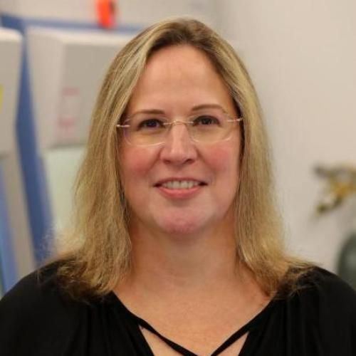 Sharon Gerecht合作论文数Department of Biomedical Engineering, Pratt School of Engineering, Duke University28Zeev Weiner合作论文数25
 Ofer Binah合作论文数21Anna Ziskind合作论文数16Zeevi-Levin Naama合作论文数16
 Lior Lowenstein合作论文数Division of Female Pelvic Medicine and Reconstructive Surgery, Loyola Medical Center13
 Ron Beloosesky合作论文数13Atara Novak合作论文数11Peter Jakobi合作论文数11Abraham Lightman合作论文数11Bettina Fishman合作论文数11Barad Lili合作论文数10
 Nissim Benvenisty合作论文数The Alexander Silberman Institute of Life Science, Hebrew University of Jerusalem10Hanna Segev合作论文数10D Manor合作论文数10Shtrichman Ronit合作论文数9
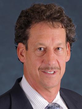 Michael Ross合作论文数The Lundquist Institute;David Geffen School of Medicine, University of California, Los Angeles;Fielding School of Public Health, University of California, Los Angeles9
 Yuval Ginsberg合作论文数8judith chebat合作论文数8
 Cohen Smadar合作论文数Department of Biotechnology Engineering, Ben Gurion University of the Negev8Sivan Osenberg合作论文数8
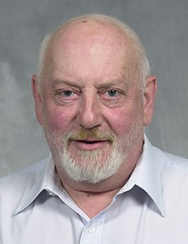 Michel Revel合作论文数Department of Molecular Genetics of the Weizmann Institute of Science8Israel Thaler合作论文数8
 Jacob Levron合作论文数8
 Oliver Brüstle合作论文数Institute of Reconstructive Neurobiology, University of Bonn8N. Lewit合作论文数7
 Ilana Laevsky合作论文数7
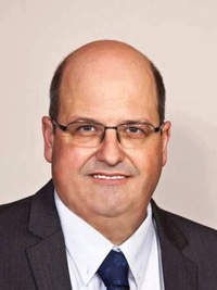 Lior Gepstein合作论文数Department of Cardiology, Rambam Health Care Campus;Faculty of Medicine, Technion - Israel Institute of Technology7Eyal Schiff合作论文数7Arie Drugan合作论文数7
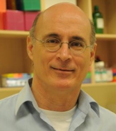 Gideon Rechavi合作论文数Sheba Cancer Research Center;Sackler Faculty of Medicine, Tel Aviv University;Cancer Biology Research Center, Tel Aviv University;Wohl Institute for Translational Medicine7
 Izhak Kehat合作论文数Cardiology Department, Rambam Medical Center7Etan Z Zimmer合作论文数7
 Nibal Awad合作论文数7Germanguz Igal合作论文数1 Ruth and Bruce Rappaport Faculty of Medicine, Sohnis and Forman Families Center for Stem Cell and Tissue Regeneration Research6Shahar Cohen合作论文数6Liron Eldor合作论文数6Ayelet Dar合作论文数Department of Immunology;Weizmann Institute of Science6
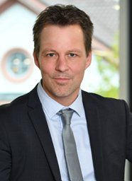 Philipp Koch合作论文数University of Bonn and Hertie Foundation Institute of Reconstructive Neurobiology6
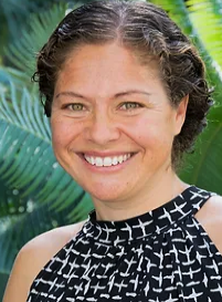 Maya Schuldiner合作论文数Department of Molecular Genetics, Weizmann Institute of Science6
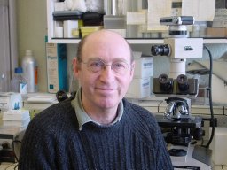 Daniel Aberdam合作论文数INSERM U.385, Biologie et Physiopathologie de la Peau, Faculté de Médecine, F-06107 Nice, France6Michal Izrael合作论文数6Katya Dolnikov合作论文数5Leshansky Lucy合作论文数INSERTECH Stem Cell Center, Ruth and Bruce Rappaport Faculty of Medicine, Technion, Haifa, Israel;Stem Cell Center, Ruth and Bruce Rappaport Faculty of Medicine, Technion, Haifa, Israel.5Domev Hagit合作论文数5Steinberg Maya合作论文数5




