Stem cell differentiation is central to development and regenerative medicine, but the complex underlying processes hinder our ability to control it experimentally. Computational models can help form hypotheses and generate predictions. There are many types of computational models available to aid in understanding stem cell differentiation-related processes, but for an experimental biologist, it might be hard to select the modelling approach that matches their research question, and to know what data would be needed to make use of the model. This review is aimed at experimental biologists to introduce various modelling types, relate them to the types of questions these models can help answer and outline the necessary data to gain new insights. The review discusses mechanistic dynamic models, both ordinary differential equation (ODE) and abstract, multiscale models and data-driven deep learning approaches. Each model class is introduced with what the model represents, the insights it can provide, validation strategies and limitations. With this review, we want to make it easier to incorporate modelling within experimental workflows for stem cell differentiation-related research, to aid experimentation and accelerate discovery.
Fluorescence recovery after photobleaching (FRAP) is a widely used technique for investigating protein dynamics in live cells. FRAP enables researchers to monitor protein mobility and binding interactions in real time, providing valuable insights into cellular regulatory mechanisms. Initially developed to explore membrane fluidity, FRAP has evolved to enable study of nuclear processes, including transcription factor (TF) dynamics crucial for gene regulation. In this review, we focus on how FRAP has advanced our understanding of TF dynamics in health and disease. TFs exhibit complex interactions with DNA that are essential for cellular function. FRAP helps to quantify these interactions, revealing how TF mobility and chromatin binding influence gene expression patterns. We further explore the role of FRAP in studying TFs in disease. Research on TF dynamics in cancer, diabetes, and osteoarthritis underscores the method's potential to identify disease-related regulatory mechanisms. By providing a nuanced understanding of TF behavior, FRAP presents a promising avenue for developing targeted therapies across diverse pathologies.
Mechanistic ordinary differential equation models of gene regulatory networks are a valuable tool for understanding biological processes that occur inside a cell, and they allow for the formulation of novel hypotheses on the mechanisms underlying these processes. Although data-driven methods for inferring these mechanistic models are becoming more prevalent, it is often unclear how recent advances in machine learning can be used effectively without jeopardi zing the interpretability of the resulting models. In this work, we present a framework to leverage neural networks for the identification of data-driven models for time-dependent intracellular processes, such as cell differentiation. In particular, we use a graph autoencoder model to suggest novel connections in a gene regulatory network. We show how the improvement of the graph suggested using this neural network leads to the generation of hypotheses on the dynamics of the resulting identified dynamical system.
Understanding time-dependent intracellular processes, such as cell differentiation, is key to developing new therapies for a wide range of diseases. Models that connect transcription factor activity to dynamic expression patterns are rare, despite the increased availability of time-series data. To identify key regulators of time-dependent biological processes, we present the pipeline DANSE: Dynamics inference Algorithm on Networks Specified by Enhancers. Starting from multi-omics data, our pipeline constructs a data-driven mechanistic transcription factor (TF) network and subsequently defines a dynamic model based on this TF network. The combination of a TF network and a mechanistic model allows for the identification of a small set of key transcription factors predicted to drive the modelled biological process. We showcase the result of our pipeline by applying DANSE to two different datasets that describe iPSC differentiation. Models constructed using DANSE suggest testable hypotheses for the perturbation of gene expression, for example, knockdown or overexpression, that influence cell fate. In this way, DANSE is a powerful tool for generating novel hypotheses in a data-driven manner that take into account the dynamic nature of multi-omics time series data.
The transcription factor SOX9 is integral to tissue homeostasis and is implicated in skeletal malformation, campomelic dysplasia, and osteoarthritis (OA). Despite extensive research, the complete regulatory landscape of SOX9 transcriptional activity, interconnected with signaling pathways (TGFβ, WNT, BMP, IHH, NFκB, and HIF), remains challenging to decipher. This study focuses on elucidating SOX9 signaling in OA pathology using Fluorescence Recovery After Photobleaching (FRAP) to assess SOX9 activity directly in live human primary chondrocytes (hPCs). Single cell FRAP data revealed two distinct subpopulations with differential SOX9 dynamics, showing varied distribution between healthy and OA hPCs. Moreover, inherently elevated SOX9-DNA binding was observed in healthy hPCs compared to preserved and OA counterparts. Anabolic factors (BMP7 and GREM1) and catabolic inhibitors (DKK1 and FRZb) were found to modulate SOX9 transcriptional activity in OA-hPCs. These findings provide valuable insights into the intricate regulation of SOX9 signaling in OA, suggesting potential therapeutic avenues for modulating SOX9 activity in diseased states.
Purpose (the aim of the study): Osmolarity and fixed charge density of the joint are known to decrease during the progression of osteoarthritis (OA) and are traditionally considered consequences of the disease. In cancer, Alzheimer's, and diabetes, changes to extracellular osmolarity are considered integral to disease etiology. We observed differences in synovial fluid osmolarity (OA ~300 mOsm, healthy ~400 mOsm) and chondrocyte cell size (OA: larger, healthy: smaller) between OA and healthy joints.
Tissue engineering protocols achieve building miniature hearts but mechanisms determining cell differentiation still need to be fully understood and optimized. In this study, we present a gene regulatory network (GRN) that describes the differentiation of committed cardiomyocytes towards ventricular or atrial cardiomyocytes. The GRN is coupled with Boolean dynamics and steady state analysis shows steady states which agree with the experimental expression of marker genes. Our Boolean model extends earlier work on a model describing the first and second heart field formation to include atrial and ventricular cardiomyocytes. Thus, our study paves the way for the generation of heart field-specific cardiomyocytes located in specific chambers of the fully developed heart. The Boolean model is validated through simulations and by its ability to reproduce known knockouts. ### Competing Interest Statement The authors have declared no competing interest.
Purpose (the aim of the study): hMSCs are of great interest in tissue engineering and regenerative medicine to treat cartilage and bone defects due to their multi-lineage differentiation potential. Decades of research has uncovered multiple factors including signaling pathways, biomaterials, microenvironments that steer the differentiation of hMSCs into a particular lineage. Although several different protocols are in place to differentiate hMSCs into chondrocytes, in vitro differentiations are still sub-optimal and donor variation poses an additional challenge in stem cell-based therapies.
Osteoarthritis (OA) is a multifactorial degenerative joint disease of which the underlying mechanisms are yet to be fully understood. At the molecular level, multiple factors including altered signaling pathways, epigenetics, metabolic imbalance, extracellular matrix degradation, production of matrix metalloproteinases, and inflammatory cytokines, are known to play a detrimental role in OA. However, these factors do not initiate OA, but are mediators or consequences of the disease, while many other factors causing the etiology of OA are still unknown. Here, it is revealed that microenvironmental osmolarity can induce and reverse osteoarthritis-related behavior of chondrocytes via altered intracellular molecular crowding, which represents a previously unknown mechanism underlying OA pathophysiology. Decreased intracellular crowding is associated with increased sensitivity to proinflammatory triggers and decreased responsiveness to anabolic stimuli. OA-induced lowered intracellular molecular crowding could be renormalized via exposure to higher extracellular osmolarity such as those found in healthy joints, which reverse OA chondrocyte's sensitivity to catabolic stimuli as well as its glycolytic metabolism.
The formation of fibrocartilage during articular cartilage regeneration remains a clinical problem affecting adequate restoration of articular cartilage in joints. To stimulate chondrocytes to form articular cartilage, we investigated the use of amyloid fibril-based scaffolds. The proteins α-synuclein, β-lactoglobulin, and lysozyme were induced to self-assemble into amyloid fibrils and, during dialysis, formed micrometer scale amyloid networks that resemble the cartilage extracellular matrix. Our results show that lysozyme amyloid micronetworks supported chondrocyte viability and extracellular matrix deposition, while α-synuclein and β-lactoglobulin maintained cell viability. With this study, we not only confirm the possible use of amyloid materials for tissue regeneration but also demonstrate that the choice of protein, rather than its amyloid-fold per se, affects the cellular response and tissue formation.
The multi-lineage differentiation capacity of human mesenchymal stem cells (hMSCs) enables its potential for tissue engineering and regenerative medicine. Master transcription factors play a key role during development, differentiation, homeostasis and disease pathology. RUNX2 is the master transcription factor for bone development, and it regulates several important signaling pathways during chondrogenic and osteogenic differentiation of hMSCs. However, modulation of RUNX2 activity during hMSC differentiation into various lineages is not yet fully described. We differentiated hMSCs into chondro-, osteo-, and adipogenic lineages and studied RUNX2 protein dynamics using Transcription Factor - Fluorescence Recovery After Photobleaching (TF-FRAP) at different time points. The TF-FRAP method can capture the dynamic changes of RUNX2 protein mobility at the single cell level resolution, and cluster analysis shows how RUNX2 dynamics change at subpopulation level in proliferating and differentiating hMSCs. Our data show that although whole hMSC population is exposed to differentiation stimuli, some subpopulations in hMSCs do not respond to environmental cues.
In recent years, the mathematical and computational sciences have developed novel methodologies and insights that can aid in designing advanced bioreactors, microfluidic setups or organ-on-chip devices, in optimizing culture conditions, or predicting long-term behavior of engineered tissues in vivo. In this review, we introduce the concept of computational models and how they can be integrated in an interdisciplinary workflow for Tissue Engineering and Regenerative Medicine (TERM). We specifically aim this review of general concepts and examples at experimental scientists with little or no computational modeling experience. We also describe the contribution of computational models in understanding TERM processes and in advancing the TERM field by providing novel insights. Impact Statement Although in recent years the use of mathematical and computational sciences has increased in the Tissue Engineering and Regenerative Medicine (TERM) field, we believe that a further integration of experimental and computational approaches has a huge potential for advancing the field due to the ability of models to explain and predict experimental results and efficiently optimize TERM product and process designs. By providing an overview of existing computational models, how they have contributed to the field, as well as a future perspective, this review represents an important step to help realize TERM's ultimate goal: a cure instead of care.
Here we show how to measure the mobility of transcription factors using fluorescence recovery after photobleaching (FRAP). Transcription factors are DNA-binding proteins that, upon binding to specific DNA motifs, regulate transcription of their target genes. FRAP is a simple, fast, and cost-effective method, and is a widely used quantitative method to measure the dynamics of fluorescently labeled molecules in solution, membranes, and inside living cells. Dynamics, specified by the immobile fraction, recovery half-time, diffusion constant, and ratio of molecules contributing to different phases of FRAP recovery, can be quantified by FRAP. This can be useful to understand their function in gene regulation. This tutorial is intended to familiarize the reader with the FRAP procedure to quantify transcription factor dynamics using a standard confocal microscope and analysis using MATLAB (MathWorks). This article will guide the reader through the preconditions of FRAP, and a detailed and step-by-step procedure of preparing cells, bleaching protocol, data analysis in MATLAB, and visualization of the FRAP data.
Computational modeling of biological networks is increasing in popularity due to the increased demand for understanding biological processes. This understanding requires integration of a variety of experimental data that allows understanding of complex mechanisms regulating cell and tissue function. However, the mathematical complexity of many modeling tools have thusfar prevented broad adaptation and effective use by molecular biologists. In this chapter, we show by example how one can start building a model in ANIMO and how to adapt the model to experimental data. We show how this model can be used for simulating network activities, testing hypotheses, and how to improve the model using wet-lab data.
Co-culture of chondrocytes and mesenchymal stromal cells (MSCs) has been shown to be beneficial in engineering cartilage tissue in vitro. In these co-cultures, MSCs increase the proliferation and matrix deposition of chondrocytes. The MSCs accomplish this beneficial effect by so-called trophic actions. Thus, large cartilage constructs can be made with a relatively small number of chondrocytes. In this chapter, we describe different methods for making co-cultures of MSCs and chondrocytes. We also provide detailed protocols for analyzing MSC-chondrocyte co-cultures with cell tracking, proliferation assays, species-specific polymerase chain reactions (PCR), rheological analysis, compression analysis, RNA-sequencing analysis, short tandem repeats analysis, and biochemical examination.
A fundamental question in cartilage biology is: what determines the switch between permanent cartilage found in the articular joints and transient hypertrophic cartilage that functions as a template for bone? This switch is observed both in a subset of OA patients that develop osteophytes, as well as in cell-based tissue engineering strategies for joint repair. A thorough understanding of the mechanisms regulating cell fate provides opportunities for treatment of cartilage disease and tissue engineering strategies. The objective of this study was to understand the mechanisms that regulate the switch between permanent and transient cartilage using a computational model of chondrocytes, ECHO. To investigate large signaling networks that regulate cell fate decisions, we developed the software tool ANIMO, Analysis of Networks with interactive Modeling. In ANIMO, we generated an activity network integrating 7 signal transduction pathways resulting in a network containing over 50 proteins with 200 interactions. We called this model ECHO, for executable chondrocyte. Previously, we showed that ECHO could be used to characterize mechanisms of cell fate decisions. ECHO was first developed based on a Boolean model of growth plate. Here, we show how the growth plate Boolean model was translated to ANIMO and how we adapted the topology and parameters to generate an articular cartilage model. In ANIMO, many combinations of overactivation/knockout were tested that result in a switch between permanent cartilage (SOX9+) and transient, hypertrophic cartilage (RUNX2+). We used model checking to prioritize combination treatments for wet-lab validation. Three combinatorial treatments were chosen and tested on metatarsals from 1-day old rat pups that were treated for 6 days. We found that a combination of IGF1 with inhibition of ERK1/2 had a positive effect on cartilage formation and growth, whereas activation of DLX5 combined with inhibition of PKA had a negative effect on cartilage formation and growth and resulted in increased cartilage hypertrophy. We show that our model describes cartilage formation, and that model checking can aid in choosing and prioritizing combinatorial treatments that interfere with normal cartilage development. Here we show that combinatorial treatments induce changes in the zonal distribution of cartilage, indication possible switches in cell fate. This indicates that simulations in ECHO aid in describing pathologies in which switches between cell fates are observed, such as OA.
 H.B.J. (Marcel) Karperien合作论文数Faculty of Science and Technology, University of Twente72
 Stefano Schivo合作论文数19
 Zhong Leilei合作论文数19
 Kannan Govindaraj合作论文数Dept Dev Bioengn, Univ Twente17
 Jeroen Leijten合作论文数University of Twente17
 XiaoBin Huang合作论文数Chongqing University, Chongqing University15
 Jetse Scholma合作论文数14
 Jaco van de Pol合作论文数Department of Computer Science, Aarhus University;University of Twente13
 C.A. Van Blitterswijk合作论文数Maastricht University12
 Rom Langerak合作论文数Faculty of EEMCS, Formal Methods and Tools, University of Twente11
 Diane S. Lidke合作论文数Department of Molecular Biology, Max Planck Institute for Biophysical Chemistry10
 Thomas Jovin合作论文数Max Planck Institute for Biophysical Chemistry9
 Nicole Georgi (Raiss)合作论文数8
 Gert B Eijkel合作论文数7Mireille M. A. E. Claessens合作论文数Department of Physics;Technische Universit;Department of Physics, Technische Universit6
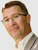 Ron M.A. Heeren合作论文数Institute for Advanced Study, Technische Universitat Munchen;University of Maastricht6Sakshi Khurana合作论文数5
 Andre Van Wijnen合作论文数The Stein/Lian Laboratory, Department of Biochemistry, Larner College of Medicine, University of Vermont5Kerkhofs Johan合作论文数5
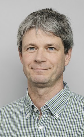 Rainer Heintzmann合作论文数Institut fur Physikalische Chemie, Friedrich Schiller Universitat Jena5
 B. Cillero-Pastor合作论文数5
 Liesbet Geris合作论文数Department of Mechanical Engineering, Katholieke Universiteit Leuven5M.C.E. van Dalen合作论文数4
 Jan Hendriks合作论文数4
 Ling Wu合作论文数4
 Bin Ma (马斌)合作论文数School of Biomedical Engineering, Shanghai Jiao Tong University4Donna J. Arndtjovin合作论文数4Marieke Meteling合作论文数3Sakshi Kannan合作论文数3
 Ellie B.M. Landman合作论文数3
 Boudewijn Bakker合作论文数3Kiss Andras合作论文数3
 Jan Maarten Wit合作论文数Department of Pediatrics, Leiden University Medical Center3
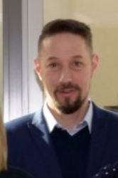 Peter Nagy合作论文数Department of Molecular Immunology and Toxicology, National Institute of Oncology, Hungary;Debrecen University;Department of Pathology, University of Otago3
 s. Khurana合作论文数3
 Laura Gillespie合作论文数3
 Donna J. Arndt-Jovin合作论文数3
 Paul Van Der Vet合作论文数University of Twente3Theo Verrips合作论文数3
 Bernd Rieger合作论文数Department of Imaging Physics, Faculty of Applied Sciences, Delft University of Technology3Gary D. Paterno合作论文数3Srirang Manohar合作论文数2Jacqueline Plass合作论文数2
 Ricardo A. Urquidi Camacho合作论文数2Maurice C E van Dalen合作论文数2
 Ingrid Meulenbelt合作论文数Sect Mol Epidemiol, Leiden Univ2Joyce Emons合作论文数2Cillero-Pastor Berta合作论文数2
 Nathalie Groen合作论文数2




