Alemtuzumab, fludarabine, and melphalan reduced-intensity conditioning (RIC) regimen is associated with a high incidence of mixed chimerism and secondary graft failure in patients undergoing allogeneic hematopoietic stem cell transplant (HSCT) for inborn errors of immunity (IEI). We hypothesized that addition of thiotepa to this regimen would reduce secondary graft failure without increasing toxicity in children and young adults undergoing allogenic HSCT for IEI. We retrospectively reviewed charts of patients undergoing allogenic HSCT for IEI who received alemtuzumab (starting on day -14), fludarabine (150 mg/m2 or 5 mg/kg over days -8 to -4), and melphalan (140 mg/m2 or 4.7 mg/kg on day -3) containing RIC regimen with the addition of thiotepa (200 mg/m2 on day -2). Mixed chimerism and low-level mixed chimerism were defined as whole blood donor chimerism <95% and <50%, respectively, on two consecutive measures within a 2-week period. Thirty-one patients underwent allogeneic HSCT at a median age of 1.7 years (0.3-21 yrs). Patient demographics are outlined in Table 1. All patients engrafted at a median of 12 days (range, 8-17 days). The incidence of mixed chimerism was 0% (0/31), 20% (5/25), and 46% (7/15) upon engraftment at day +100 and one-year post-HSCT, respectively. Incidence of low-level mixed chimerism was 0% (0/25) and 6.6% (1/15) at day +100 and one-year post-HSCT, respectively. Three (9.6%) patients required a secondary intervention (2 CD34 selected boost and 1 second HSCT). Two (6.4%) patients developed sinusoidal obstruction syndrome, and 11 (35%) patients developed thrombotic microangiopathy. Six (19%) patients developed grade I-II skin GVHD, with none developing grade III-IV GVHD. One (3.2%) patient developed limited chronic GVHD. Thirteen of 31 (42%) patients had viral reactivation requiring treatment, with CMV (54%), EBV (54%), and adenovirus (15%) being the most common. Twenty-six (84%) patients are surviving at median follow-up of 10 months (range 2m-4y 2m) post-HSCT. Table 1.Patient demographics (n = 31).Median age at transplant (range)1y 8m (4m-21y 11m)Median Follow up (range)10 m (2m-4y 2m)GenderMale19 (61%)Female12 (39%)DiagnosisHLH19 (61%)sJIA5 (16%)XLP1 (3.2%)XIAP1 (3.2%)LRBA deficiency1 (3.2%)XMEN1 (3.2%)SCID1 (3.2%)Griscelli1 (3.2%)Early onset IBD1 (3.2%)Donor RelationshipRelated21 (68%)Unrelated10 (32%)HLA Match10/1021 (68%)9/106 (19%)7/101 (3%)6/103 (10%)Stem cell sourceBM23 (74%)PBSCs, CD34 selected5 (16%)PBSCs, alpha beta depleted3 (10%)GVHD prophylaxisCSA/MMF11 (35.5%)CSA/MMF/Abatacept10 (32%)CD34 selection5 (16%)Alpha-beta T cell depletion3 (10%)CSA/MMF/PT-Cy2 (6.5%) Addition of thiotepa to a RIC regimen containing fludarabine, alemtuzumab, and melphalan reduces secondary graft failure without increasing toxicity in children and young adults undergoing allogenic HSCT for IEI. Longer duration of follow-up is needed to assess durability of donor chimerism.
Hemophagocytic lymphohistiocytosis (HLH) is a rare clinical syndrome caused by severe systemic hyperinflammation. HLH can be rapidly fatal if unrecognized or inadequately treated. It is important that clinicians are able to utilize diagnostic testing to assess for HLH and determine the underlying causes including possible inborn errors of immunity (IEI). This article summarizes many of the tools available to aid with the diagnostic evaluation of patients with possible HLH and underlying IEI.
Most pathogenic variants in the BIRC4/XIAP gene lead to absent or very low XIAP protein expression distinguishable via protein staining, but missense variants may allow expression of reduced or normal levels of XIAP. Thus, normal protein levels alone cannot fully exclude possible XIAP deficiency. As such, an assay examining function is needed to complement XIAP protein expression. We present clinical and analytical validation data of a NOD2 stimulation assay for the diagnosis of XIAP functional deficiency. Peripheral blood mononuclear cells (PBMCs) were isolated from heparinized whole blood. Cells were stimulated for 2 h with either LPS as a positive control (signaling is independent of XIAP) or L18-MDP, which signals through NOD2 and requires XIAP for normal signaling. Cells were stained for viability and lineage markers before being fixed and permeabilized, followed by staining for TNF-α and IL-8. Results were acquired on a BD FACSLyric flow cytometer. The increase in TNF-α and IL-8 levels (delta) was calculated for LPS and MDP stimulations by subtracting levels found in untreated cells (Figure 1A). Figure 1. Functional characterization of an XIAP deficiency cohort. (A) Histograms depict monocyte responses to either PBS for mock stimulation, LPS as a positive control, and MDP as the test stimulation. Shown for a healthy adult and an XIAP patient bearing c.1141C>T, p.Arg381Ter. Percent positive cells gated displayed above each marker for both TNF-α and IL-8. (B) Tukey box plots comparing control and patient NOD2 functional readouts. Dotted lines represent calculated cutoffs to obtain 100% accuracy by ROC analysis. A total of 19 male patients were collected, of which there were 4 sets of siblings. Age at sampling ranged between 1 month and 56 years old. For controls, we ran 50 samples between 9 and 69 years of age. XIAP expression was normal in 11% of this cohort. All patients had defective TNF-α and IL-8 upregulation upon MDP stimulation (Figure 1B). The results were reproducible upon patent resampling over several years. No trends in age or sex were noted among controls. Our data demonstrate the importance of a functional NOD2 assay for the functional confirmation of XIAP deficiency. LPS stimulation produces robust TNF-α and IL-8 response, while response to MDP in XIAP-deficient cases is close to null. The test returned excellent accuracy and typically requires only 4-6 mL whole blood.
Abnormal lymphocyte homeostasis underly several Inborn Errors of Immunity (IEoI). In vitro assessment of lymphocyte homeostasis is achieved by specific apoptosis assays reflective of specific homeostasis programs and pathways that are mediated through specific proteins. This review discusses those programs, pathways and proteins and describes the development and use of the in vitro Fas-mediated apoptosis assay, as it relates to the IEoI Autoimmune Lymphoproliferative Syndrome (ALPS) and describes other disorders of lymphocyte homeostasis in the context of other forms of in vitro apoptosis assessment.
Type I interferons have wide-ranging impact on the immune system in response to viruses, bacteria, and parasites. However, excessive and uncontrolled levels could lead to various autoimmunity and autoinflammatory conditions, a subset of which have been linked to monogenetic mutations in a growing number of genes and termed type I interferonopathies (monogenic autoinflammatory diseases). The gold standard for diagnosis remains genetically determining a disease-causing mutation, but the IFN signature has emerged as a useful and relatively rapid diagnostic screening tool. Traditionally performed by RT-PCR, more recent RNA-hybridization technologies promise to significantly improve patient diagnosis. The Nanostring nCounter platform was selected as it can be multiplexed and directly quantifies the number of RNA transcripts without the need for prior reverse transcription and amplification, avoiding bias. Mirroring previous NIH studies, 29 genes associated with the IFN I pathway were picked. For clinical accuracy, we reanalyzed samples previously tested on a similar platform at NIH and recruited known affected patients locally that were freshly collected into PAXgene tubes. On the other hand, target oligos were synthesized for the entire panel and pooled for analytical validation. Scores obtained from 16 patients and 9 controls through NIH highly correlated between centers. Reference ranges were estimated from 82 individuals aged 1-69 years old. We found scores do not trend with either age or sex, thus not requiring brackets. An ROC comparing heathy controls against 31 known patients pooled from both NIH and local sources returned a 99% accuracy for type I interferonopathies (Figure 1). Using the pooled oligos, precision studies were preformed, including repeatability, reproducibility, interlot, instrument linearity, and inter-operator. Additionally, whole blood samples were used to test for sample stability and processed sample stability, all giving results within acceptable bias or variation. Figure 1. Type I interferon scores of patients with known elevated or normal levels performed at Cincinnati Children’s. Positive samples from NIH and Cincinnati were pooled (n = 31) and compared against all controls (n = 98), as shown as blue open circles in an ROC curve (right plot). Red horizontal lines represent median and interquartile range. Dotted black horizontal line represents the 97.5th percentile of the reference cohort. We report data on both analytical and clinical validation of an interferon score test using the Nanostring nCounter platform performed at the Cincinnati Children’s Hospital Diagnostic Immunology Laboratory. This shall soon be the first clinically available test in North America for the diagnosis and treatment monitoring of various interferon-mediated diseases.
BACKGROUND:Biallelic loss-of-function mutations in the lipopolysaccharide-responsive and beige-like anchor (LRBA) gene lead to a severe syndrome of early-onset immune dysregulation called LRBA deficiency. Monoallelic CTLA4 mutations lead to a similar phenotype. In both conditions, cytotoxic T lymphocyte-associated protein 4 (CTLA-4) levels are significantly decreased. In previously reported cases of symptomatic disease associated with LRBA pathogenic variants, patients usually have severely decreased or absent LRBA protein levels. OBJECTIVE:We describe 5 patients with biallelic missense variants in the LRBA gene presenting predominantly with Evans syndrome or colitis. METHODS:LRBA and CTLA-4 levels were investigated in LRBA missense, "classic" LRBA and in CTLA-4 insufficiency samples. RESULTS:Surprisingly, all 5 LRBA missense patients had normal expression of LRBA protein. However, CTLA-4 intracellular expression was reduced to similar levels as those seen in patients with CTLA-4 insufficiency at resting state. Lower levels of surface CTLA-4 are seen on cell activation, indicating that these LRBA variants lead to reduced CTLA-4 cell surface expression. Several of the missense variants are shared between unrelated patients in the cohort, suggesting a mutational hot spot or founder effect for those with shared ancestry. CONCLUSION:Novel LRBA deficiency variants result in quantitative or qualitative LRBA defects, leading to reduced intracellular resting levels and induced surface levels of CTLA-4.
X-linked severe combined immunodeficiency (SCID-X1) is caused by mutations of interleukin-2 receptor γ chain (IL2RG), resulting in a lack of response to common γ-chain (γc, IL-2Rγ, or CD132)–dependent cytokines and T- B+ NK- SCID. For patients who lack matched related donors, autologous gene therapy transducing CD34+ hematopoietic progenitors with a viral vector expressing the IL2RG cDNA is a promising intervention that avoids graft-versus-host disease. Whether all IL2RG variants, particularly those that express a mutant protein, can be corrected through gene therapy is not clear. Here, we report a novel variant in exon 8 of IL2RG (c.961_962insC, p.Leu321fsX327), an insertion at position 961-962, that causes a frameshift and premature stop codon in the cytoplasmic tail of IL2RG. This mutation disrupts the Box 2 JAK3–binding motif essential for cytokine-induced signaling. The patient (XSCID05) presented with typical SCID and underwent two infusions autologous CD34+ stem cells transduced with a self-inactivating gammaretroviral vector without chemotherapy conditioning, resulting in poor T cell reconstitution despite sustained gene marking in CD3+ T cells (Figure 1A, 1B). Figure 1. Summary of abstract. A) The vector copy number (VCN) for the patient (#5) compared with other patients in the trial. B) The CD3 T cell count in patient (#5) after two rounds of gene therapy is shown compared with other patients in the trial. C) HEK-Blue (HB) reporter assay cell lines that express IL2RG, IL-2/-15, or IL-7 receptors, and a STAT5-sensitive reporter gene were transduced with lentiviral vectors expressing the patient’s IL2RG mutation (XSCID05), WT IL2RG, or empty vector control, then treated with increasing concentrations of IL-2, IL-15, or IL-7. pSTAT5-sensitive reporter gene activity was quantified using spectrophotometry. Representative data showing that overexpression of the patient mutation interfered with signaling of the endogenous IL2RG. D) Results of multiple experiments plotted as area under the curve (AUC) normalized to the empty vector control of that experiment. IL2RG mutants lacking surface expression (p.Ser94X, p.Cys62Ser) were used as additional controls. n.s. = not significant, **p = 0.01-0.001, ***p ≤ 0.001. We utilized HEK-Blue reporter assay cell lines to study the function of this mutant. HEK-Blue cells stably overexpress components of the relevant cytokine receptor and signaling pathway, including IL2RG, and a STAT5-sensitive reporter protein. We hypothesized that overexpression of WT IL2RG would render cells more sensitive to lower amounts of cytokine, shifting the curve to the left, and transduction with empty vector or mutants lacking surface expression (p.Ser94X, p.Cys62Ser) would have no effect, while the putative XSCID05 dominant negative mutant would interfere with endogenous IL2RG signaling, shifting the curve to the right. Lentiviral transduction of these constructs followed by treatment with increasing concentrations of IL-2, IL-15, or IL-7 revealed interference of mutant IL2RG with endogenous IL2RG signaling (Figure 1C, 1D). These findings suggest this mutant exerts a dominant negative effect on transgene function, providing an explanation for the poor T cell reconstitution observed in the patient and demonstrating that some IL2RG variants may not be correctable by lentiviral gene therapy. Our data highlight the need for further investigation into IL2RG variants that may mitigate otherwise successful curative use of gene therapy.
Reduced intensity and reduced toxicity conditioning regimens offer low rates of toxicities and superior survival for patients with inborn errors of immunity undergoing allogeneic HCT. However, these approaches can be associated with increased rates of mixed chimerism and secondary graft loss. Day 0 alemtuzumab levels of 0.15-0.9 μg/mL minimize the risks of acute GVHD, clinically significant mixed chimerism, and secondary graft failure. We hypothesized that model-informed precision dosing of alemtuzumab with therapeutic concentration intervention (precision alemtuzumab dosing) could achieve target Day 0 alemtuzumab levels of 0.15-0.9 ug/mL in >80% of patients. Prospectively enrolled patients were given model-informed initial alemtuzumab dosing of 10 mg/m2 divided over Days -14 to -12. Alemtuzumab levels were measured through Day -5 or -4. Individual PK profiles were estimated using MW/Pharm software (version 2.4) based on our previously reported population PK model. Patients who were projected to clear alemtuzumab by Day 0 to <0.15 ug/mL were given additional model-informed individualized “top-up” alemtuzumab dosing on Day -3 or -2 (Figure 1). Figure 1. Twenty patients of median age 1.5 years (range 3 months-23 years) were treated. The underlying diagnoses included HLH (n = 10), sJIA (n = 5), WAS (n = 2), XMEN (n = 1), CGD (n = 1), and SCID (n = 1). Sixteen patients received fludarabine, melphalan, and thiotepa, and 4 patients received busulfan and fludarabine in addition to alemtuzumab. Sixteen patients (80%) achieved Day 0 alemtuzumab levels within the optimal therapeutic range of 0.15-0.9 μg/mL. Fifteen patients were evaluable for Day +100 outcomes. One patient developed acute grade I GVHD. Six patients (40%) experienced mixed chimerism, predominantly in the T cell lineage as is typical of alemtuzumab-containing RTC preparative regimens. Five of these patients maintained myeloid chimerism >97% and one patient who had reactivated HLH during conditioning experienced clinically significant mixed chimerism in all lineages and subsequent secondary graft failure. Precision alemtuzumab dosing is feasible and results in target range achievement in 80% of patients compared with only approximately 25% of patients treated with standard-of-care alemtuzumab dosing regimens. Use as part of reduced toxicity conditioning approaches appears to result in low rates of acute GVHD, clinically significant mixed chimerism, and secondary graft failure at Day +100.
Autoimmune lymphoproliferative syndrome (ALPS) is a rare genetic disorder featuring chronic lymphadenopathy, splenomegaly, cytopenias, and increased lymphoma risk. Differentiating ALPS from immunodeficiencies with overlapping symptoms is challenging. This study evaluated the performance and the diagnostic yield of a 15-gene NGS panel for ALPS at Cincinnati Children’s Hospital Medical Center. Samples from 802 patients submitted for ALPS NGS panel were studied between May 2014 and January 2023. A total of 62 patients (7.7%) had a definite diagnosis: 52/62 cases (84%) showed 37 unique pathogenic/likely pathogenic germline FAS variants supporting ALPS diagnosis (6.5%, 52/802). The ALPS diagnostic yield increased to 30% in patients who additionally fulfilled abnormal ALPS immunology findings criteria. 17/37 (46%) diagnostic FAS variants were novel variants reported for the first time in ALPS. 10/802 cases (1.2%) showed diagnostic findings in five genes (ADA2, CTLA4, KRAS, MAGT1, NRAS) which are related to autoimmune lymphoproliferative immunodeficiency (ALPID). Family studies enabled the reclassification of variants of unknown significance (VUS) and also the identification of at-risk family members of FAS-positive patients, which helped in the follow-up diagnosis and treatment. Alongside family studies, complete clinical phenotypes and abnormal ALPS immunology and Fas-mediated apoptosis results helped clarify uncertain genetic findings. This study describes the largest cohort of genetic testing for suspected ALPS in North America and highlights the effectiveness of the ALPS NGS panel in distinguishing ALPS from non-ALPS immunodeficiencies. More comprehensive assessment from exome or genome sequencing could be considered for undefined ALPS-U patients or non-ALPS immunodeficiencies after weighing cost, completeness, and timeliness of different genetic testing options.
Background The CDC and ACIP recommend COVID-19 vaccination for patients with inborn errors of immunity (IEI). Not much is known about vaccine safety in IEI, and whether vaccination attenuates infection severity in IEI. Objective To estimate COVID-19 vaccination safety and examine effect on outcomes in patients with IEI. Methods We built a secure registry database in conjunction with the US Immunodeficiency Network to examine vaccination frequency and indicators of safety and effectiveness in IEI patients. The registry opened on January 1, 2022, and closed on August 19, 2022. Results Physicians entered data on 1245 patients from 24 countries. The most common diagnoses were antibody deficiencies (63.7%). At least one COVID-19 vaccine was administered to 806 patients (64.7%), and 216 patients received vaccination prior to the development of COVID-19. The most common vaccines administered were mRNA-based (84.0%). Seventeen patients were reported to seek outpatient clinic or emergency room care for a vaccine-related complication, and one patient was hospitalized for symptomatic anemia. Eight hundred twenty-three patients (66.1%) experienced COVID-19 infection. Of these, 156 patients required hospitalization (19.0%), 47 required ICU care (5.7%), and 28 died (3.4%). Rates of hospitalization (9.3% versus 24.4%, p < 0.001), ICU admission (2.8% versus 7.6%, p = 0.013), and death (2.3% versus 4.3%, p = 0.202) in patients who had COVID-19 were lower in patients who received vaccination prior to infection. In adjusted logistic regression analysis, not having at least one COVID-19 vaccine significantly increased the odds of hospitalization and ICU admission. Conclusion Vaccination for COVID-19 in the IEI population appears safe and attenuates COVID-19 severity.
Alemtuzumab, fludarabine, and melphalan containing-reduced intensity conditioning (RIC) is commonly used in patients undergoing allogeneic hematopoietic cell transplantation (HCT) for definitive treatment of high-risk inborn errors of immunity (IEI). Although survival is favorable, there is an increased risk of mixed chimerism leading to secondary graft failure. This study evaluated factors associated with the risk of developing mixed chimerism, particularly the influence of age in patients undergoing allogeneic HCT for non-severe combined immune deficiency (SCID) IEI who received a uniform RIC regimen that included intermediate schedule alemtuzumab, fludarabine, and melphalan. We hypothesized that age would impact the incidence of mixed chimerism. We retrospectively reviewed records of patients who underwent HCT for non-SCID IEI with a uniform RIC regimen that included intermediate schedule alemtuzumab (1 mg/kg divided over days -14 to -10), fludarabine (150 mg/m2 or 5 mg/kg if weight <10 kg divided over days -9 to -4), and melphalan (140 mg/m2 or 4.7 mg/kg if weight <10 kg on day -3) between 2010 and 2020 at our institution. Mixed chimerism was defined as <95% donor chimerism on 2 or more consecutive occasions in whole blood. Ninety-three patients who underwent RIC-HCT for non-SCID IEI using intermediate schedule alemtuzumab, fludarabine, and melphalan were categorized into 3 groups: age <1 year, age 1 to 5 years, and age >5 years. Forty-nine patients (52.7%) developed mixed chimerism, at a median of 34 days post-HCT (range, 10 to 1396 days). Mixed chimerism developed in 88.9% (n = 16/18) of the age <1 year group, in 57.1% (n = 20/35) of the age 1 to 5 years group, and in 35% (n =14/40) of the age >5 years group. Patients age <5 years were significantly more likely to develop mixed chimerism (χ2 (3, N = 93) = 14.8; P = .001). We observed a significantly increased cumulative incidence of developing mixed chimerism associated with age <1 year (P = .0002). Competing risk regression analysis showed a 3-fold higher risk of developing mixed chimerism for age <1 year (subdistribution hazard ratio (HR), 3.05; 95% confidence interval [CI], 1.11 to 8.38; P = .031,) compared to age >5 years and a significantly decreased risk of mixed chimerism in patients who developed acute GVHD prior to any intervention (OR, .24; 95% CI, .09 to .65; P = .005) There were no significant associations between mixed chimerism and graft source, graft type, CD34+ or CD3+ cell dose, HLA match, or underlying disease (hemophagocytic lymphohistiocytosis [HLH] versus non-HLH). Additionally, the need for secondary intervention was evaluated; 27 patients (29.0%) required 1 or more secondary interventions (donor lymphocyte infusion, CD34 boost, or second HCT). Patients age <1 year with mixed chimerism were significantly more likely than patients age >5 years to require secondary intervention for mixed chimerism (P = .004). Our study demonstrates that age <5 years, especially age <1 year, is associated with an increased risk of developing mixed chimerism in patients undergoing RIC-HCT for non-SCID IEI using intermediate-schedule alemtuzumab, fludarabine, and melphalan. Our data suggest tailoring regimen intensity based on age to reduce the incidence of mixed chimerism. Children age <5 years, particularly those age <1 year, require a higher-intensity regimen. Possible strategies include adding thiotepa or using a busulfan-based reduced toxicity regimen.
Background: P47phox (neutrophil cytosolic factor -1) deficiency is the most common cause of autosomal recessive chronic granulomatous disease (CGD) and is considered to be associated with a milder clinical phenotype. Allogeneic hematopoietic cell transplantation (HCT) for p47phox CGD is not well described. Objectives: We sought to study HCT for p47phox CGD in North America. Methods: Thirty patients with p47phox CGD who received allogeneic HCT at Primary Immune Deficiency Treatment Consortium centers since 1995 were included. Results: Residual oxidative activity was present in 66.7% of patients. In the year before HCT, there were 0.38 CGD-related infections per person -years. Inflammatory diseases, predominantly of the lungs and bowel, occurred in 36.7% of the patients. The median age at HCT was 9.1 years (range 1.5-23.6 years). Most HCTs (90%) were performed after using reduced intensity/toxicity conditioning. HCT sources were HLAmatched (40%) and -mismatched (10%) related donors or HLA-matched (36.7%) and -mismatched (13.3%) unrelated donors. CGD-related infections after HCT decreased significantly to 0.06 per person -years ( P 5 .038). The frequency of inflammatory bowel disease and the use of steroids also decreased. The cumulative incidence of graft failure and second HCT was 17.9%. The 2 -year overall and event -free survival were 92.3% and 82.1%, respectively, while at 5 years they were 85.7% and 77.0%, respectively. In the surviving patients evaluated, >= 95% donor myeloid chimerism at 1 and 2 years after HCT was 93.8% and 87.5%, respectively. Conclusions: Patients with p47phox CGD suffer from a significant disease burden that can be effectively alleviated by HCT. Similar to other forms of CGD, HCT should be considered for patients with p47phox CGD.
Background: The clinical features of patients with recombination activating gene (RAG1 and 2) deficiency are diverse, ranging from severe combined immunodeficiency to combined immunodeficiency with granulomatous disease and/or autoimmunity (CID-G/AI). The latter phenotype has been typically associated with hypomorphic mutations, resulting in residual RAG enzymatic activity and frequent autoimmune manifestations. Autoimmune hemolytic anemia (AIHA) is most common, and its development is thought to be due to the breakdown of T/B-cell tolerance, usually triggered by an environmental factor (e.g., viral infection). Its therapeutic response to standard treatment is often unsatisfactory. However, our understanding of this condition is still limited to case reports and small series. Herein, we systematically present clinical, immunological features, treatments, and outcomes of the largest case series of AIHA in RAG deficiency. Methods: To assemble a highly annotated and curated patient database, we retrospectively collected detailed clinical information of AIHA in patients with RAG deficiency through multi-national collaboration and reviewed all published cases with RAG deficiency and concurrent AIHA from PubMed between 2001 and 2021. Results: We identified 74 patients with RAG deficiency who developed AIHA during their disease course, and characteristics of the cohort are shown in the Table. Of our own case series (n = 20), the median age at clinical and molecular diagnosis was 1.6 and 2.3 years, respectively. There was a slight predominance of female patients (53.7%). RAG1 mutations were found in 75.0% of cases, and CID-G/AI (55.0%) was the predominant clinical phenotype with a median RAG1 activity of 19.3%. AIHA was typically preceded by viral infections (55.0%) and presented with severe symptomatic anemia (median hemoglobin 6.4 g/dL), with recurrent episodes (median 2), and required multiple units of transfusion support (median 5 units). Most cases were warm AIHA with IgG autoantibodies (92.9%) detected by Coombs tests. In parallel, anti-cytokine autoantibodies against IFNα (85.7%), IFNω (57.1%), and IL12 (28.6%) were frequently found in these patients. Despite the use of high-dose corticosteroid (85.0%) and intravenous immunoglobulin (68.8%), more than half of them required additional immunosuppressive treatment (52.6%), and ultimately, 15 (75%) patients underwent allogeneic stem cell transplant. Among them, refractory cytopenias were the indication of transplant in seven patients. Cases reported in the literature (n = 54) had a similar clinical profile to our cohort; therefore, they were combined for further analysis (n = 74). We found a unique immunological profile of patients fulfilled the diagnosis of Evans syndrome (n = 34, 45.9%), including a trend of more female patients (65.5 vs. 42.9%, p = 0.062), a higher frequency of positive Coombs test (96.7 vs. 75.0%, p = 0.023), lower total lymphocyte (median 1009 vs. 2120/µL, p = 0.015), CD8+ (median 102 vs. 226/µL, p = 0.004), CD56+ cell counts (median 209 vs. 382/µL, p = 0.005), and more cases underwent allogeneic stem cell transplant (75.0 vs. 45.9%, p = 0.014). In the combined cohort, the 10-year overall survival was 55.4%, and allogeneic stem cell transplant was associated with reduced mortality (31.7 vs. 57.1%, p = 0.036). Conclusion: Patients with RAG deficiency often experience severe, recurrent, and treatment-refractory cytopenias, including AIHA during their disease course. Those with Evans syndrome demonstrated a unique immunological profile with reduced numbers of cytotoxic cells, which required further mechanistic investigations. Multiple lines of AIHA-directed therapies are typically required, and allogeneic stem cell transplant, if indicated, may improve the long-term outcomes in this population. Figure 1View largeDownload PPTFigure 1View largeDownload PPT Close modal
Toll-like receptor 8 gain-of-function (TLR8 GOF) is a recently described inborn error of immunity due to germline or somatic mutations in the TLR8 gene and characterized by severe neutropenia, infections, lymphoproliferation, humoral immune defects, and in some cases, bone marrow failure. Treatment of TLR8 GOF has been challenging to date, and guidance for optimal therapy is lacking. We describe the clinical characteristics and management of 10 patients with TLR8 GOF variants, including the first female patient and further clinical outcome on the 6 patients initially reported with this disease with a focus on the role of allogeneic hematopoietic cell transplantation (HCT). All but one patient were male ranging from 0.5 months to 28 years at age of symptom onset. Eight patients had somatic mutations with variant allele frequencies of 7-26%; two patients had germline mutations. The 9 male patients all had severe neutropenia and varying degrees of anemia and/or thrombocytopenia. Oral ulcers, recurrent infections, hepatosplenomegaly, and hypogammaglobulinemia were common. Immune phenotype was variable, ranging from severe lymphopenia to marked T cell lymphoproliferation. Several patients had an inverted CD4:CD8 T cell ratio and skewing of T cell subsets to the terminal effector memory phenotype. Increased double-negative T cells were also observed. All patients had low class switched memory B cells. Bone marrows were hypo- to hypercellular with marked myeloid hypoplasia, and in most cases, with increased T cells, large granular lymphocytes, and/or lymphoid aggregates. Neutropenia was initially responsive to granulocyte colony stimulating factor (GCSF) +/- steroids in several patients but ultimately become refractory to therapy. Immunomodulatory agents and chemotherapy were unsuccessful. Six patients underwent allogeneic HCT; two patients died at 8 and 18 years without definitive therapy; and one patient was lost to follow-up. The female patient had germline disease and presented with pure red blood cell aplasia at 2 weeks of life and is scheduled to undergo HCT. Transplant data were available for 5 of 6 patients who underwent HCT. Age at transplant was 3 to 21 years. One patient who received a graft from a 7/8 HLA-matched unrelated donor developed severe veno-occlusive disease and secondary graft failure. The patient underwent a second transplant and ultimately died. Another patient received a matched sibling donor HCT while critically ill from Candida lusitaniae and mucormycosis infection with associated multisystem organ failure. The patient engrafted with 100% donor chimerism but died on day +15 post-HCT. The remaining 3 patients are alive and well 8 months to 2 years post-HCT. Two patients received grafts from HLA-matched donors (1 sibling and 1 unrelated) with reduced intensity conditioning and cyclophosphamide, mycophenolate mofetil and vorinostat for graft-versus-host disease (GVHD) prophylaxis. The third patient underwent a haploidentical HCT with myeloablative conditioning and post-transplant cyclophosphamide. Post-HCT complications included acute skin and/or gastrointestinal GVHD that was responsive to therapy in all patients, and severe lung GVHD in one patient. There were no major infectious complications. All three patients had full donor chimerism in myeloid and T cells with complete resolution of disease phenotype at last follow-up. The patient for whom detailed transplant data is not available received a haploidentical HCT. He developed mixed myeloid chimerism for which he received multiple donor lymphocyte infusions and was alive at last contact >3 years post-HCT. TLR8 GOF should be considered in male and female patients with severe, unexplained neutropenia refractory to GCSF, and particular care should be taken when investigating for mutations given the disease-causing variant allele frequencies as low as 7%. Neutropenia and immune dysregulation were refractory to all therapies in our cohort, and TLR8 GOF was fatal in two patients who did not receive definitive therapy. Allogeneic HCT is curative and should be considered without delay for medically suitable patients.
Autoimmune lymphoproliferative syndrome (ALPS) is an inherited disorder of lymphocyte homeostasis classically due to mutation of FAS, FASL, and CASP10 genes (ALPS-FAS/CASP10). Despite recent progress, about one-third of ALPS patients does not carry classical mutations and still remains gene orphan (ALPS-U, undetermined genetic defects). The aims of the present study were to compare the clinical and immunological features of ALPS-FAS/CASP10 versus those of ALPS-U affected subjects and to deepen the genetic characteristics of this latter group. Demographical, anamnestic, biochemical data were retrieved from medical record of 46 ALPS subjects. An enlarged panel of genes (next-generation sequencing) was applied to the ALPS-U group. ALPS-U subjects showed a more complex phenotype if compared to ALPS-FAS/CASP10 group, characterized by multiorgan involvement ( P = 0.001) and positivity of autoimmune markers ( P = 0.02). Multilineage cytopenia was present in both groups without differences with the exception of lymphocytopenia and autoimmune neutropenia that were more frequent in ALPS-U than in the ALPS-FAS/CASP10 group ( P = 0.01 and P = 0.04). First- and second-line treatments were able to control the symptoms in 100% of the ALPS-FAS/CASP10 patients, while 63% of ALPS-U needed >2 lines of treatment and remission in some cases was obtained only after target therapy. In the ALPS-U group, we found in 14 of 28 (50%) patients 19 variants; of these, 4 of 19 (21%) were known as pathogenic and 8 of 19 (42%) as likely pathogenic. A characteristic flow cytometry panel including CD3CD4-CD8-+TCRαβ+, CD3+CD25+/CD3HLADR+, TCR αβ+ B220+, and CD19+CD27+ identified the ALPS-FAS/CASP10 group. ALPS-U seems to represent a distinct entity from ALPS-FAS/CASP10; this is relevant for management and tailored treatments whenever available.
 Rebecca A. Marsh合作论文数108
 Alexandra (Lisa) H. Filipovich合作论文数Department of Pediatrics, UC;Cincinnati Children’s Hospital Medical Center83Stella M. Davies合作论文数82Michael B. Jordan合作论文数Division of Bone Marrow Transplantation/Immune Deficiency, Cincinnati Children's Hospital Medical Center76Parinda A. Mehta合作论文数53
 Sonata Jodele合作论文数Division of Bone Marrow Transplantation and Immune Deficiency, Cincinnati Children's Hospital Medical Center52
 Sharad Chandra合作论文数49
 Michael S. Grimley合作论文数Cincinnati Children's Hospital Medical Center49
 Kasiani C. Myers合作论文数Division of Bone Marrow Transplantation and Immune Deficiency, Cincinnati Children's Hospital Medical Center45
 Ashish Kumar合作论文数42
 Luigi D. Notarangelo合作论文数Laboratory of Clinical Immunology & Microbiology, National Institute of Allergy and Infectious Diseases, National Institutes of Health;Immune Deficiency Genetics Section, National Institute of Allergy and Infectious Diseases, National Institutes of Health30
 Pooja Khandelwal合作论文数Division of Bone Marrow Transplantation and Immune Deficiency, Cincinnati Children's Hospital Medical Center26Chandrakasan Shanmuganathan合作论文数Children's Hospital of Michigan, Wayne State University22
 Kejian Zhang合作论文数21
 Jennifer Puck合作论文数School of Medicine, University of California, San Francisco21
 Morton Cowan合作论文数School of Medicine, University of California, San Francisco17
 Sung-Yun Pai合作论文数Center for Cancer Research, National Cancer Institute17Fleisher Thomas A合作论文数15
 Harry Lewis Malech合作论文数Laboratory of Clinical Immunology & Microbiology, National Institute of Allergy and Infectious Diseases, National Institutes of Health;Genetic Immunotherapy Section, National Institute of Allergy and Infectious Diseases, National Institutes of Health14
 Christopher C. Dvorak合作论文数Division of Pediatric Gastroenterology, Hepatology and, Clinical Nutrition14Leiding Jennifer W合作论文数13Chen Karin合作论文数Department of Pediatrics, Division of Allergy, Immunology &Rheumatology, University of Utah;Department of Pediatrics, University of Utah13
 Elie Haddad合作论文数Department of Pediatrics, Faculty of Medicine, University of Montreal;Department of Microbiology, Immunology et Infectiology, Faculty of Medicine, University of Montreal;Immuno-Allergy and Rheumatology Division, CHU Sainte-Justine13
 Kathleen E. Sullivan合作论文数Department of Pediatrics, Perelman School of Medicine, University of Pennsylvania;Division of Allergy and Immunology, Children's Hospital of Philadelphia13
 Mi-Ok Kim合作论文数12
 Andrew Gennery合作论文数Faculty of Medical Sciences, Newcastle University;Institute of Translational and Clinical Research, Paediatric Immunology Department, Great North Children's Hospital12
 Donald Kohn合作论文数Department of Microbiology, Immunology and Molecular Genetics, David Geffen School of Medicine, University of California, Los Angeles;Broad Stem Cell Research Center, University of California, Los Angeles12
 Denise Bellman合作论文数12
 Neena Kapoor合作论文数12
 Brent R. Logan合作论文数Division of Biostatistics, Department of Data Science Institute, Medical College of Wisconsin;Health Research Center, Medical College of Wisconsin;Clinical & Translational Science Institute, Medical College of Wisconsin12
 Suhag Parikh合作论文数12Jolan E Walter合作论文数11
 Aleksandra Petrovic合作论文数11
 Troy Torgerson合作论文数Allen Institute for Immunology;University of Washington11
 Samuel C C Chiang合作论文数Center for Infectious Medicine, Karolinska Institutet;Center for Infectious Medicine, Karolinska Institutet11El-Bietar Javier合作论文数11Davila-Saldana Blachy合作论文数11
 Linda G. Griffith合作论文数Department of Biological Engineering, School of Engineering, Massachusetts Institute of Technology11Lauri Burroughs合作论文数11Avni Y. Joshi合作论文数11Jennifer Heimall合作论文数11Michael A. Pulsipher合作论文数10Risma Kimberly A合作论文数10
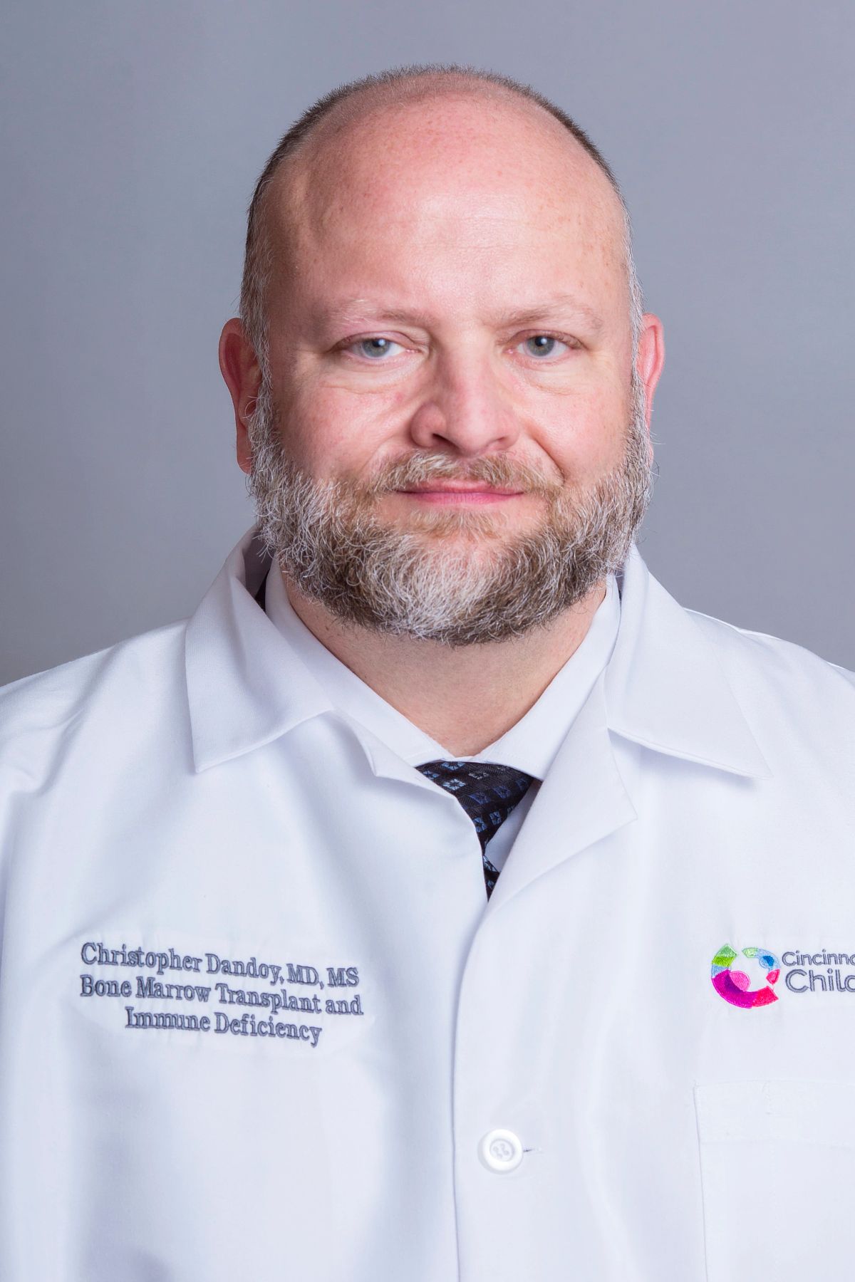 Christopher E. Dandoy合作论文数Cincinnati Children's Hospital Medical Center;University of Cincinnati Cancer Center;University of Cincinnati College of Medicine10Joyce Villanueva合作论文数10Benjamin L. Laskin合作论文数Division of Nephrology and Hypertension, Cincinnati Children’s Hospital Medical Center10Jeffrey J Bednarski合作论文数10Susan E. Prockop合作论文数Bone Marrow Transplant Service, Memorial Sloan-Kettering Cancer Center10
 Deepak Chellapandian合作论文数Blood & Marrow Transplant Program, Johns Hopkins All Childrens Hosp9




